The Heart lec 2 (pt 1)

0.0(0)
Studied by 0 people
call kaiCall Kai
Locked
learnLearn
examPractice Test
spaced repetitionSpaced Repetition
heart puzzleMatch
flashcardsFlashcards
GameKnowt Play
Card Sorting

1/64

encourage image

There's no tags or description

Looks like no tags are added yet.

Last updated 4:55 AM on 9/18/26
Name
Mastery
Learn
Test
Matching
Spaced
Call with Kai
Chat

No analytics yet

Send a link to your students to track their progress

65 Terms

1
New cards
Where is the heart located and what is its approximate size and weight?

-Located in the thoracic cavity near the anterior chest wall; posterior to the sternum

.

-Base of heart lies at the 3rd costal cartilage; posterior to sternum

.

-Apex of heart reaches the 5th intercostal space


2
New cards

Heart location relative to mediastinum

Anterior portion of mediastinum

3
New cards
How is the heart positioned within the thorax?

The Centre of the base is slightly left of the midline

,

and the heart is rotated toward the left with the right atrium and right ventricle dominating the anterior view

.

A line from the base to the apex points further to the left.

4
New cards
What are the three layers of the heart wall?

The three layers are the epicardium ,myocardium and endocardium.
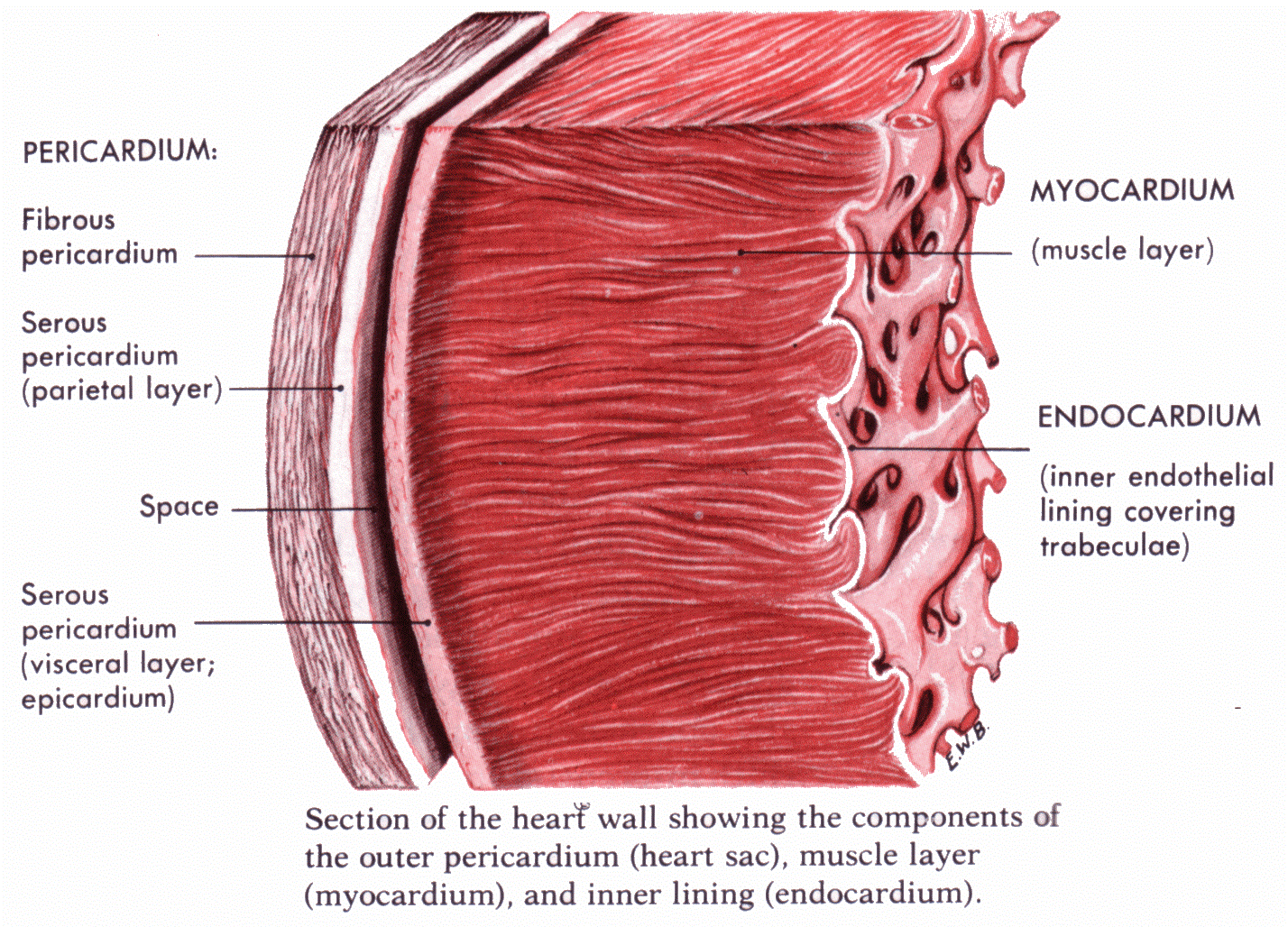
<p>The three layers are the epicardium ,myocardium and endocardium.</p>
5
New cards
What is the pericardium and what are its two main layers?

-The pericardium is a serous membrane associated with the pericardial cavity

.

- Divided into:

epicardium (inner visceral layer) and pericardium (outer parietal layer)
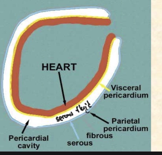
<p>-The pericardium is a serous membrane associated with the pericardial cavity</p><p>.</p><p>- Divided into:</p><p>epicardium (inner visceral layer) and pericardium (outer parietal layer)</p>
6
New cards

Fibrous pericardium

Contains dense network of collagen fibers that stabilize the position of the heart and associated vessels within the mediastinum

<p>Contains dense network of collagen fibers that stabilize the position of the heart and associated vessels within the mediastinum </p>
7
New cards

Fluid filled space between two serous layers

Pericardial cavity containing 15-50ml of pericardial fluid

8
New cards

Function of pericardial fluid

Acts as a lubricant reducing friction between the opposing visceral and parietal surfaces as the heart beats

9
New cards

What is the epicardium and what is its structure and composition?

- outer layer of the heart wall covering the heart

.

-Visceral layer of serous pericardium (epicardium)

.

- It consists of exposed mesothelium and underlying layer of loose areolar connective tissue attached to the myocardium

10
New cards

Parietal layer of serous pericardium composition

outer dense fibrous layer

.

Areolar layer

.

inner mesothelium

11
New cards

What is the myocardium and what is its function and composition ?

- The thick muscular layer of the heart composed mainly of cardiac muscle with blood vessels and nerve.

.

-Forms the atria and ventricles

.

-Has concentric layers of cardiac muscle tissue for efficient pumping of blood


<p>- The thick muscular layer of the heart composed mainly of cardiac muscle with blood vessels and nerve.</p><p>.</p><p>-Forms the atria and ventricles </p><p>.</p><p>-Has concentric layers of cardiac muscle tissue for efficient pumping of blood</p><p></p>
12
New cards
What is the endocardium and where is it found?

-The endocardium is the inner lining of the heart chambers and valves

.

-It consists of simple squamous epithelium with underlying areolar connective tissue.

<p>-The endocardium is the inner lining of the heart chambers and valves</p><p>. </p><p>-It consists of simple squamous epithelium with underlying areolar connective tissue.</p>
13
New cards
What are the major external features seen on the surface of the heart?

-Two ventricles

.

- Two atria with auricles extending from the atria

.

-The coronary sulcus(horizontal) separates the atria from the ventricles

.

- Anterior and posterior interventricular sulci separate the ventricles.

14
New cards

Sulci

grooves on the surface of the heart dividing it into 4 chambers

<p>grooves on the surface of the heart dividing it into 4 chambers </p>
15
New cards

Auricle of heart

Formed when atria deflate when not filled with blood becoming a lumpy, wrinkled flap
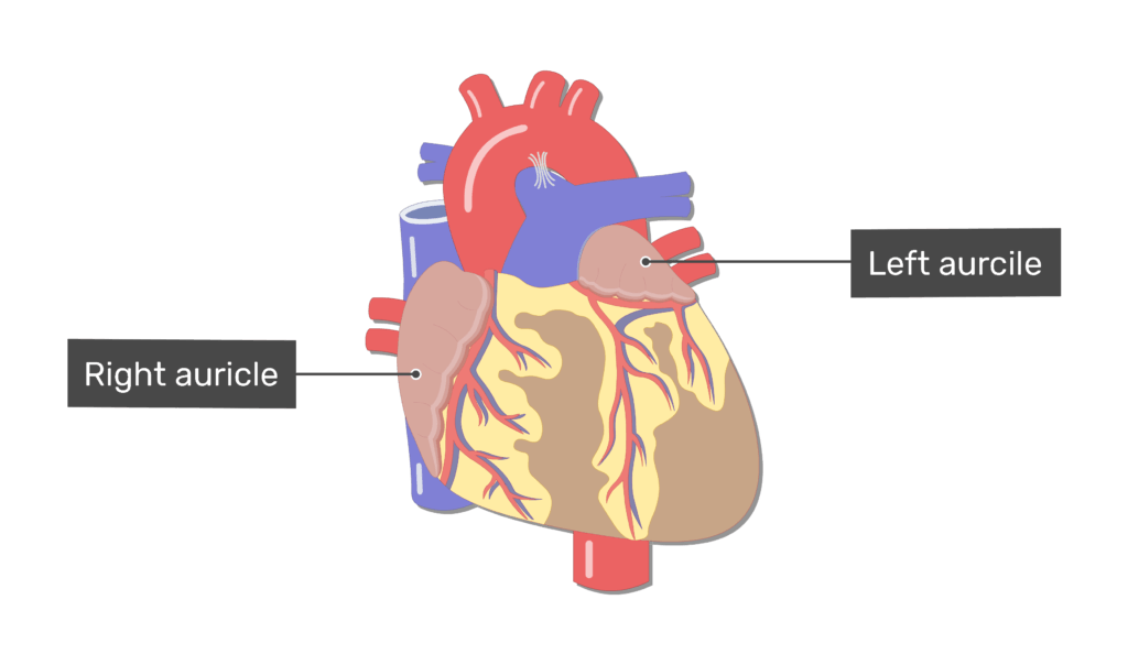
<p>Formed when atria deflate when not filled with blood becoming a lumpy, wrinkled flap </p>
16
New cards

What is the coronary sulcus, another name and explain.

-The coronary sulcus or atrioventricular groove

.

-transverse groove around the heart that represents dividing the atria and ventricles

<p>-The coronary sulcus or atrioventricular groove </p><p>.</p><p>-transverse groove around the heart that represents dividing the atria and ventricles</p>
17
New cards
What are the anterior and posterior interventricular sulci?

They are vertical grooves or shallow depressions on the heart separating the right and left ventricles.

<p>They are vertical grooves or shallow depressions on the heart separating the right and left ventricles.</p>
18
New cards

What are the major great vessels associated with the heart

-Superior & Inferior Vena Cavae

.

-Pulmonary Trunk

.

-Pulmonary Veins

.

-Aorta


The superior and inferior venae cavae return deoxygenated blood to the right atrium; the pulmonary trunk carries blood from the right ventricle to the pulmonary arteries; pulmonary veins return oxygenated blood to the left atrium; and the aorta carries oxygenated blood from the left ventricle to the body.
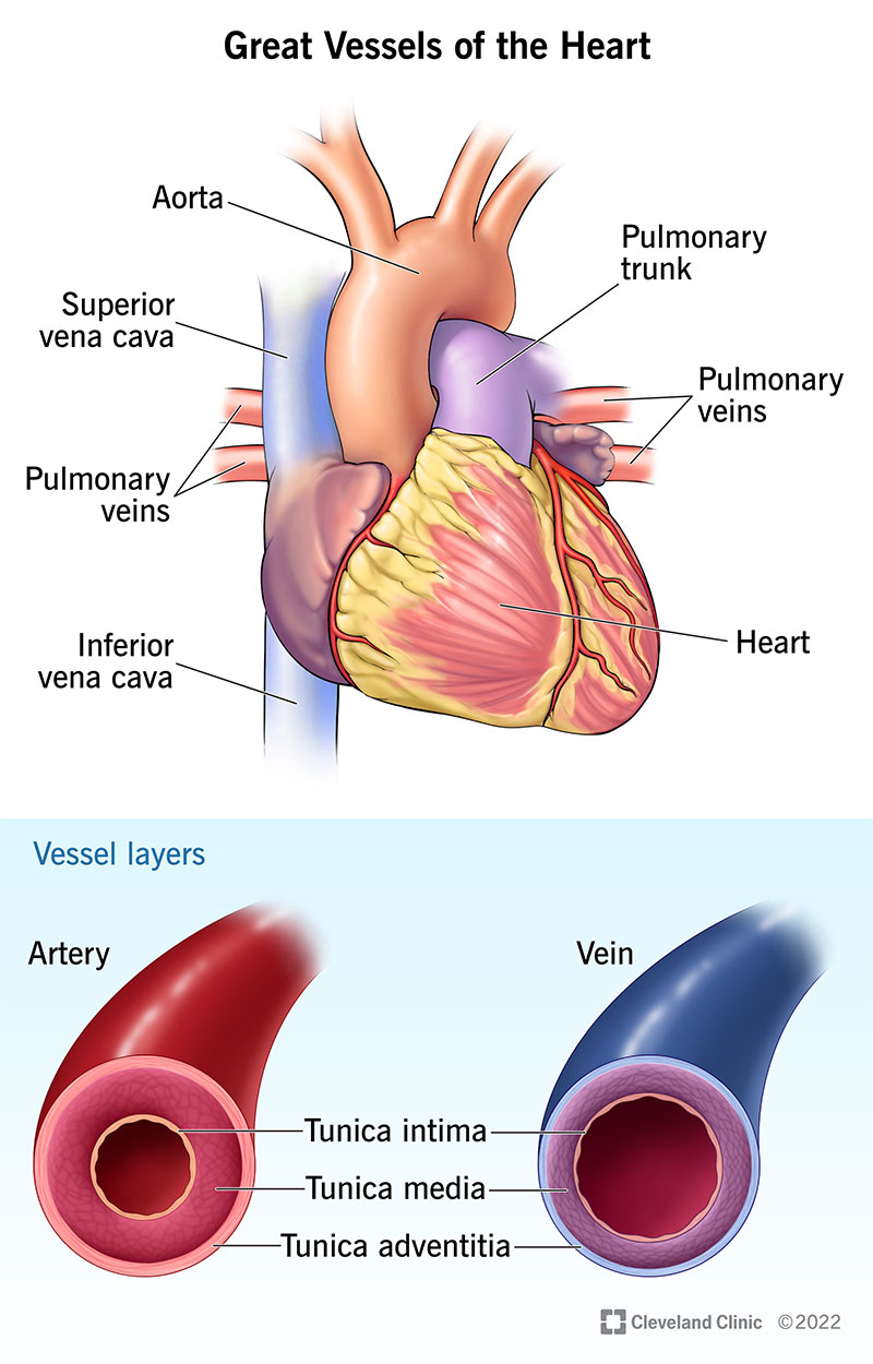
<p>-Superior &amp; Inferior Vena Cavae</p><p>.</p><p>-Pulmonary Trunk</p><p>.</p><p>-Pulmonary Veins</p><p>.</p><p>-Aorta</p><p></p><p></p><p>The superior and inferior venae cavae return deoxygenated blood to the right atrium; the pulmonary trunk carries blood from the right ventricle to the pulmonary arteries; pulmonary veins return oxygenated blood to the left atrium; and the aorta carries oxygenated blood from the left ventricle to the body.</p>
19
New cards

Superior & Inferior Vena Cavae function

-Superior vena cavae: brings deoxygenated blood from head, neck, upper limbs and chest to the atrium

.

-Inferior vena cavae: carries deoxygenated blood form lower limbs and from the right atrium to the rest of the trunk


20
New cards

Pulmonary Trunk

eaves right ventricle bifurcates (pulmonary arteries) and goes to lungs

21
New cards

Pulmonary Veins

return oxygenated blood to the left atrium

22
New cards
What are the three major parts of the aorta?

-Ascending aorta

.

-aortic arch

.

- descending aorta.

23
New cards
What is the function of the ductus arteriosus during fetal circulation?
The ductus arteriosus is a fetal opening between the pulmonary trunk and proximal descending aorta that allows blood to bypass the fluid-filled nonfunctioning fetal lungs.
24
New cards
What happens to the ductus arteriosus after birth?
It normally closes after birth and becomes the ligamentum arteriosum.
25
New cards
What is patent ductus arteriosus?
Patent ductus arteriosus occurs when the ductus arteriosus fails to close after birth. The lecture associates it with pulmonary hypertension and potentially congestive heart failure and arrhythmias.
26
New cards
What are the two major septa of the heart?
The interatrial septum separates the right and left atria while the interventricular septum separates the right and left ventricles.
27
New cards
What is the foramen ovale and what happens to it after birth?
The foramen ovale is a fetal opening in the interatrial septum that allows blood to pass between the atria. After birth a flap closes the opening leaving the fossa ovalis.
28
New cards
What is patent foramen ovale?
Patent foramen ovale occurs when the fetal foramen ovale fails to close completely after birth.
29
New cards
What is the function of the right atrium and what blood vessels enter it?
The right atrium receives deoxygenated blood and pumps it into the right ventricle. It receives blood from the superior vena cava inferior vena cava and coronary sinus.
30
New cards
Where is the SA node located and what is its significance?
The sinoatrial SA node is located in the right atrium and is involved in initiating the heart's electrical activity.
31
New cards
What is the function of the left atrium?
The left atrium receives oxygenated blood from the pulmonary veins and pumps it into the left ventricle.
32
New cards
Why are the atrial walls thinner than the ventricular walls?
The atria only need to pump blood a short distance into the ventricles so they require less muscular force than the ventricles.
33
New cards
What are auricles and what is their function?
Auricles are ear-like extensions of the atria that increase the internal volume of the atria.
34
New cards
What are pectinate muscles and what is their function?
Pectinate muscles are ridges of muscle in the atria. Their wave-like fibres allow stronger atrial contraction while requiring relatively little muscle mass.
35
New cards
What is the function of the right ventricle?
The right ventricle receives deoxygenated blood from the right atrium and pumps it into the pulmonary trunk toward the lungs.
36
New cards
What is the function of the left ventricle?
The left ventricle receives oxygenated blood from the left atrium and pumps it into the aorta for distribution to the body.
37
New cards
Why is the wall of the left ventricle thicker than the wall of the right ventricle?
The left ventricle must generate higher pressure to pump blood throughout the systemic circulation whereas the right ventricle only pumps blood through the pulmonary circulation.
38
New cards
What are trabeculae carneae and what is their function?
Trabeculae carneae are irregular muscular ridges inside the ventricles that help prevent the ventricular walls from sticking together or being suctioned inward during contraction.
39
New cards
What is the moderator band?
The moderator band is a muscular structure in the right ventricle that transmits impulses from the right bundle branch of the conducting system.
40
New cards
What is the main function of the heart valves?
Heart valves ensure one-way blood flow by preventing blood from flowing backward.
41
New cards
What are atrioventricular AV valves and what do they prevent?
AV valves lie between the atria and ventricles and prevent blood from flowing back into the atria when the ventricles contract.
42
New cards
What are the two AV valves?
The tricuspid valve is between the right atrium and right ventricle while the bicuspid mitral valve is between the left atrium and left ventricle.
43
New cards
What is the difference between the tricuspid and bicuspid valves?
The tricuspid valve has three leaflets and is located between the right atrium and right ventricle. The bicuspid/mitral valve has two leaflets and is located between the left atrium and left ventricle.
44
New cards
What are chordae tendineae and papillary muscles and how do they support the AV valves?
Chordae tendineae attach the ends of the AV valve leaflets to papillary muscles. Together they help prevent the valve leaflets from being forced backward into the atria during ventricular contraction.
45
New cards
What are semilunar valves and what do they prevent?
Semilunar valves prevent blood from flowing backward from the great arteries into the ventricles after ventricular contraction.
46
New cards
What are the two semilunar valves and where are they located?
The pulmonary semilunar valve lies between the right ventricle and pulmonary trunk while the aortic semilunar valve lies between the left ventricle and ascending aorta. Each has three cusps.
47
New cards
What causes the first and second heart sounds?
The first heart sound "lub" is produced by closure of the AV valves. The second heart sound "dup" is produced by closure of the semilunar valves.
48
New cards
What is the pulmonary circuit?
The pulmonary circuit carries deoxygenated blood from the right side of the heart to the lungs for oxygenation and returns oxygenated blood to the heart.
49
New cards
What is the systemic circuit?
The systemic circuit carries oxygenated blood from the left side of the heart to the body and returns deoxygenated blood from the body to the heart.
50
New cards
What is coronary circulation?
Coronary circulation is the circulation that supplies the heart muscle with blood and removes metabolic wastes.
51
New cards
Where do the right and left coronary arteries originate?
Both coronary arteries originate at the base of the ascending aorta at the aortic sinuses.
52
New cards
When does most coronary blood flow occur and why?
Coronary blood flow occurs mainly between heart contractions because the elastic rebound of the arteries allows blood to flow through the coronary vessels during ventricular relaxation.
53
New cards
What areas does the right coronary artery supply?
The right coronary artery supplies the right atrium portions of both ventricles and parts of the cardiac conducting system including the SA and AV nodes.
54
New cards
What are the major branches of the right coronary artery and what do they supply?
The marginal arteries extend over the surface of the right ventricle while the posterior interventricular artery runs in the posterior interventricular sulcus and supplies the interventricular septum and adjacent portions of the ventricles.
55
New cards
What are the major branches of the left coronary artery?
The left coronary artery gives rise to the circumflex artery and anterior interventricular artery.
56
New cards
What areas does the left coronary artery supply?
The left coronary artery supplies the left ventricle left atrium and interventricular septum.
57
New cards
What is the pathway and function of the anterior interventricular artery?
The anterior interventricular artery runs in the anterior interventricular sulcus and supplies the interventricular septum and adjacent ventricular regions. It forms anastomoses with the posterior interventricular artery.
58
New cards
What is the function of the circumflex artery?
The circumflex artery supplies regions of the left side of the heart and extends around the heart toward the posterior surface where it can meet or fuse with small branches of the right coronary artery.
59
New cards
What is the coronary sinus and where does it drain?
The coronary sinus collects blood from several cardiac veins and drains into the right atrium near the base of the inferior vena cava.
60
New cards
Which cardiac vein drains the area supplied by the anterior interventricular artery?
The great cardiac vein drains the area supplied by the anterior interventricular artery and empties into the coronary sinus.
61
New cards
Which cardiac vein drains the anterior right ventricle?
The anterior cardiac vein drains the anterior right ventricle directly into the right atrium.
62
New cards
Which cardiac vein drains the area supplied by the circumflex artery?
The posterior cardiac vein drains the area served by the circumflex artery and empties into the great cardiac vein.
63
New cards
Which cardiac vein drains the area supplied by the posterior interventricular artery?
The middle cardiac vein drains the area supplied by the posterior interventricular artery and empties into the coronary sinus.
64
New cards
Which cardiac vein drains the posterior right atrium and right ventricle?
The small cardiac vein drains the posterior right atrium and right ventricle and empties into the coronary sinus.
65
New cards
What is anastomosis in coronary circulation?
Anastomosis is a connection between blood vessels that provides an alternate route for blood flow. In coronary circulation branches of different arteries can form connections with one another.



