Option A: Neurobiology
1/197
Earn XP
Description and Tags
Name | Mastery | Learn | Test | Matching | Spaced | Call with Kai |
|---|
No analytics yet
Send a link to your students to track their progress
198 Terms
What is embryogenesis?
The formation of a fully-formed organism from a fertilised egg
What are the initial germ layers from which all tissues are developed?
The ectoderm, mesoderm, and endoderm
What process forms the germ layers?
Gastrulation
During gastrulation, what will develop for chordates?
A flexible notochord, eventually forming the neural tube
What process forms the neural tube?
Neurulation
What are the steps to neurulation?
Notochord forms from mesoderm cells soon after gastrulation is complete
Signals from notochord cause inward folding of ectoderm at the neural plate
Ends of the neural plate fuse and disconnect to form an autonomous neural tube
The neural tube elongates as the embryo develops, forming the central nervous system
The cells of the neural crest will differentiate to form the components of the peripheral nervous system
Why are Xenopus often used to model neurulation?
They possess robust embryos that can tolerate extensive manipulation
The developmental stages of embryogenesis can be easily investigated
What are the main structures visible during neurulation?
Three germ layers
Hollow cavity called the archenteron (will become digestive tract)
Notochord (flexible rod that stimulates neurulation)
Neural tube (developed from the infolding of the neural plate)
Why can spina bifida occur?
Closure of the neural tube is not simultaneous - the area where the brain forms is advanced over the caudal region, where closure occurs more slowly
What is spina bifida?
A birth defect resulting in the incomplete closure of the neural tube and incomplete fusing of the vertebrae, leaving the spinal cord nerves exposed and prone to damage
Where is spina bifida most common?
The lumbar and sacral areas, as these are regions where closure is slowest
What are the two types of spina bifida?
Occulta and cystica
What are the two types of spina bifida cystica?
Meningocele and myelomeningocele
What is spina bifida occulta?
This is when splits in the vertebrae are so small that the spinal cord does not protrude (mild)
What is spina bifida meningocele?
This is a case of spina bifida where a meningeal cyst forms (more severe)
What is spina bifida myelomeningocele?
This is a case of spina bifida where a meningeal cyst forms including spinal elements
What are the effects of spina bifida?
Degrees of paralysis, bowel dysfunction, and bladder dysfunction
What factors influence spina bifida?
Genetic and environmental
What diet aspect in pregnancy can cause spina bifida?
Not enough folate
What aspect of the neural tube allows for differentiation of nerve cells?
The multipotent neuronal stem cells that can differentiate
What are the two main nerve cells that come from the neural tube?
Neurons → nerve cells that conduct messages (sensory, motor, or relay)
Glial cells → provide physical and nutritional support for neurons (make up 90% of brain’s nerve cells)
How are neurons produced?
By progenitor neuroblasts via a process called neurogenesis
What are the results of neurogenesis
Neurons that (mostly) survive for the lifetime of the individual and do not proliferate following embyogenesis (they are post-mitotic), however certain brain regions may be capable of adult neurogenesis
Why do immature neurons migrate?
To adopt precise final positions that allow for the formation of neural circuitries, which is critical for the development of brain and spinal architecture
What are the two processes that cause neural migration?
Glial guidance and somal translocation
What is glial guidance?
When glial cells provide a scaffolding network along which an immature neuron can be directed to its final location
What is somal translocation?
When the neuron forms and extension at the cell’s perimeter and then translocates its soma along this length
What is the structure of an immature neuron?
A cell body (soma) with a nucleus and cytoplasm
What happens to the structure of an immature neuron as it matures?
Axons and dendrites grow in response to chemical signals from surrounding cells
In which parts of the nervous system are the axons short and long?
Shorter axons are typically within the central nervous system while longer ones stretch to the peripheral nervous system
How do axons grow?
They have a growth cone at their tip containing growth filaments called filipodia
How does filipodia achieve axon growth?
Extension of filipodia causes expansion of the internal cytoskeleton within the growth cone, causing growth
How is the direction of axon expansion determined?
Chemical stimuli from surrounding cells:
Chemoattractant (grow towards)
Chemorepellant (grow away)
What is a synapse?
A junction at which a neuron transmits a signal to another cell (relay neuron or effector), usually through chemical signals (sometimes electrical signals)
How is a vast array of communication pathways developed?
Through a developing neuron forming multiple synapses
Where can neurons form synapses in the CNS?
With other axons, dendrites, or soma
Where can neurons form synapses in the PNS?
A muscle fibre (neuromuscular) or gland (neuroglandular)
Why do embryos and babies have more synapses than adults?
Because neurons will form multiple synapses to maximise available connections
As organism matures, some synapses are strengthened through frequent sue
Others are not used and the connections are weakened and do not persist
This concept is central to how organisms learn
What is neural pruning?
The loss of unused neurons by removing excess axons and eliminating their synaptic connections
What is the purpose of neural pruning?
To reinforce complex wiring patterns associated with learned behaviour
What influences neural pruning?
Environmental factors mediated by the release of chemical signals from glial cells
What is neuroplasticity?
The capacity for the nervous system to change and rewire its synaptic connections
What is the purpose of neuroplasticity?
To allow individuals to reinforce certain connections (learning) or circumvent damaged regions
What are the two primary mechanisms of neuroplasticity?
Rerouting and sprouting?W
What is rerouting?
Creating an existing nervous connection through and alternative neural pathway
What is sprouting?
The growth of a new axon or dendrite fibres to enable new neural connections to be formed
What is a stroke?
A sudden death of brain cells in a localised area due to inadequate blood flow, leading to improper functioning of the brain due to the loss of neural connections in the affected area
What are the two main types of stroke?
Ischemic - clot within the blood restricting oxygenation to an associated region of the brain
Hemorrhagic - ruptured blood vessel causing bleeding within a section of the brain
When may a stroke be temporary?
If the brain is able to reorganise its neural architecture to restore function, allowing healthy areas of the brain to adopt the functionality of damaged regions
What components of the nervous system does the neural tube become?
Anterior part → forms the brain during cephalisation (development of the brain)
Remainder → develops into the spinal cord
Neural crest → forms the peripheral nervous system
What are the three primary structures of the embryonic brain?
The forebrain, midbrain, and hindbrain
What is the function of the human brain?
The brain acts as an integration and coordination system for the control of body systems, processing sensory information and relaying motor responses
What are the major external structures of the brain?
The cerebral hemisphere, the cerebellum, and the brainstem
What are the major internal structures of the brain?
The hypothalamus, pituitary gland, and corpus callosum
What are the four lobes of the cerebral cortex?
Frontal lobe - controls motor activity and dopamine-related tasks
Parietal lobe - touch sensation and spatial navigation
Temporal lobe - auditory processing and language comprehension
Occipital lobe → visual processing and sight perception
What is the function of the cerebellum?
Coordination of unconscious motor functions including balance, movement, and coordination
What is the function of the brainstem?
Control of automatic/involuntary activites (breathing, swallowing, heart rate, etc.)
What are the three structures of the brainstem?
The ponds, medulla oblongata, and the midbrain
What is the function of the hypothalamus?
To maintain homeostasis through coordination of the nervous and endocrine systems, also producing some hormones secreted through the posterior pituitary
What are the two lobes of the pituitary gland?
The anterior lobe, the adenohypophysis, and the posterior lobe, the neurohypophysis
What is the function of the adenohypophysis?
Secretion of hormones such as FSH, LH, growth hormone, and prolactin
What is the function of the neurohypophysis?
Secretion of hormones such as ADH and oxytocin
What is the corpus callosum?
A bundle of nerve fibres connecting the two cerebral hemispheres
How can animal models indicate brain function?
Function can be identified by stimulating regions with electrodes or removing via lobotomy
How can lesions indicate brain function?
Lesions can be seen though post-mortem analysis or scans
Loss of function paired with knowledge of the location of the lesion can indicate the proper function of the damaged area
What is an autopsy?
A post-mortem examination of a corpse via dissection in order to evaluate causes of death
What is an fMRI?
A functional magnetic resonance image, recording changes in blood flow within the brain to identify activated areas that is non-invasive
What does the visual cortex do?
Receives neural impulses from light-sensitive cells in the eyes
What is Broca’s area responsible for?
Speech production: if it is damaged, meaningful speech cannot be produced
What is the nucleus accumbens?
The pleasure reward pathway, secreting neurotransmitters responsible for pleasure such as dopamine and satiety like serotonin, as well as communicating with other mechanisms of pleasure
How does the cerebral cortex of humans compare to that of other animals?
It is larger and much more highly developed, responsible for our capacity for more cognitive though
How has the surface area of the human brain increased while still fitting within the cranium?
The folding, gyrification, of the cerebral cortex to form wrinkled peaks, gyrus, and troughs, sulcus, increasing the SA:V ratio
What can predict the cognitive capacity of an organism?
The extent of gyrification in the cerebral cortex
What is the function of the left cerebral hemisphere?
Processing tactile information from the right side of the body in the spinal cord or brain stem
Processing visual information from the right side of each eye in the optic chiasma
Muscular contractions for the right side from the motor cortex
This is called contralateral processing (as opposed to ipsilateral processing)
What is the function of the right cerebral hemisphere?
Processing tactile information from the left side of the body in the spinal cord or brain stem
Processing visual information from the left side of each eye in the optic chiasma
Muscular contractions for the left side from the motor cortex
This is called contralateral processing (as opposed to ipsilateral processing)
How can the nervous system be divided?
Nervous system
Central nervous system
Peripheral nervous system
Sensory (afferent) pathway
Motor (efferent) pathway
Voluntary (somatic)
Involuntary (autonomic)
What is the function of the autonomic nervous system?
To control involuntary processes in the body using centres in the brainstem
What are the sympathetic nerves used for?
The release of norepinephrine to mobilise body systems
What are the parasympathetic nerves used for?
The release of acetylcholine to relax body systems and conserve energy
What is the role of the medulla oblongata?
Coordination of involuntary activities such as swallowing, breathing, and heart rate
What happens during sympathetic responses?
Decrease of salivation and blood flow to the gut
Increased ventilation rate and dilation of airways due to reduction in blood pH (increased CO2)
Increased heart rate by increasing sinus rhythm of sinoatrial node
What happens during parasympathetic responses?
Increased salivary release and blood flow to the gut
Lowered ventilation and constriction of airways due to increased blood pH
Reduced heart rate (through vagus nerve) by lowering normal sinus rhythm of sinoatrial node
What is the pupil reflex?
An involuntary response from the brainstem due to the ANS, resizing the iris to minimize light and protect the retina
What is brain death?
The permanent absence of measurable activity in both the cerebrum and brainstem
How is brain death tested for?
The pupil reflect → won’t happen in those who are brain dead
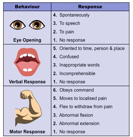
Glasgo Coma Scale → determines neurological health of someone with brain injury
What is the correlation between body size and brain size in different animals?
There is a positive correlation between body size and brain size that is roughly linear, but not directly proportional
How does the brain : body ratio occur in larger animals?
It decreases; body mass increases disproportionately to an increase in brain mass as most tasks only require fixed brain capacity
What is the correlation between brain size and intelligence?
None
What is encephalization?
The amount of brain mass relative to an animal’s body mass
What figure for mammals predicts potential cognition?
The encephalization quotient (EQ)
How much of the body’s energy does the brain consume?
~20%, despite only making up ~2% of the body’s mass
Why does the brain require so much energy?
Energy is needed to maintain a resting potential when neurons are not firing (sodium-potassium pumps use ATP)
Energy is used to synthesise large numbers of neurotransmitters to facilitate neuronal communication
What is sensitivity?
The ability of an organism to detect external and internal changes and respond accordingly
How do organisms achieve sensitivity?
Receptors detect changes in stimuli, generating nerve impulses to be relayed to the brain and effector organs
Different receptors recognise different stimuli
What sensory organ is responsible for sight perception?
The eye
What is the structure of the eye?
Two cavities separated by a lens (anterior = aqueous humour, posterior = vitreous humour)
Lens attached to ciliary muscles to change the focus of the lens
Pupil where light enters
Iris that constricts and dilates to change light entering the pupil
Exposed portion is coated by layer called cornea, lubricated by conjunctiva
Internal surface has three layers → outer = sclera, middle = choroid, and inner = retina
Retina’s fovea centralis is responsible for sharpest vision
Nerve signals are sent from the retina through the optic nerve
What are the components of the retina?
Pigment epithelium
Photoreceptors (rods and cones) - convert light stimuli into electrical nerve impulses
Bipolar cells - transmit photoreceptors’ nerve impulses to ganglion cells
Ganglion cells - fibres form the optic nerve tract
What sensory organ is responsible for sound perception?
The ear
What are the structures in the ear?
The pinna → external ear
The auditory canal → channels sound waves
The tympanic membrane (eardrum)
Ossicles → transfer vibrations to oval window
Oval window → transmits signals to cochlea
Cochlea → converts sound stimuli into electrical nerve impulses
Semicircular canals → detect movement and balance
Round window → dissipates vibrations
Auditory nerve → transmits signals to brain
What is photoreception?
The mechanism of light detection (by the eyes) that leads to vision when interpreted by the brain