non-traumatic joint pain: upper extremity
 Call Kai
 Learn
Learn Practice Test
 Spaced Repetition
 Match
Match Flashcards
 Knowt Play
1/73

There's no tags or description
Looks like no tags are added yet.
Name | Mastery | Learn | Test | Matching | Spaced | Call with Kai | Chat |
|---|
No analytics yet
Send a link to your students to track their progress
74 Terms
Repetitive strain → muscle imbalance, fascial restriction, impaired lymphatic/venous return.
how do overuse disorders contribute to somatic dysfunctions?
gentle techniques (indirect MFR, BLT, lymphatics), avoid high-force manipulation.
OMT focus of arthritic conditions (RA, OA)
facilitated segments model
Repeated irritation (somatic or visceral) at a spinal segment lowers the threshold for activation → persistent hyperexcitability → pain, altered reflexes, tissue texture changes.
T2-8
sympathetics of the upper extremities
chronic cuff tear
supraspinatus/infraspinatus muscle wasting may indicate
rotator cuff injury
DDx of shoulder ROM where active is more limtied than passive
adhesive capsulitis or arthritis
DDX if both active and passive shoulder ROM are limited
neer, hawkins-kennedy
special tests for subacromial impingement
neer test
impingment test of subscalpular n/supraspinatus
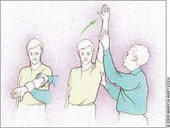
hawkins-kennedy test
compression of the supraspinatus tendon against the coracoacromial ligament to assess the possibility of impingement of the subacromial bursa
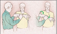
drop arm test, empty can (Jobe's) test, external rotation lag
tests for rotator cuff tear
drop arm test
identifies tear and/or full rupture of rotator cuff
Empty Can Test/ Supraspinatus Test
Purpose: Assess supraspinatus involvement with shoulder injury.
Method: Pt. raises arms in scaption to shoulder height with thumps up. Pt rotates thumbs down ( "empty can" ). PT applies resistance.
Positive Test: Pain or weakness
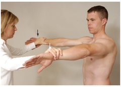
apprehension test, relocation test, O'Brien's
tests for labral injury/instability
apprehension test
movement of a joint while observing the patient for signs of pain and increased joint laxity, to assess joint stability
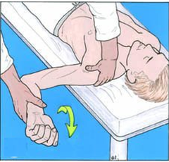
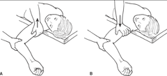
Relocation Test
tests for anterior glenohumeral instability
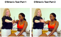
O'Brien Test
tests for SLAP lesion
speed's, yergason's
test for biceps tendon
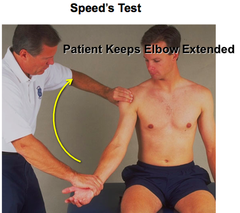
speed's test
stretching or lengthening of the biceps tendon to assess the possibility of tenosynovitis
Yergason's Test
pt in sitting with shoulder in neutral/stabilized against trunk, elbow flexed 90 deg and forearm pronated. PT places one hand on patient's forearm and other over bicipital groove. pt instructed to actively supinate and laterally rotate against resistance. (+) tendon of biceps long head will "pop out" of groove, (+) pain or tenderness in bicipital groove. Identifies bicipital tendonitis and integrity of transverse ligament.
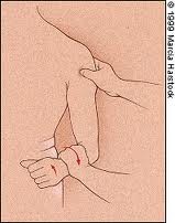
cross arm adduction
test for AC joint
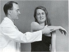
anterior dislocation
most common type of shoulder dislocation
anterior shoulder dislocation
Arm held abducted and externally rotated.
Loss of normal rounded deltoid contour ("squared-off" appearance).
Humeral head palpable anteriorly.
Apprehension test positive (patient resists abduction/external rotation).
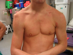
anterior: abducted and externally rotated
posterior: adducted and internally rotated
how is the arm held in anterior vs posterior dislocation
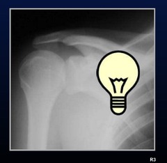
light bulb sign on AP view
XRay sign of posterior dislocation
inferior dislocation
what type of dislocation?
Arm locked in abduction, humeral head inferiorly displaced.
rotator cuff tendonitis
Pain with overhead activity, often gradual onset; worse at night lying on shoulder
rotator cuff tendonitis
Painful arc of abduction (70-120°), pain with resisted cuff testing (empty can, external rotation), strength usually intact
Compression of supraspinatus tendon & bursa under acromion; insidious pain with overhead motion
what causes impingement syndrome?
impingement syndrome
Positive Neer and Hawkins-Kennedy tests; painful arc; no true weakness unless tear develops
subacromial area
name area of palpation
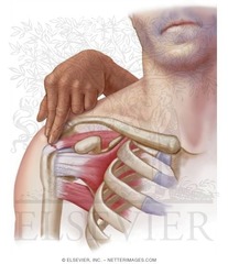
subacromial bursitis
-Acute or subacute pain after overuse or trauma; diffuse lateral shoulder pain
-Pain with passive ROM and palpation of subacromial area; often pain throughout motion, not just arc; weakness due to pain inhibition, not tendon tear
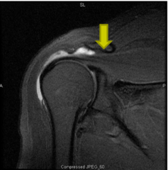
subacromial bursitis
diffuse tenderness, pain with passive + active ROM, weakness from pain not structural tear.
spencer technique
A 7-step articulatory technique for glenohumeral joint dysfunction; can be applied in arthritis, adhesive capsulitis, or restricted ROM.
lateral recumbent, physician behind
position of patient for spencer technique
1. Extension - move humerus posteriorly.
2. Flexion - move humerus anteriorly.
3. Circumduction with compression - small circles while pressing humerus into joint.
4. Circumduction with traction - small circles while distracting humerus.
5. Abduction & adduction - move arm away from and toward body.
6. Internal rotation - elbow flexed, hand behind back, move further posterior.
7. Pump (lymphatic technique) - apply gentle springing pressure to humeral head.
what are the steps of spencer technique?
Every Fancy Cat Takes An Indoor Dump
Extension: Move the arm into extension while stabilizing the shoulder girdle.
Flexion: Move the arm into flexion while stabilizing the shoulder girdle.
Circumduction with Compression: Apply gentle compression through the elbow while moving the arm in small, rhythmic circumduction circles.
Circumduction with Traction: Apply traction to the arm and perform small circumduction circles.
Adduction with External Rotation: Guide the patient's hand to their opposite shoulder, inducing adduction and external rotation.
Abduction: Move the arm into abduction while stabilizing the shoulder girdle.
Internal Rotation: Have the patient place their hand behind their back and bring the arm into internal rotation.
what is the mnemonic for the steps of spencer's technique?
spencer technique
this technique helps improve range of motion, especially in pts with adhesive capsulitis
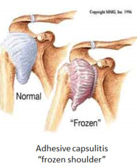
adhesive capsulitis
extreme stiffness of the shoulder joint (a.k.a. frozen shoulder)
Lateral Epidondylitis (tennis elbow)
elbow pain with resisted wrist extension indicates
medial epicondylitis
elbow pain with resisted wrist flexion indicates
radial tunnel syndrome
elbow pain with resisted supination indicates
UCL injury MOI
elbow valgus stress test (medial elbow instability) indicates
Lateral Epidondylitis
elbow pain caused by overuse of wrist extensors
medial epicondylitis
elbow pain due to overuse of wrist flexors/pronators
radial head fracture
post-trauma, pain with pronation/supination.
cubital tunnel syndrome
numbness/tingling 4th-5th digits, worsens with elbow flexion.
extensor carpi radialis brevis
most likely muscle involved with lateral epidondylitis
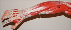
radial head often restricted in posterior motion
-prefers supination, anterior radial head
osteopathic findings (radial head) typical of lateral epicondylitis
common flexor tendon, pronator teres, flexor carpi radialis
tendons/muscles associated with medial epidondylitis
radial head often restricted in anterior motion (prefers pronation, posterior radial head)
radial head findings associated with medial epicondylitis
posterior radial head
from FOOSH (fall on outstretched hand) → difficulty with supination.
anterior radial head
from fall backward on extended arm → difficulty with pronation.
Physician pronates and flexes wrist to barrier.
Patient gently tries to extend/supinate against resistance (isometric).
Hold 3-5 seconds, relax, repeat → new barrier.
muscle energy for lateral epicondylitis
Patient supine/seated.
Physician supinates and extends wrist to barrier.
Patient gently tries to flex/pronate against resistance.
Hold 3-5 seconds, relax, repeat → new barrier.
muscle energy for medial epicondylitis
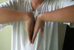
Phalen's, Tinel's, carpal compression test.
special tests for carpal tunnel
Phalen's test
test for carpal tunnel syndrome
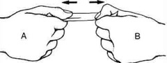
Froment's sign
test for cubital tunnel syndrome
radial nerve entrapment
wrist drop, sensory loss dorsum hand indicates this nerve injury
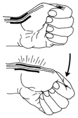
Finkelstein's test
special test for de Quervain's tenosynovitis
carpal tunnel syndrome
numbness/tingling in thumb, index, middle fingers; worse at night; thenar weakness/atrophy.
cervical radiculopathy (C6-8)
neck pain with radiation, dermatomal distribution.
Wrist splint in neutral, especially at night.
NSAIDs, corticosteroid injection if severe.
Ergonomic adjustments.
medical management of carpal tunnel syndrome
-MFR of transverse carpal ligament
-thenar/hypothenar release
-cervical/thoracic/somatic dysfunction
OMM techniques for carpal tunnel syndrome
Thumb spica splint, NSAIDs, corticosteroid injections if refractory.
Avoid repetitive thumb abduction/ulnar deviation.
medical management of de Quervain's tenosynovitis
abductor pollicis longus + extensor pollicis brevis tendons
de Quervain's tenosynovitis is inflammation of what tendons?
MFR of wrist
counterstrain tenderpoint near radial styloid
muscle energy
forearm prontation/supination
osteopathic treatment for dQT
near radial styloid → position thumb into flexion/adduction with slight ulnar deviation.
tenderpoint associated with dQ tenosynovitis
Place wrist into extension barrier.
Patient gently flexes wrist against resistance.
Hold 3-5 sec, relax, move to new barrier. Repeat.
muscle energy technique for carpal tunnel
Stabilize wrist in mild adduction.
Patient gently abducts thumb against resistance.
Repeat cycles of isometric contraction and relaxation.
muscle energy technique for de Quervain's
Rest: prevents further injury, allows initial healing.
Ice: vasoconstriction → reduces blood flow, inflammation, and pain. Apply 15-20 min every 2-3 hrs for first 48 hrs.
Compression: decreases swelling/edema; use elastic bandage or sleeve (ensure circulation intact).
Elevation: above heart level → promotes venous/lymphatic drainage, decreases edema.
role of RICE therapy
Diclofenac gel (Voltaren)
topical NSAID for localized pain
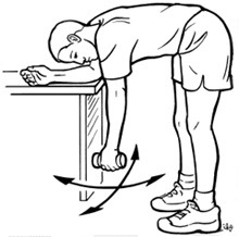
Pendulum swings (Codman's exercises).
Wall crawls (finger walk up wall).
Resistance band external/internal rotation.
shoulder home exercises for rotator cuff tendonitis, impingement
Codman's exercises
Finger tip wall walking exercises to restore shoulder movements