Animal cells ! final pt. 2
1/238
There's no tags or description
Looks like no tags are added yet.
Name | Mastery | Learn | Test | Matching | Spaced |
|---|
No study sessions yet.
239 Terms
unicellular organisms
single-celled eukaryotic organisms that can change shape and move by extending pseudopodia.
Amoebas
multicellular organisms
organisms composed of multiple cells that work together to perform various functions and maintain homeostasis.
humans, animals, plants, fungi, and some algae
Four terms correctly in different levels of biological organization
multicellular organism → organ systems → organs → tissues → cells → organelles → molecules/atoms
what are the organ systems?
digestive
circulatory
respiratory
immune and lymphatic
excretory
endocrine
reproductive
nervous
integumentary
skeletal
muscular
the four main categories of animal tissues?
epithelial
connective
muscle
nervous
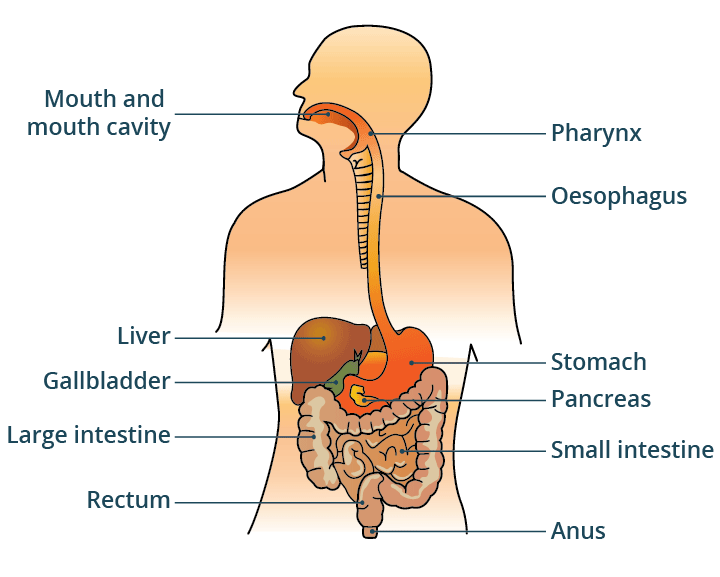
digestive (components/functions?)
components: mouth, pharynx, esophagus, stomach, intestines, liver, pancreas, anus
Functions: Food processing (ingestion, digestion, absorption, elimination)
circulatory (components/functions?)
components: heart, blood vessels, blood
Functions: internal distribution of materials
respiratory (components/functions?)
components: lungs, trachea, other breathing tubes
functions: gas exchange (uptake of oxygen; disposal of carbon dioxide)
immune and lymphatic (components/functions?)
components: bone marrow, lymph nodes, thymus, spleen, lymph vessels, white blood cells
functions: body defence (fighting infections and cancer)
excretory (components/functions?)
components: kindeys, ureters, unrinary bladder, urethra
skin: water + electrolytes lost through sweat glands
lungs: in charge of removing carbon dioxide
liver: breaks down toxic substances in blood + produces urea
large intestine (colon): removes solid waste + some water in feces
function: disposal of metabolic wastes; regulation of osmotic balance of blood
endocrine (components/functions?)
component: pituitary, thyroid, pancreas, adrenal, and other hormone-secreting glands
function: coordination of body activities; detection of stimuli and formulation of responses to them (such as digestion and metabolism)
reproductive (components/functions?)
components: ovaries or testes, and associated organs
function: protection against mechanical injury, infection, drying out; thermoregulation and support of developing embryos.
nervous (components/functions?)
components: brain, spinal cord, nerves, sensory organs
functions: body support, protection of internal organs, movement locomotion and other movement.
integumentary (components/functions?)
components: skin and its derivatives (hair, claws, and skin glands)
skeletal (components/functions?)
components: skeleton (bones, tendons, ligaments, cartilage)
muscular (components/functions?)
components: skeletal muscles
epithelial tissue
tightly packed cells that cover the outside of the body and lines the organs and cavities within body
tissue layers are:
simple ( one layer but can have any epithelial cell-geometry shape )
stratified ( multiple layers but can have any epithelial-cell geometry shape)
pseudostratified ( single layer of cells but looks like multiple layers so its THICK; nuclei are oriented a different way ) CANT HAVE GEOMETRY SHAPE LIKE EPITHELIAL CELLS

epithelial cells
the individual cells that make up epithelial tissue, providing protection, secretion, and absorption functions.
shapes may be
cuboidal ( like a dice )
columnar ( rectangular like bricks )
squamous ( flat like tiles )
vertebrates
animals with a backbone, including mammals, birds, reptiles, amphibians, and fish.
connective tissue
mainly binds and supports other tissues
contains cells that are loosely arranged in a liquid, jellylike solid matrix
5 major connective tissues…
Loose connective tissue
Adipose tissue
Fibrous (dense) connective tissue
Blood
Cartilage
BBLFC*
loose connective tissue
BBLFC*
type of connective tissue that binds epithelia to underlying tissues and holds organs in place (common type of tissue in vertebrates)
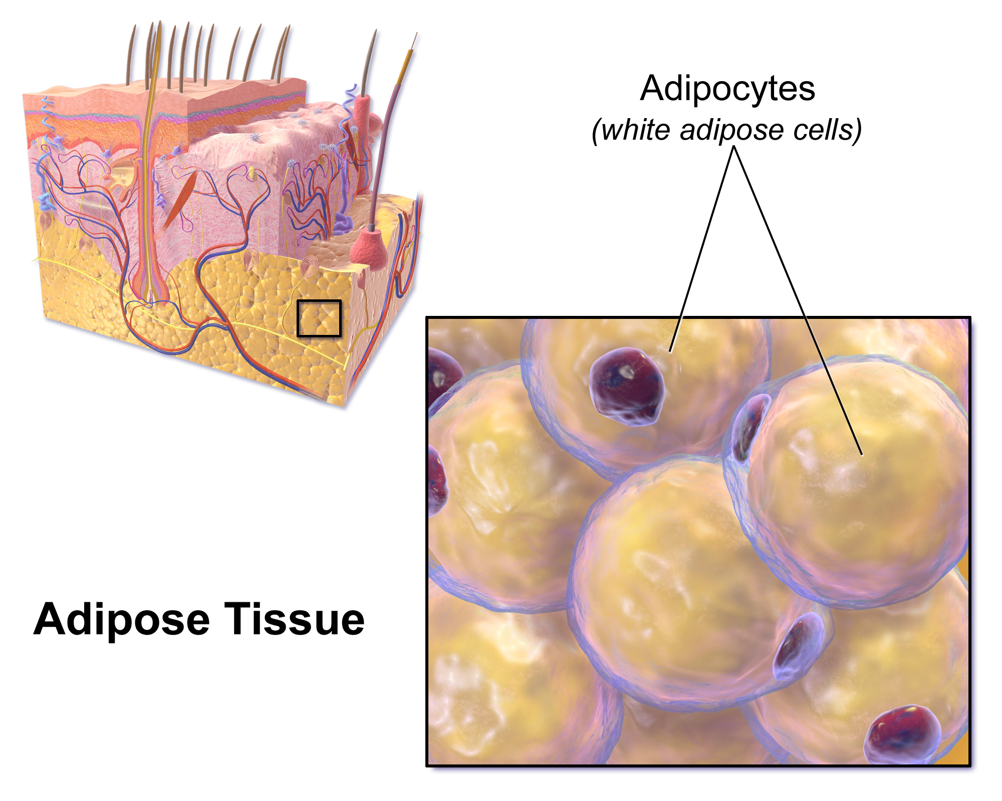
adipose tissue
BBLFAC*
type of connective tissue that stores fat for insulation and fuel
each adipose cell contains a large fat droplet that swells when fat is stored and shrinks when the body uses fat as fuel
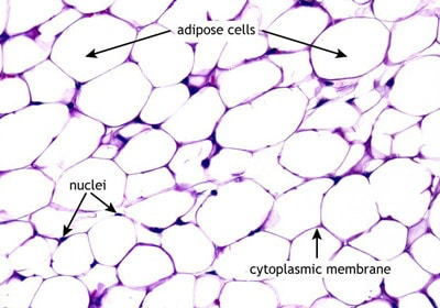
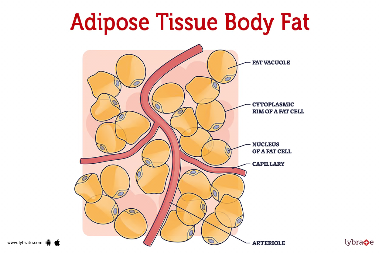
adipocyte composition
cells in adipose tissue that primarily consist of a large lipid droplet, cytoplasm, and a nucleus, enabling them to store fat efficiently.
"Adipose" refers to the tissue itself (body fat), while "adipocyte" refers to the specialized cells that make up that tissue (fat cells)
fibrous (or dense) connective tissue
BBLFC*
type of connective tissue found in tendons that attach muscles to bones and ligaments, which connect bones to joints.
contains large amounts of collagen fibres
Blood
BBLFC*
type of connective tissue composed of blood cells and cell fragments in blood plasma
matrix is a liquid called plasma, consists of water, salts, and a variety of dissolved proteins
suspended(spread throughout) in the plasma are erythrocytes (red blood cells), leukocytes (white blood cells) and cell fragments called thrombocytes (platelets).
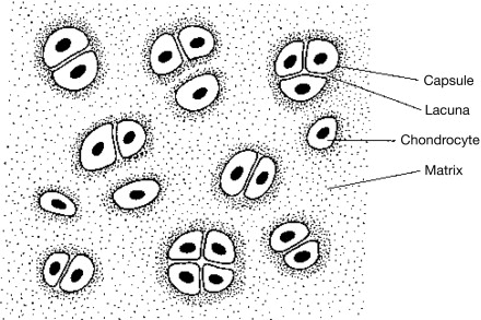
Cartilage
BBLFC*
a type of connective tissue that are strong and flexible support material (found in nose, ears, intervertebral discs, and part of ribcage)
abundance of collagenous fibres embedded in a rubbery matrix made of a substance called chondroitin sulfate. (protein-carbohydrate complex).
composed of chondrocytes

chondrocytes
secretes collagen and chondroitin sulfate and are the primary cells in cartilage.
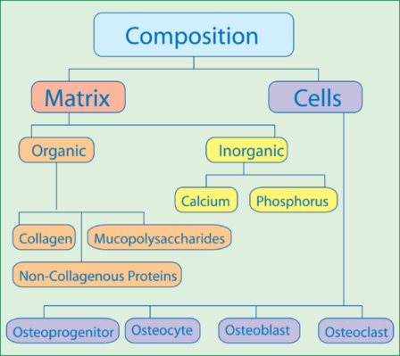
Bone
BBLFC*
a type of connective tissue that are mineralized and forms the skeleton
large amounts of two types of matrix material
organic matrix: similar to matrix material found in other connective tissues (some amount of collagen and elastic fibres) → provides strength and flexibility to the tissue
inorganic matrix: consists of mineral salts; mostly calcium salts, that give the tissue hardness
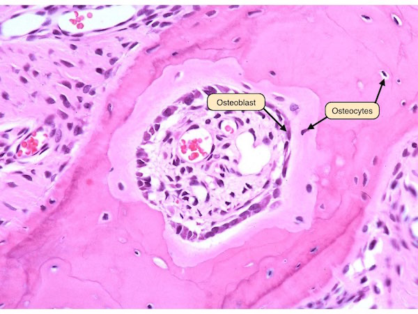
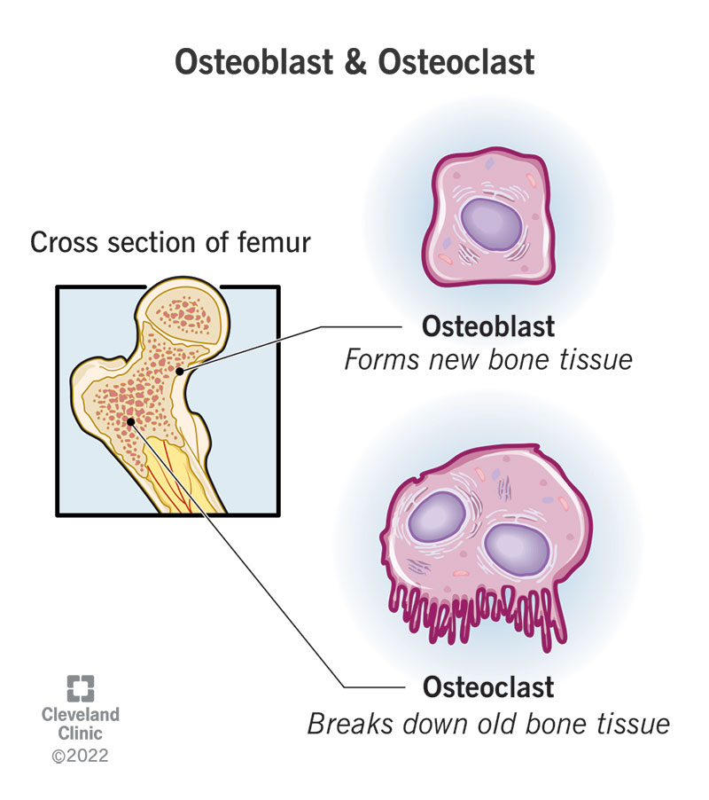
THREE TYPES OF CELLS IN BONES: oseteoblasts, osteocytes, and osteoclasts.
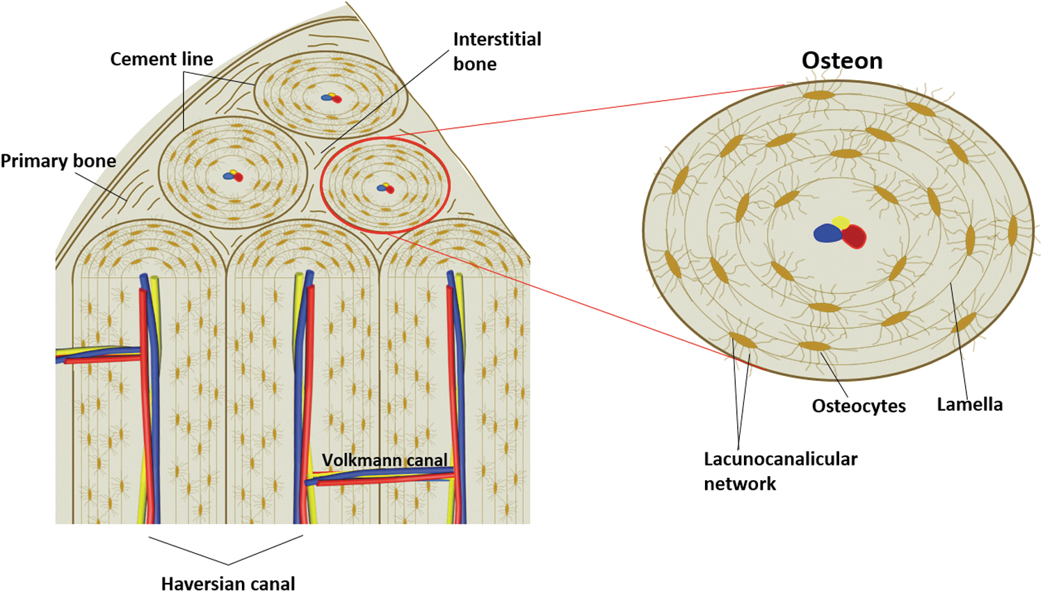
Haversian system (osteons)
The Haversian system is the structural unit of compact bone. It consists of:
Central (Haversian) canal: Contains blood vessels and nerves.
Concentric lamellae: Layers of mineralized matrix surrounding the central canal.
Lacunae: Small spaces housing osteocytes (bone cells).
Canaliculi: Tiny channels connecting lacunae, allowing communication and nutrient exchange between osteocytes.
Function: Provides strength and support while enabling nutrient delivery and waste removal via the central canal.
osteoblasts
Immature cells responsible for creating new bone during growth and repair. They deposit bone material into the matrix. Once surrounded by this matrix, they transform into osteocytes, which are less active but help maintain the bone structure.
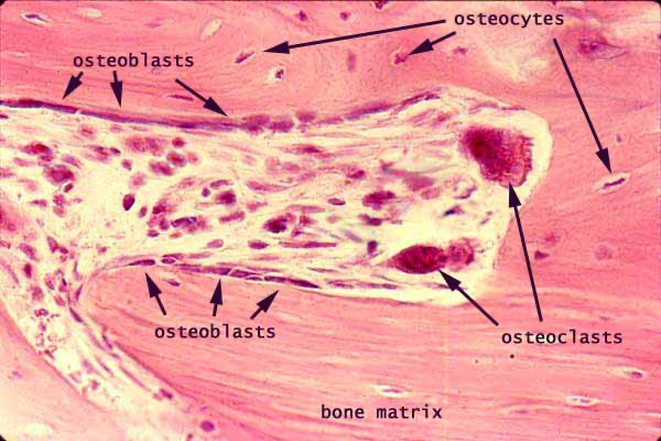
osteocytes
mature bone cells found in lacunae of the bone interconnected by the canaliculi.
cells are spider-shaped and responsible for maintaining bone tissue
osteoclasts
multi-nucleated cells active in breaking down bone tissue and resorbing minerals, for bone re-modeling
provide access to calcium stored in tissues
secrete acids and proteolytic enzymes to dissolve collagen and mineral coating (in order to raise calcium levels in blood if they are too low)
Muscle tissue
consists of long cells called muscle fibres, which contract in response to nerve signals
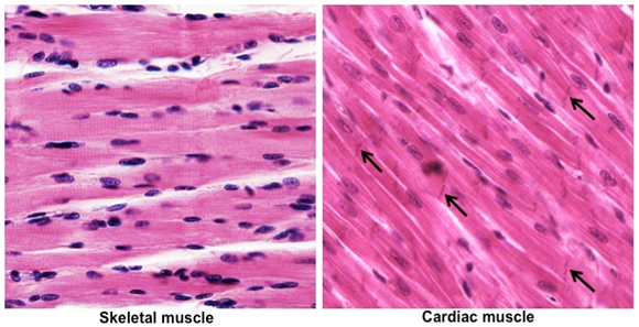
skeletal muscle
smooth muscle
cardiac muscle
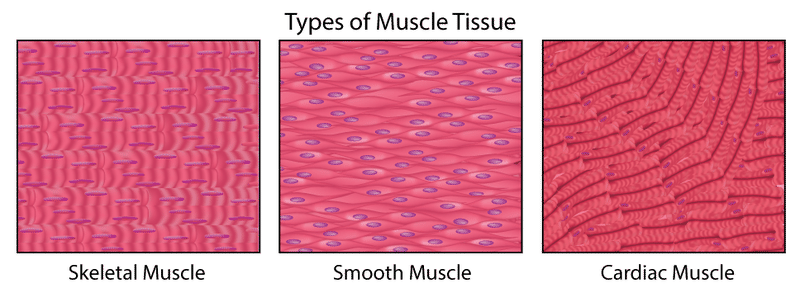
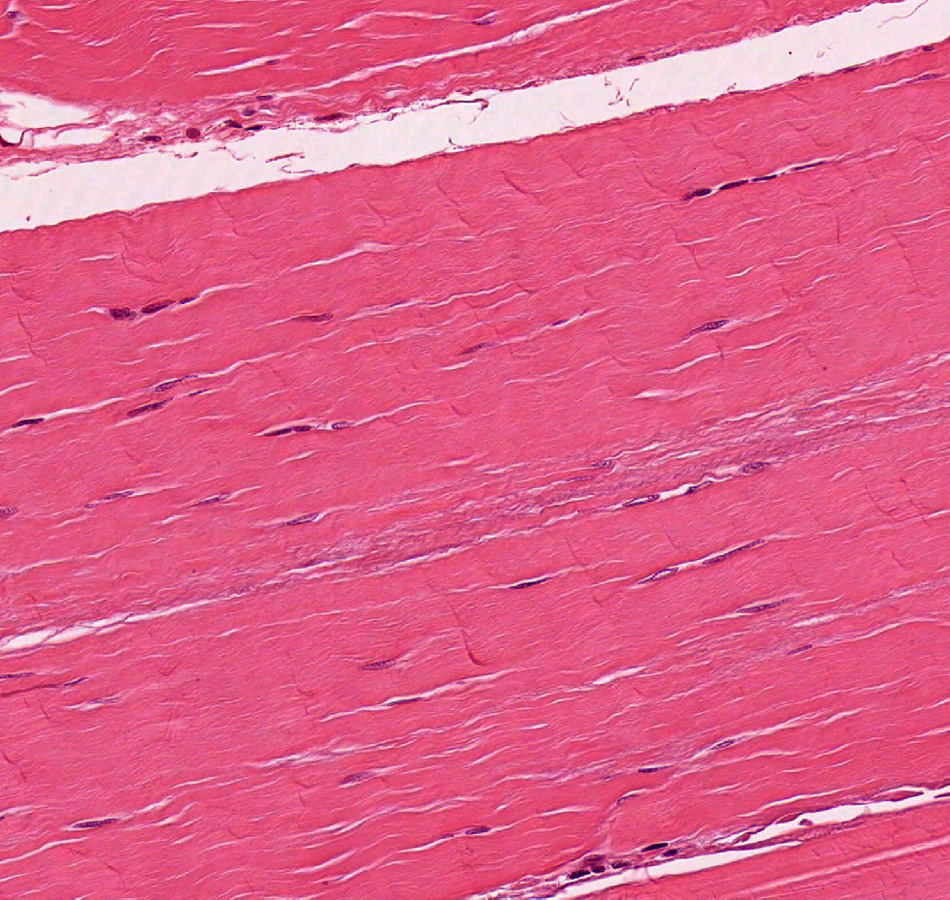
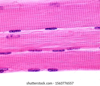
skeletal muscle
is attached to bones and is responsible for voluntary body movement
description: single, very long, cylindrical, multinucleate cells with very obvious striations
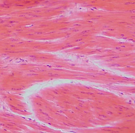
smooth muscle
mainly lines internal organs (i.e., digestive tract, urinary tract, reproductive tract, blood vessels, etc.) and is responsible for involuntary movements such as peristalsis and regulating blood flow.
description: single, fusiform, uninucleate cells; no striations. (fusiform = spindle-shape)
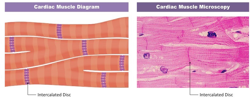
cardiac muscle
is responsible for heart contraction to help pump blood throughout body (under involuntary control)
description: branching chains of cells; unicleate, striations; intercalated discs.
intercalated disks
gap junctions that provide direct electrical coupling between cardiac cells
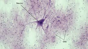
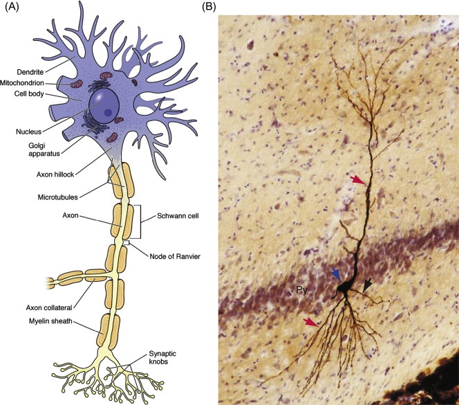
Nervous tissue
senses stimuli and transmits electrical signals throughout the animal
nervous tissues contains:
neurons, or nerve cells, that transmit nerve impulses
Glial cells, or glia, that help nourish, insulate , and replenish neurons (i.e., provide support and protection for neurons)
Most neurons have two distinct types of projections from the cell body, where the nucleus is located called axons and dendrites.
axons
are long, slender projections of neurons that transmit electrical impulses away from the cell body.
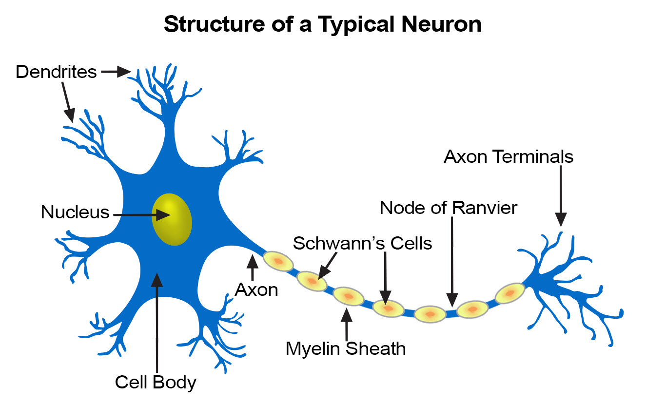
dendrites
short, branching projections that receive signals from other neurons and transmit them to the cell body.
herbivores
are animals that primarily consume plants as their main source of food.
carnivores
are animals that primarily eat meat or the flesh of other animals.
omnivores
animals that eat both plants and other animals.
ingestion
The process of taking in food and nutrients through the mouth for digestion.
digestion
mechanical digestion: (chewing + churning) increases surface area of food for faster chemical digestion
chemical digestion: enzymatic hydrolysis which splits bonds in molecules with addition of water
process of breaking down food into soluble molecules that are small enough to absorb.
absorption
uptake of nutrients by body cells
elimination
passage of undigested material out of digestive compartment and removal of waste from the body.
Mammalian digestive system (gastrointestinal tract)
pathway of how food enters body and then solid wastes are expelled
tongue
salivary glands
oral cavity
pharynx
esophagus
sphincter, stomach, sphincter → duodenum of small intestine
small intestine, large intestine, rectum, then anus
(digestion unit)
peristalsis
Rhythmic contractions of smooth muscles
aids in mechanical digestion
(digestion)
First stage of digestion
Takes place in the oral cavity (mouth) where food is mechanically broken down (chewing/churning) by teeth and mixed with saliva, starting the chemical digestion process.
mechanical digestion increases surface area and allows enzymes in saliva to access nutrients quicker
Tongue shapes food into a BOLUS (round-ball)and pushes it to the pharynx for swallowing.
pharynx
part of the first stage of digestion after chewing and salivating
a junction that opens to both the esophagus and the trachea (windpipe)
swallowing causes the epiglottis to block entry to the trachea
(digestion)
epiglottis
A flap of tissue that covers the trachea during swallowing, preventing food from entering the windpipe.
(digestion)
salivary glands
Glands in the mouth that produce saliva, which contains enzymes that initiate the digestion of carbohydrates and helps lubricate food for easier swallowing.
salivary amylase: carbs
lingual lipase: fats

(digestion)
Enzymes in saliva?
include salivary amylase (produced by salivary glands for carbohydrate digestion)
lingual lipase (produced by cells in tongue that start triglyceride digestion)
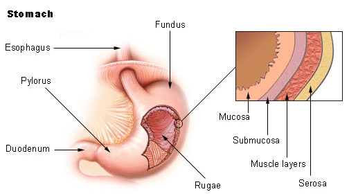
stomach
(accordion-like folds w/ very elastic wall)
stores food and secretes gastric juice, which converts a meal to acidic chyme
coordinated contraction and relaxation of stomach smooth muscle churn the stomach’s contents
churning = mixes/breaks down food (a type of mechanical digestion)
(2nd part of digestion)
gastric juice
stomach secretes this and it’s composed of hydrochloric acid and the enzyme pepsin.
(digestion)
sphincters
prevents chyme from entering the esophagus and regulates entry into small intestine (won’t go too quickly)
(sphincter, stomach, sphincter)
(digestion)
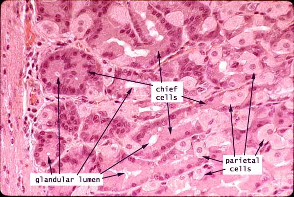
parietal cells
secretes hydrogen (H+) and chloride (Cl-) ions to form hydrochloric acid in the stomach.
(digestion)
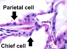
chief cells
secretes inactive pepsinogen, activated to pepsin when mixed w/ hydrochloric acid in the stomach.
They are responsible for protein digestion.
They come from the gastric mucosa in the stomach.
(digestion)
gastric mucosa
The mucous membrane layer of the stomach that contains glands and cells responsible for secreting gastric juices, including hydrochloric acid and digestive enzymes.
(digestion)
mucous cells (or goblet cells)
secretes mucus, protecting stomach lining from gastric juice

(digestion)
small intestine
(3rd part of digestion)
longest section of alimentary canal (gastrointestinal tract)
major organ of digestion and absorption
first portion of small intestine is the duodenum where chyme from the stomach mixes w/ digestive juices from the pancreas, liver, gallbladder, and the small intestine itself
(digestion)
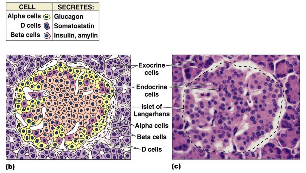
Pancreas
An organ that produces digestive enzymes and hormones.
secretes zymogens (pro-enzymes) to prevent the enzymes from digesting the cells of the pancreas itself.
zymogens or pro-enzymes are dormant (non-functional) enzyme activated in the small intestine to aid in digestion.
Beta cells produce insulin
Alpha cells produce glucagon

(part 3 of digestion)
pro-enzymes
inactive forms of digestive enzymes that are activated in the small intestine (produced by pancreas)
WHY? to prevent self-digestion of the pancreas
secretions are alkaline as a result of
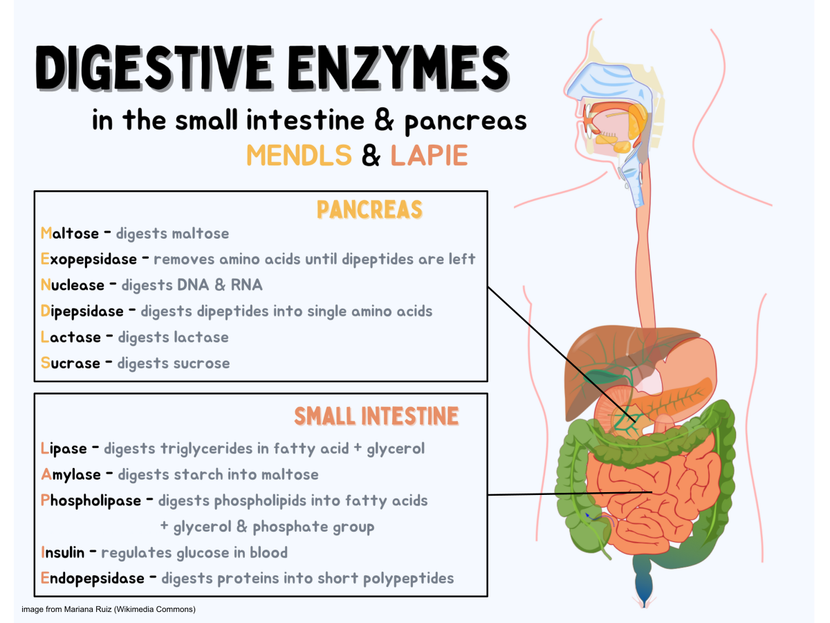
trypsinongen and chymotrypsinogen = activated into trypsin and chymotrypsin (breaks down small polypeptides [protein polymers]) chymotrypsin into amino acids
Procarboxypeptidase = activated into carboxypeptidase (breaks down smaller polypeptides)
Prolipase = activated into lipase (breaks down fats)
Proamylase and Pronucleases = activated to amylase (breaks down carbohydrates) and nucleases (break down nucleic acids)
(digestion)
Bile
A digestive fluid produced by the liver and stored in the gallbladder
helps in the emulsification and digestion of fats in the small intestine.
type of mechanical digestion
(digestion)
emulsification
transformation of large lipid droplets into small lipid droplets
increasing the surface area for chemical digestion of fats by lipases
Liver
An organ that
produces bile
detoxifies blood to get rid of harmful substances (alcohol + drugs)
stores vitamins / iron
stores simple sugar glucose as glycogen
converts glycogen to usable sugar when the body/s sugar (glucose) levels fall below normal
break down hemoglobin + insulin and other hormones
converts ammonia to urea
destroys old red blood cells
(digestion)
hemoglobin in regards to liver
A protein in red blood cells that carries oxygen from the lungs to the body's tissues and returns carbon dioxide from the tissues back to the lungs.
liver breaks this down to recycle iron and produce bile.
(digestion)
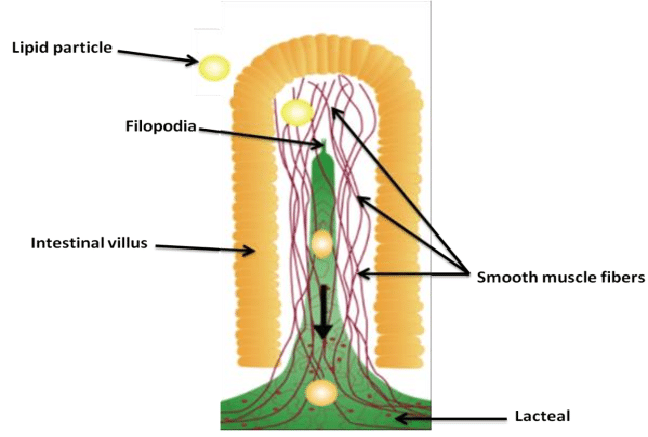
Villi and microvilli
present in small intestines that increases surface area for absorption (microvilli and villi exposed to intestinal lumen = space/cavity)
large microvillar surface area greatly increases rate of nutrient absorption
Villi are in the small intestinal lining, while micro villi are tiny projections on each villus that further enhance absorption.
each villus contains network of blood vessels and small lymphatic vessels
(digestion)
lacteal
A lymphatic vessel in the villi of the small intestine.
Function:
Absorbs dietary fats and fat-soluble vitamins from digested food.
Transports chylomicrons (fat-protein particles) into the lymphatic system, which eventually delivers them to the bloodstream.
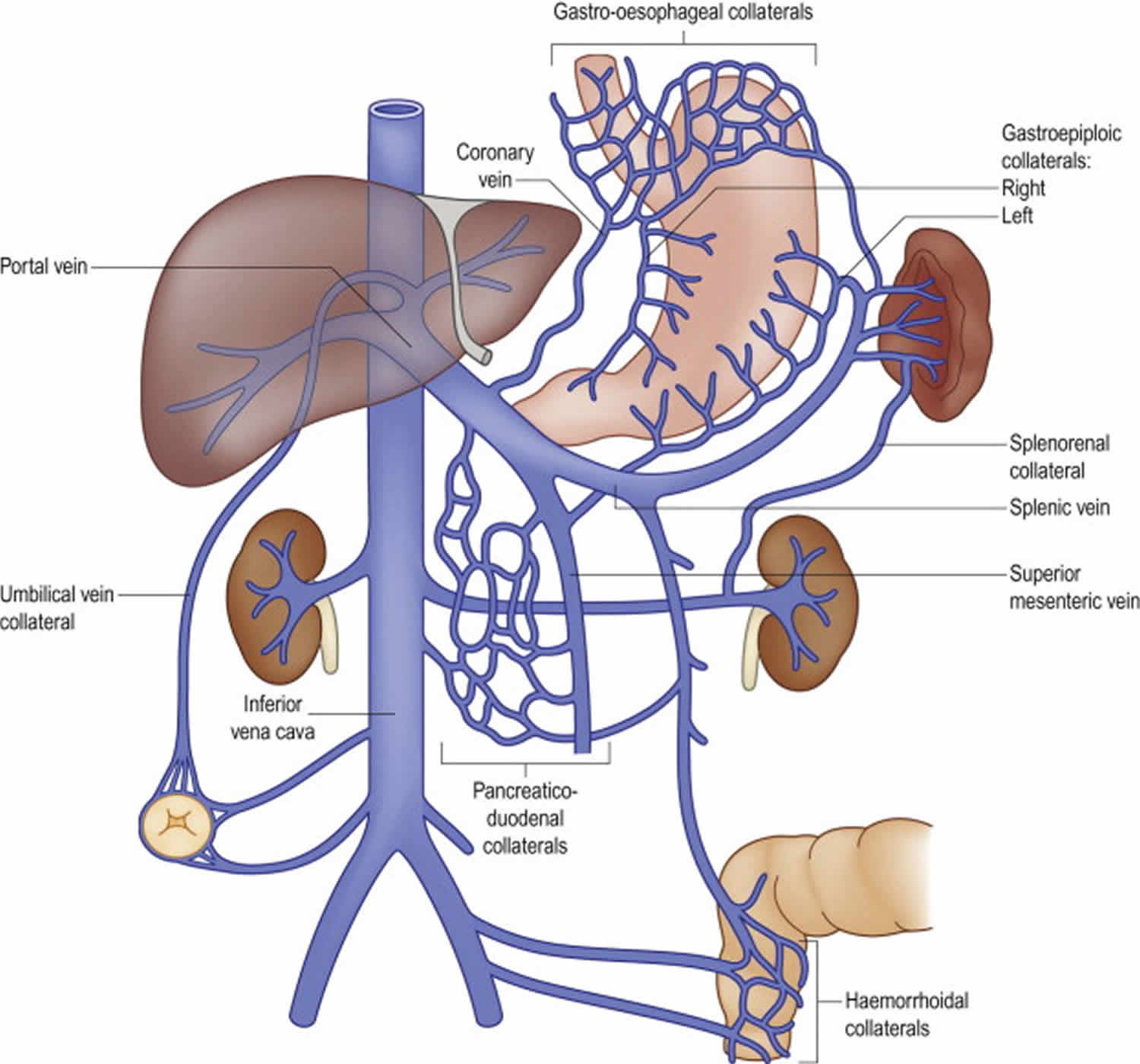
Connection to Hepatic Portal System:
Capillaries and veins from lacteals merge with blood vessels from the large intestine, stomach, and spleen to form the hepatic portal vein, which carries nutrient-rich blood to the liver.

(digestion)
hepatic portal vein
formed when capillaries and veins from the lacteals (in the small intestine) and blood vessels from the large intestine, stomach, and spleen merge.
system delivers nutrient-rich blood to the liver for processing before returning it to the heart.
main job is to transport nutrients absorbed from the digestive tract to the liver; where they are filtered, stored, or distributed to the rest of the body.
(digestion)
epithelial cells in small intestines
Structure:
Simple columnar epithelial cells line the small intestine.
Functions:
Absorb glycerol and fatty acids, recombining them into fats.
Package fats into chylomicrons (coated with phospholipids, cholesterol, and proteins) for transport.
Absorb amino acids and sugars, which enter the bloodstream directly.

(digestion)
chylomicrons
tiny fat-lipoprotein particles that carry dietary fats and fat-soluble vitamins from the intestines to the rest of the body. They enter a lacteal (a lymphatic vessel in the intestinal villi) for transport.

(digestion)
large intestine
(part four of digestion)
also known as a colon, is connected to the small intestine and is responsible for absorbing water and electrolytes, forming and storing feces before elimination.
water reabsorption, recovering water that enters the alimentary canal
reabsorption of mineral ions (sodium and chlorine)
wastes become more solid (faces) as they move through the colon and temporarily stored in the rectum UNTIL person uses the toilet
houses strains of bacterium ESHERICHIA COLI, which produce vitamins (K, biotin and folic acid) that are reabsorbed into the bloodstream
(digestion)
cecum
The beginning of the large intestine, connecting the ileum of the small intestine to the colon.
aids in fermentation (breaks down carbohydrates and other organic substances) of plant material
(digestion)
fermentation
A process where microorganisms (like bacteria or yeast) break down carbohydrates and other organic substances.
Produces gases (e.g., carbon dioxide) and acids (e.g., lactic acid) as byproducts.
Used in food production (e.g., yogurt, bread, beer) and waste decomposition.
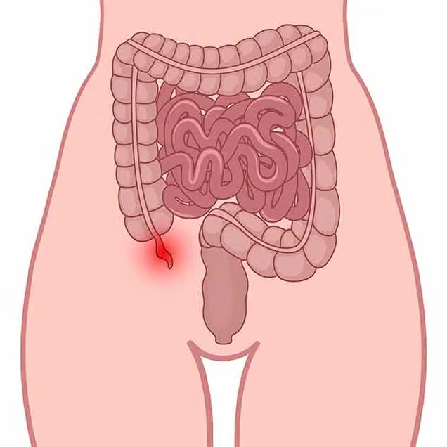
appendix
a small, tube-like structure attached to the cecum plays a MINOR role in immunity
(digestion)
gastrin
a hormone that’s secreted by stimulus of food arriving in stomach and stretching the stomach walls.
GASTRIN circulates in bloodstream back to stomach to stimulate gastric acid secretion.
(digestion)
rectum
The final section of the large intestine, where feces are stored before being expelled from the body during defecation.
(digestion)
hormones
chemical signals that regulate various physiological processes in the body.
glucose
a simple sugar that is an important energy source in living organisms and is a component of many carbohydrates.
glucagon
a hormone produced by the pancreas that raises blood glucose levels by promoting the conversion of glycogen to glucose in the liver.
glycogen
stored form of glucose in animals, primarily found in liver and muscle cells.
chyme
acidic mixture of partially digested food that is formed in the stomach and enters the small intestine via duodenum
triggers cells lining wall of duodenum to release hormones cholecystokinin (CCK) and secretin into the blood stream.
if chyme is rich in fats = high levels secretin and CCK released to inhibit peristalsis in stomach and secretion of gastric juices to slow down digestion
(digestion)
CCK (cholecystokinin)
a hormone that triggers ..
the gallbladder to contract, secreting bile into the duodenum by the bile duct
pancreas to secrete digestive pancreatic juices into duodenum
(digestion)
secretin
a hormone that triggers..
the pancreas to secrete HCO3- ions into duodenum so that chyme is neutralized.
(digestion)
accessory organs in digestion
salivary glands, liver, gallbladder, and pancreas that aid in the digestive process by producing enzymes and other substances.
glucose homeostasis *
(digestion)
the regulation of blood glucose levels to maintain a stable internal environment.
hormones insulin and glugagon regulate the break down of glycogen into glucose
LIVER IS PRIMARY SITE FOR GLUCOSE HOMEOSTASIS
carbohydrate-rich meals raise insulin, triggering synthesis of glycogen
low blood sugar causes glucagon to stimulate the break down of glycogen and release glucose

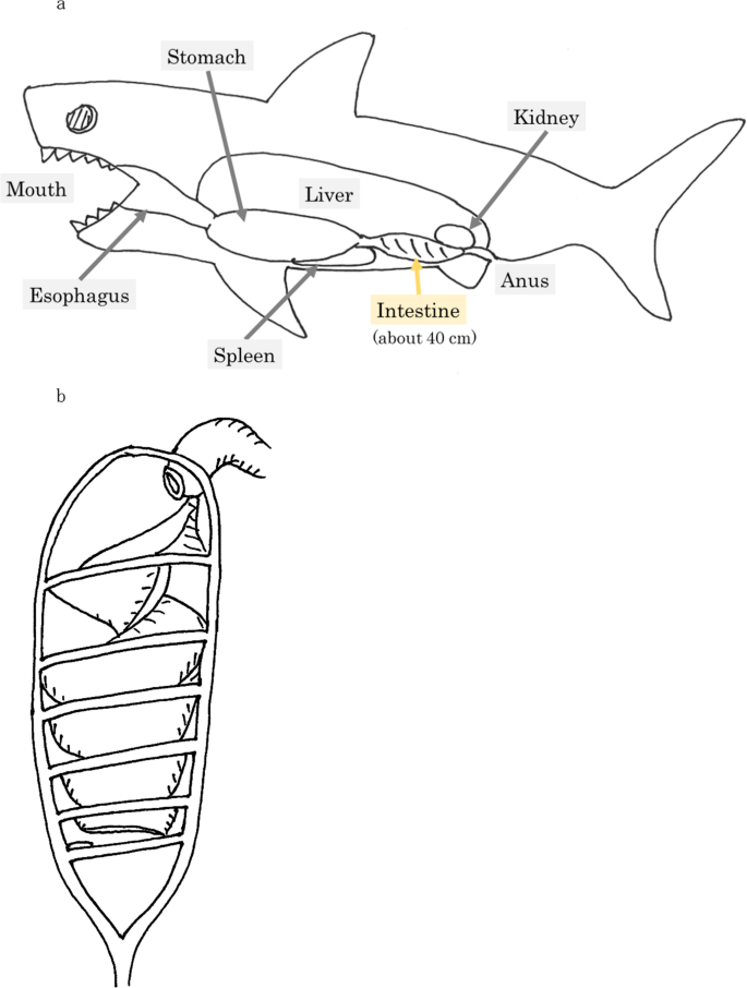
spiral valve
a structure in some fish that increases the surface area for nutrient absorption in the intestines.
internally twisted/coiled
typhlosole
a fold in the intestine of some invertebrates that increases the surface area for nutrient absorption.
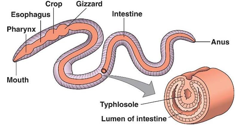
(seen in bivalve mollusks, lampreys, some annelids, and starfishes)
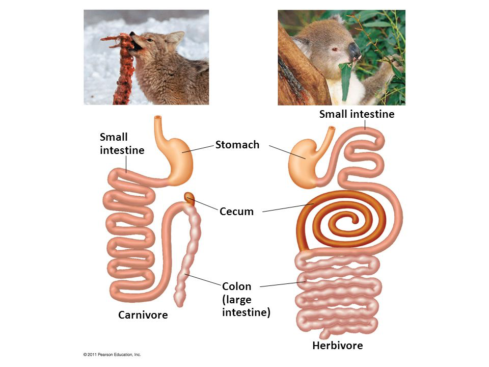
digestive tract differentiation in herbivores and carnivores
The variations in the structure and function of the digestive system between herbivorous and carnivorous animals, reflecting their dietary needs.
Herbivores typically have longer intestines and specialized chambers for fermentation, while carnivores have shorter, simpler digestive tracts.
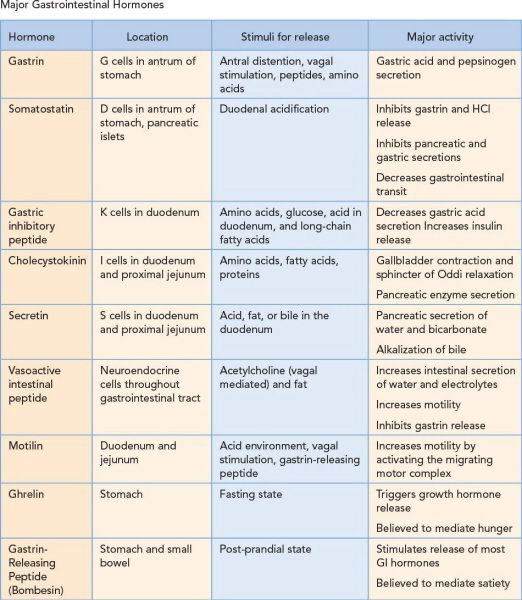
Summary of all hormones involved in digestion that secretes enzymes + sources *
osmoregulation
the process by which organisms regulate the balance of water and solutes in their bodies to maintain homeostasis.
avoids bodily fluid from being too diluted or too concentrated
Osmosis
The movement of water across a semipermeable membrane from an area of lower solute concentration to an area of higher solute concentration. [CONCENTRATION GRADIENT]
osmolarity
the measure of total solute concentration in a solution, expressed as the # of osmoles of solute per liter of solution. (mOSm)
human blood = 300 mOSm/L
[total solute] determines the movement of water
iso-osmotic
a solution that has the same osmolarity as another solution (net movement of water across semi-permeable membrane = zero).
hypo-osmotic
a solution that has a lower osmolarity than another solution, (net movement of water into the hypo-osmotic solution)
Lower [solute], but Higher [free water]
hyper-osmotic
a solution that has a higher osmolarity than another solution (net movement of water out of the hyper-osmotic solution and into the hypo-osmotic sol’n)
Higher [solute], but Lower [free water].
Kidney
two bean-shaped organs located below ribcage that are the centre of a mammals’ excretory system.
play largest role in osmoregulation of body’s internal environment.
they regulate the balance of water and electrolytes, filtering blood to produce urine.
blood supplied by renal artery, drained by renal vein
Urinary bladder
a hollow muscular organ that stores urine before it is excreted from the body. It expands as it fills and contracts during urination.