Neural Bases Exam 1: SUPER DECK (Ch 1-5 LOs + Picture Labeling)

0.0(0)
Studied by 0 people
call kaiCall Kai
Locked
learnLearn
examPractice Test
spaced repetitionSpaced Repetition
heart puzzleMatch
flashcardsFlashcards
GameKnowt Play
Card Sorting

1/258

encourage image

There's no tags or description

Looks like no tags are added yet.

Last updated 10:13 PM on 9/27/26
Name
Mastery
Learn
Test
Matching
Spaced
Call with Kai
Chat

No analytics yet

Send a link to your students to track their progress

259 Terms

1
New cards
<p>Subarachnoid space (the clear fluid-filled space containing cerebrospinal fluid)</p><p></p>

Subarachnoid space (the clear fluid-filled space containing cerebrospinal fluid)


Label structure A.
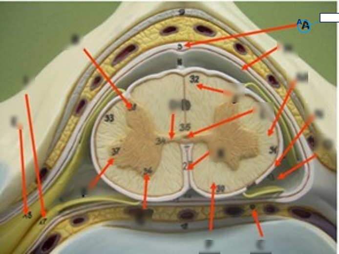
<p>Label structure A.</p>
2
New cards
<p>Label structure B, F, G</p>

Label structure B, F, G

gray matter
<p>gray matter</p>
3
New cards
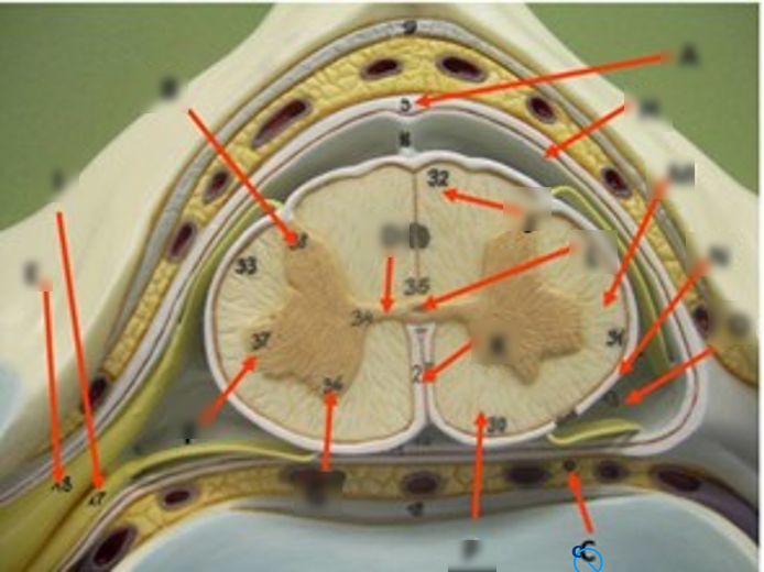
<p>Label structure C.</p>

Label structure C.

Epidural space (outermost region containing adipose tissue/fat and internal vertebral venous plexuses)

<p>Epidural space (outermost region containing adipose tissue/fat and internal vertebral venous plexuses)</p>
4
New cards
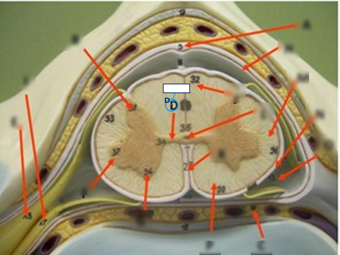
<p>Label structure D.</p>

Label structure D.

Central canal (the narrow longitudinal channel in the center containing CSF)
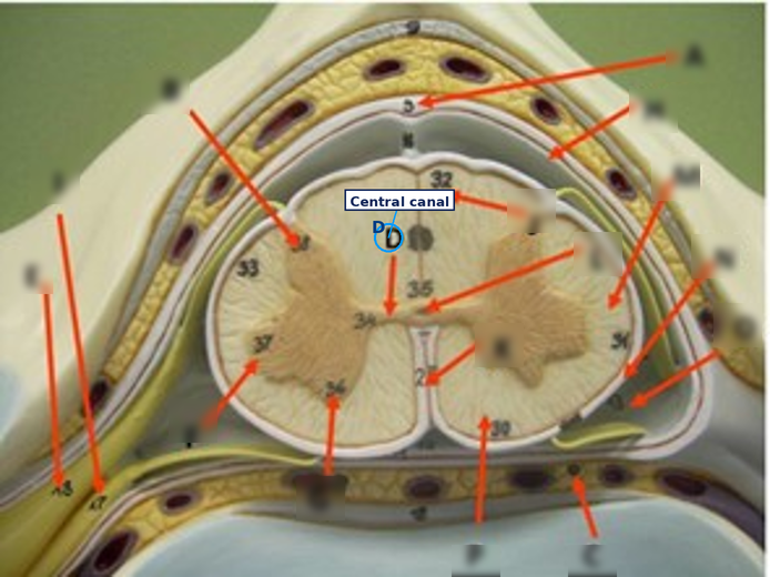
<p>Central canal (the narrow longitudinal channel in the center containing CSF)</p>
5
New cards
<p>Label structure E.</p>

Label structure E.

Dorsal (posterior) root ganglion (the sensory cell body swelling along the nerve pathway)
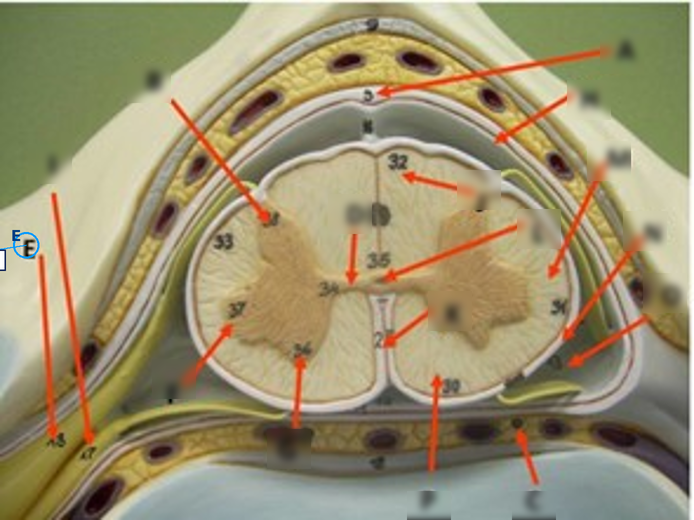
<p>Dorsal (posterior) root ganglion (the sensory cell body swelling along the nerve pathway)</p>
6
New cards
<p>Label structure H.</p>

Label structure H.

Dura mater (the thick, tough outermost meningeal layer)
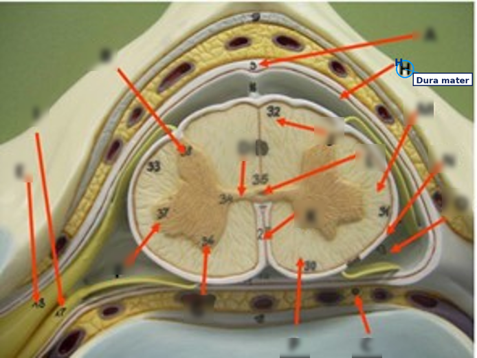
<p>Dura mater (the thick, tough outermost meningeal layer)</p>
7
New cards
<p>Label structure I.</p>

Label structure I.

Denticulate ligament (triangular extensions of the pia mater anchoring the cord laterally)

<p>Denticulate ligament (triangular extensions of the pia mater anchoring the cord laterally)</p>
8
New cards
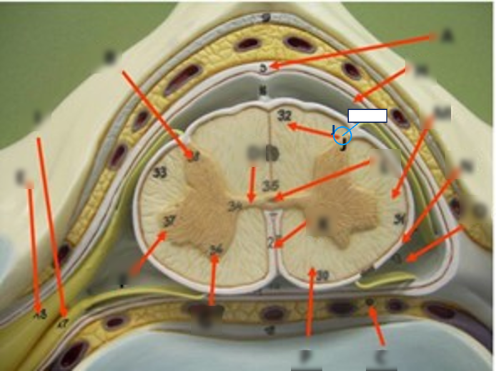
<p>Label structure J.</p>

Label structure J.

dorsal funiculus

<p>dorsal funiculus</p>
9
New cards
<p>Label structure K.</p>

Label structure K.

Pia mater


<p><span>Pia mater</span></p><p></p>
10
New cards
<p>Label structure L.</p>

Label structure L.

Gray commissure (the horizontal strip connecting the left and right gray matter halves)

<p>Gray commissure (the horizontal strip connecting the left and right gray matter halves)</p>
11
New cards
<p>Label structure M.</p>

Label structure M.

white mater

<p>white mater</p>
12
New cards
<p>Label structure N.</p>

Label structure N.

Subdural space (the narrow potential space between the dura mater and arachnoid mater)

<p>Subdural space (the narrow potential space between the dura mater and arachnoid mater)</p>
13
New cards
<p>Label structure O.</p>

Label structure O.

Arachnoid mater (the delicate, web-like middle meningeal layer)

<p>Arachnoid mater (the delicate, web-like middle meningeal layer)</p>
14
New cards
What is neurology?
The study of the anatomy, physiology, and pathology of the nervous system.
15
New cards
What does 'anatomy' mean, and what does neuroanatomy specifically study?
Anatomy = the study of structure; neuroanatomy studies the structure of the brain and spinal cord.
16
New cards
What does 'physiology' mean, and what does neurophysiology study?
Physiology = the study of function; neurophysiology studies how the nervous system works.
17
New cards
What does 'pathology' mean, and what does neuropathology study?
Pathology = the study of disease; neuropathology studies diseases of the nervous system.
18
New cards
What does a neurosurgeon do, in the simplest terms?
Removes or repairs structures that are impairing the nervous system - cutting into the system to fix it.
19
New cards
What does neuroradiology involve?
The diagnostic study of nervous system tumors and structures using radiation-based imaging techniques.
20
New cards
What does neuroembryology study?
Normal and pathological development of the nervous system.
21
New cards
What are the three major components of the nervous system, and which two make up the CNS?
Brain, spinal cord, and nerves; the brain and spinal cord together make up the CNS, while cranial and spinal nerves make up the PNS.
22
New cards
Give two of the professor's five reasons SLPs and audiologists need neurological training.
Any two of: speaking the neurologist's language; choosing the right tests based on the injury; predicting problems from a known etiology; documenting treatment change/efficacy; understanding neuroplasticity to plan treatment.
23
New cards
How does the professor define neuroplasticity?
The brain's ability to change, including in response to treatment.
24
New cards
Match each person to their neurological condition: Reagan, Fox, Reeve, Hawking, Horn.
Reagan = Alzheimer disease; Fox = Parkinson disease; Reeve = spinal cord injury (C2 separation); Hawking = ALS; Horn = stroke.
25
New cards
In the Medical model of disability, is the focus on the person's skill or on society's response?
The person's skill - what the person can't do, or a body structure that doesn't work.
26
New cards
In the Social model of disability, what is the focus?
How society interacts with the person, and how that barrier could be resolved.
27
New cards
Give the three Medical model terms and their Social model counterparts.
Impairment to function barriers; Disability to activity barriers; Handicap to participation barriers.
28
New cards
Under WHO ICD-10, which category do encephalitis and meningitis fall under?
Inflammatory diseases of the CNS.
29
New cards
Under WHO ICD-10, which category does Huntington disease fall under?
Systemic atrophies primarily affecting the CNS.
30
New cards
Under WHO ICD-10, which category does Parkinson disease fall under?
Extrapyramidal and movement disorders.
31
New cards
Under WHO ICD-10, which category does Alzheimer disease fall under, and what's happening at the cellular level?
Other degenerative diseases; involves neuron death and neuron disconnection, and is the most common degenerative disease.
32
New cards
Under WHO ICD-10, which category does multiple sclerosis fall under, and what's the underlying mechanism?
Demyelinating CNS diseases; the myelin covering that speeds motor neuron signals is what gets attacked.
33
New cards
Under WHO ICD-10, which category does epilepsy fall under?
Episodic and paroxysmal disorders.
34
New cards
Which ICD-10 category does Bell palsy fall under, and what does it look like clinically?
Nerve, nerve root, and plexus disorders; one side of the face droops.
35
New cards
Which ICD-10 category does Guillain-Barre syndrome fall under?
Polyneuropathies and other PNS disorders (demyelinating).
36
New cards
Which ICD-10 category does myasthenia gravis fall under, and what's the functional pattern?
Diseases of the myoneural junction and muscle; symptoms worsen with repeated muscle use.
37
New cards
Which ICD-10 category does anoxia fall under, and what does the brain need that anoxia disrupts?
Other disorders of the nervous system; the brain needs both oxygen and glucose, and anoxia is a lack-of-oxygen state from disrupted blood flow.
38
New cards
What are the two ends of the spectrum of belief about how the brain works?
Localization (specific functions in specific spots) at one end, holism (functions from the brain as an integrated whole) at the other.
39
New cards
Historically, what did belief about the seat of mental life shift from and to?
From cardio-centrism (mental life = the heart) to cerebro-centrism (mental life = the brain).
40
New cards
What is the difference between structural and functional imaging?
Structural imaging shows the brain's static structure/anatomy; functional imaging shows physiology - electrical activity and glucose metabolism.
41
New cards
Which imaging technique is most commonly used overall?
CT.
42
New cards
How does CT imaging work, and what is its biggest downside?
X-ray beams reflect off tissue (denser tissue reflects more); downsides are radiation exposure and showing structure only, missing new oxygen-loss damage.
43
New cards
How does MRI imaging work?
A magnetic field interacts with water in the body, flipping protons to create a clear, multi-dimensional image.
44
New cards
What are two limitations of MRI?
Cannot be used with metal in the body, and requires the patient to stay still in an enclosed space (a problem for claustrophobia).
45
New cards
How does angiography work, and what is it especially good at finding?
Iodine contrast is injected and X-ray images the vascular system; excellent at finding blood clots, with diagnosis and treatment possible in the same session.
46
New cards
How does PET imaging work?
A radioisotope tracks glucose metabolism; active brain areas need more energy, so PET shows activity by tracking where glucose use is highest.
47
New cards
What are the pros and cons of EEG/MEG?
Pro: low cost, widely available, excellent timing. Con: essentially no spatial resolution.
48
New cards
How does fMRI work, and what makes it unique?
Tracks blood oxygenation during a stimulus; unique because it gives structure and function at once, safely, with no injection needed.
49
New cards
Rank fMRI, PET, and EEG/MEG from best to worst spatial resolution.
fMRI greater than PET greater than EEG/MEG.
50
New cards
Rank EEG/MEG, fMRI, and PET from best to worst temporal resolution.
EEG/MEG greater than fMRI greater than PET.
51
New cards
What does 'spatial resolution' measure, versus 'temporal resolution'?
Spatial resolution = location; temporal resolution = timing (how quickly the brain responds to a stimulus).
52
New cards
Why is it a mistake to assume a brain area 'lighting up' on a scan is the sole area responsible for a function?
Functions typically involve networks of interacting areas - lighting up doesn't rule out other areas' involvement.
53
New cards
A patient has a pacemaker and cannot have an MRI. Which structural imaging option would you expect instead, and why?
CT (or angiography for vascular detail) - it doesn't rely on a magnetic field that would interact with implanted metal.
54
New cards
Describe anatomical position.
Body erect, arms forward, palms facing out, face forward.
55
New cards
Which plane splits the body into left and right, and what's an everyday image for it?
Sagittal (lateral) - like cutting an apple in half.
56
New cards
Which plane splits the body into front and back, and what's an everyday image for it?
Coronal (frontal) - like slicing a loaf of bread.
57
New cards
Which plane splits the body into top and bottom, and what's an everyday image for it?
Transverse (horizontal) - like hamburger layers.
58
New cards
What is a parasagittal cut?
An off-center sagittal-style cut, not exactly down the midline.
59
New cards
In an anatomical top-down view of the brain, does left stay left?
Yes - in anatomical orientation, left is left and right is right.
60
New cards
In clinical orientation, as used to read an MRI or CT slice, what happens to left and right?
They flip - left appears as right and right appears as left on the image.
61
New cards
Superior and inferior correspond to which two other directional terms?
Cranial (superior) and caudal (inferior).
62
New cards
Anterior and posterior correspond to which two other directional terms, and what do they mean?
Ventral (anterior, toward the stomach) and dorsal (posterior, toward the back).
63
New cards
What do lateral and medial mean?
Lateral = away from the midline; medial = toward the midline.
64
New cards
What do proximal and distal mean?
Proximal = nearest a limb's point of attachment; distal = farthest from it.
65
New cards
What do peripheral and central mean?
Peripheral = toward the outer surface; central = toward the center.
66
New cards
What do ipsilateral and contralateral mean?
Ipsilateral = same side; contralateral = opposite side.
67
New cards
Where do most motor pathways decussate, and what does that predict about brain-body control?
The medulla; this is why the right side of the brain generally controls the left side of the body.
68
New cards
What do supine and pronate mean?
Supine = lying with the ventral surface up (palms/face up); pronate = lying with the ventral surface down.
69
New cards
On the body, dorsal and caudal map straightforwardly. On the brain, why can these terms shift meaning?
Because of how the neural tube bends during development - on the brain, caudal can mean posterior or inferior, and dorsal can mean back or superior.
70
New cards
On the brain specifically, what do rostral, caudal, dorsal, and ventral mean?
Rostral = front; caudal = back; dorsal = top; ventral = underside.
71
New cards
Define abduct and adduct.
Abduct = moving structures apart; adduct = bringing structures together.
72
New cards
Define flexion and extension.
Flexion = bending a joint; extension = straightening a joint.
73
New cards
Do the vocal folds abduct or adduct during quiet breathing? During phonation?
They abduct during breathing and adduct during phonation.
74
New cards
What two structures must you know about the brain for the anatomical approach, per the professor's own emphasis?
The divisions and structures of the brain, plus the 12 cranial nerves.
75
New cards
In the anatomical approach, what makes up the CNS and the PNS?
CNS = brain plus spinal cord; PNS = cranial nerves plus spinal nerves.
76
New cards
In the functional approach, what's the mnemonic for efferent versus afferent?
Efferent = away from the brain (exit; motor, voluntary); afferent = to the brain (sensory).
77
New cards
In the functional approach's sensory branch, which subdivision is tied to 'rest and digest'?
Visceral sensory (versus somatosensory).
78
New cards
In the functional approach's motor branch, what are the two subdivisions?
The autonomic nervous system (involuntary) and the somatic nervous system (voluntary).
79
New cards
Is breathing controlled by the autonomic or somatic branch of the motor system?
The autonomic branch.
80
New cards
In the developmental approach, what are the three primary vesicles of the CNS?
Prosencephalon (forebrain), mesencephalon (midbrain), rhombencephalon (hindbrain).
81
New cards
The prosencephalon divides into which two secondary vesicles, and what does each become?
Telencephalon becomes the cerebral hemispheres; diencephalon becomes the thalamus and hypothalamus.
82
New cards
The rhombencephalon divides into which two secondary vesicles, and what does each become?
Metencephalon becomes the pons and cerebellum; myelencephalon becomes the medulla.
83
New cards
Does the mesencephalon divide into secondary vesicles?
No - it stays as the midbrain itself, a single structure.
84
New cards
Besides Chapter 3's developmental approach, which approach does the rest of the course follow, and in what order?
The anatomical approach, moving spinal cord to brainstem to cerebellum to diencephalon to cerebral hemispheres.
85
New cards
How many chromosome pairs does a normal somatic cell have, and how many total chromosomes?
23 pairs, totaling 46 chromosomes.
86
New cards
What is the difference between mitosis and meiosis?
Mitosis produces 2 identical daughter cells; meiosis produces 4 gametes with half the chromosome number.
87
New cards
Turner syndrome results from what kind of chromosomal defect?
A whole chromosome deletion (missing an X chromosome).
88
New cards
Cri du Chat syndrome results from what kind of defect, and what are its symptoms?
A partial chromosome deletion; round face, micrognathia, wide-spaced eyes, low tone, microcephaly, intellectual impairment, delayed speech/hearing.
89
New cards
Trisomy 21 (Down syndrome) results from what kind of defect, and what are its symptoms?
An extra chromosome; some intellectual disability, macroglossia, slanted eyes, growth failure.
90
New cards
Put these stages in order: Embryo, Zygote, Fetus, Morula, Blastocyst.
Zygote, Morula, Blastocyst, Embryo, Fetus.
91
New cards
What are the three germ layers, and what does each become?
Endoderm becomes gut/liver/lungs; mesoderm becomes skeleton/muscles/kidneys/blood/heart; ectoderm becomes skin and the nervous system.
92
New cards
Which organ system is the first to begin developing?
The central nervous system.
93
New cards
Put the six phases of CNS structural development in order.
Dorsal induction, ventral induction, neural proliferation, neuronal migration, cortical organization and synapse formation, myelination.
94
New cards
What happens during dorsal induction, and when?
GA 3-7 weeks; the neural plate forms and folds into the neural tube (neurulation), closing by about week 6.
95
New cards
What defect results if dorsal induction fails?
Neural tube defects.
96
New cards
What happens during ventral induction, and when?
GA 2-3 months; the face and brain develop from the tube's superior end, and the brain cleaves into two hemispheres.
97
New cards
What defect results if ventral induction fails?
Holoprosencephaly.
98
New cards
What happens during neural proliferation, and when?
GA 3-4 months; neurogenesis - new neurons form the gray/white matter of the cerebral hemispheres.
99
New cards
What defect results if neural proliferation fails?
Microcephaly.
100
New cards
What happens during neuronal migration, and what chemical guides it?
GA 3-5 months; neurons move to their proper location guided by reelin. By about 20 weeks, the six cortical layers are established.