Pimp1000, Podiatry Interview/PIMP/Study questions, 4th year pimp ?s
 Call Kai
 Learn
Learn Practice Test
 Spaced Repetition
 Match
Match Flashcards
 Knowt Play
1/100

There's no tags or description
Looks like no tags are added yet.
Name | Mastery | Learn | Test | Matching | Spaced | Call with Kai | Chat |
|---|
No analytics yet
Send a link to your students to track their progress
101 Terms
What is the perioperative management for patients on long-term, high-dose steroids?
Peri-op IV steroid supplementation. Stress dosing.
Hydrocortisone 100 mg IV given the night before surgery, immediately prior to surgery, and then q8h until postoperative stress relieved
What is a Gustillo Anderson type 1?
Open fracture with Clean wound <1 cm
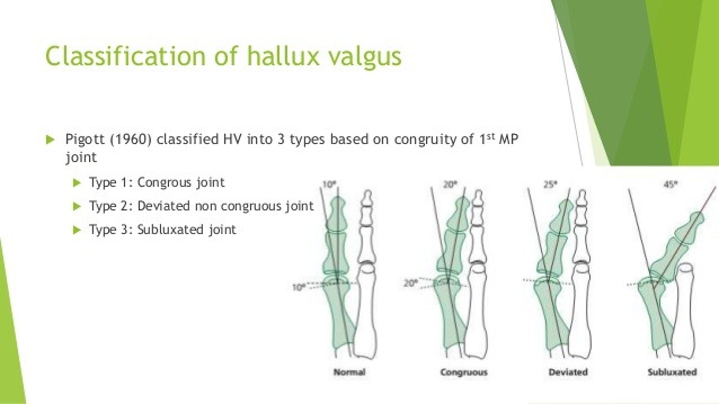
What is the Pigott Classification for and what are the stages?
Hallux Valgus Classifications (stages 1 - 4)
Stage 1
Excess pronation causes hypermobility of 1st ray. Tibial sesamoid ligament gets stretched
& fibular sesamoid ligament contracts.
Lateral subluxation of proximal phalanx occurs
Stage 2
Hallux abductus progresses, touches against 2nd digit
FHL & FHB gain lateral mechanical advantage
Crista starts to erode
Stage 3
Further subluxation at 1st MPJ, formation of IMA
IMA increases secondary to retrograde forces from abductor hallucis
Stage 4
Hallux subluxes & dislocates on 1st metatarsal
Increased crista erosion
What is the toxic dose of vicryl?(Prism pg27)
1-2 lbs
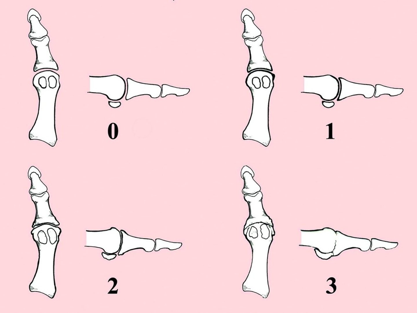
What is the Regnauld Classification for and its grades?
Hallux Limitus Classification
Grade 1 - functional hallux limitus with dorsal spurring
Intact sesamoids with no associated disease
Joint enlargement but joint space narrowing and arthrosis
<40° dorsiflexion and <20° plantarflexion
Grade 2 - broad flat metatarsal head with structural elevatus and significant spurring
Pain at rest
Osteochondral defects in metatarsal head and sesamoidal hypertrophy
Joint space hypertrophy and narrowing
75% decrease in total ROM
Grade 3 - ankylosis and articular hypertrophy with extensive peri-articular osteophytes
Osteochondral defect with joint mice and extensive 1st metatarsal-sesamoid disease
Severe loss of joint space or collapse of joint → bone on bone
FDL contracture
The "C" in CRP stands for what?
substance in serum that reacts with C- polysaccharide of pneumococcus
What is a Gustillo Anderson type 2?
Open Fx Wound 1-10 cm in diameter + soft tissue damage
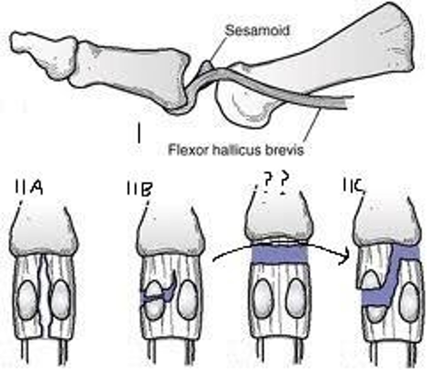
What is the Jahss Classification for and its types? (Prism p48, Watkins p464)
1st Metatarsal Dislocations:
Type l - dorsal dislocation of proximal phalanx and sesamoids with intact intersesamoid ligament
Type 2 - dorsal dislocation of proximal phalanx and sesamoids
2A - intact sesamoids with ruptured of intersesamoid ligament
2B - transverse fracture of sesamoid with intact intersesamoid ligament
What is a Gustillo Anderson type 3 A?
Open Fx Wound >10 cm + Adequate soft tissue coverage
ACE stands for what?
all cotton elastic
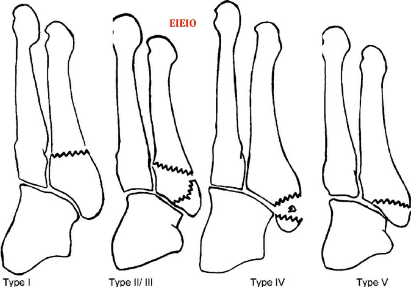
What is the Stewart Classification used for and its types? (Prism p49)
5th Metatarsal Fractures
Type 1 - extra-articular fracture at metaphyseal-diaphyseal junction (true Jones fracture)
Type 2 - intra-articular avulsion fracture of 5th metatarsal base
Type 3 - extra-articular avulsion fracture of styloid process of 5th metatarsal base
Type 4 - intra-articular comminuted fracture of 5th metatarsal base
Type 5 - extra-articular avulsion of epiphysis in children
Warfarin came from what?
Originally used for rat poison
What is a Gustillo Anderson type 3 B?
Open Fx Wound >10 cm +Periosteal stripping
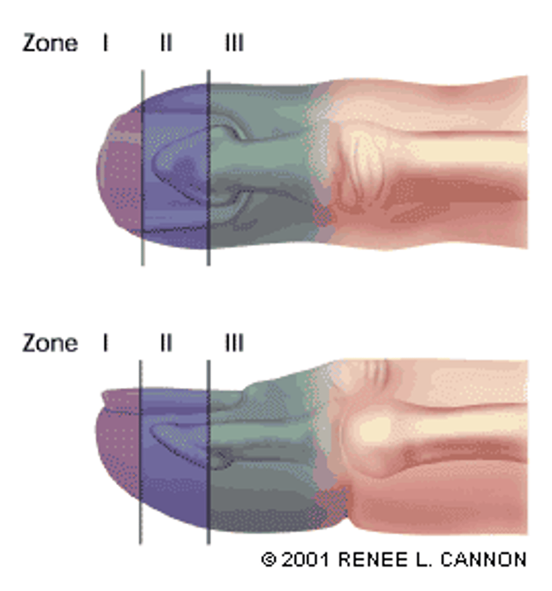
What is Rosenthal Classification used for and its zones? (Prism p47)
Nail Injury
Zone 1 - distal to bony phalanx
Zone 2 - distal to lunula
Tx: V-Y advancement
Zone 3 - proximal to distal end of lunula
If nail bed is lacerated, it is considered an open fracture
Tx: amputation
Who described >25% rule for posterior mall fx?
McDaniel- 1977
What is a Gustillo Anderson type 3C?
Open Fx Wound with arterial damage
What is a Resnick type 1?
Puncture wound. Superficial/Cutaneous w/o signs of infection
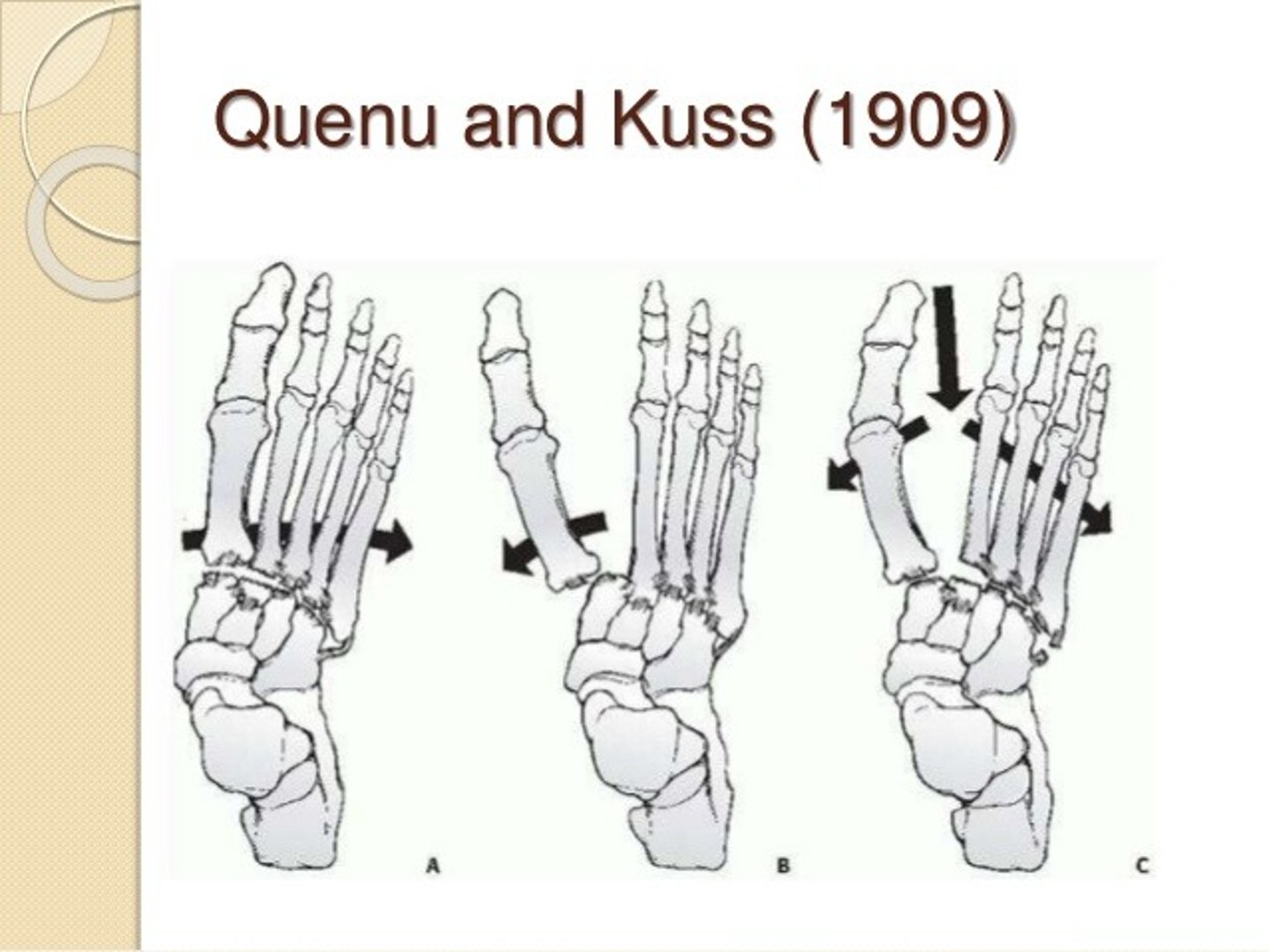
What is the Quenu & Kuss Classification used for and the three types? (Prism 49)
Lisfranc Dislocation
Convergent homolateral
All metatarsals subluxed laterally
All 5 metatarsals displaced laterally in the transverse plane
Isolateral
1st metatarsal subluxed medially or metatarsals 2-5 subluxed laterally
1 or 2 metatarsals displaced laterally in the transverse plane
Divergent
1st metatarsal subluxed medially and metatarsals 2-5 subluxed laterally
Displacement in both sagittal and transverse planes
What does TLS stand for and who invented it?
tiny little sucker
Stanley Kalish
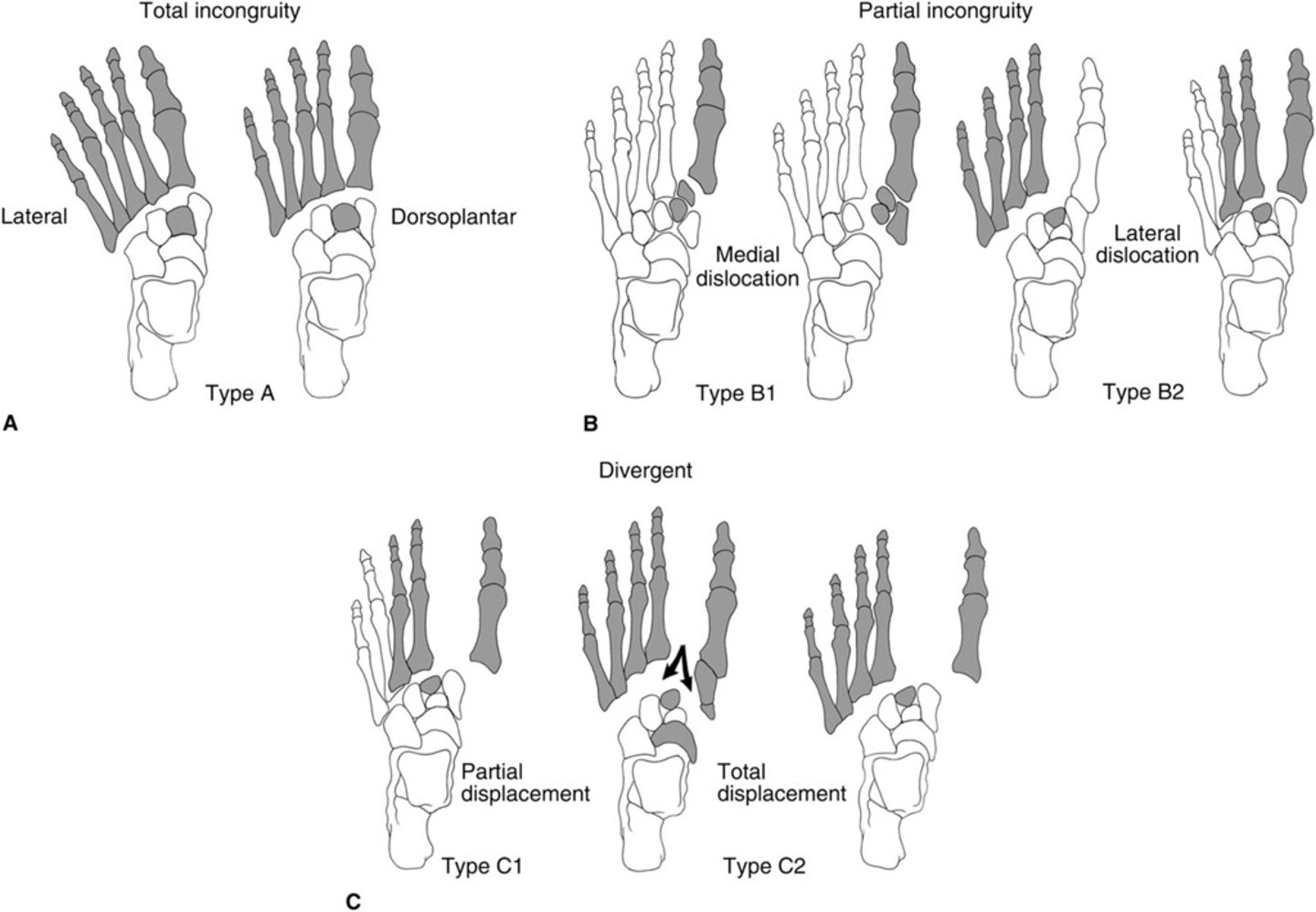
What is the Hardcastle Classification used for and its types?
Lisfranc Dislocation
A1 - homolateral
A2 - homomedial Type B - partial incongruity
Bl - partial medial displacement
1st metatarsal displaced medially and/or in combination with metatarsals 2-4
B2 - partial lateral displacement
Lateral displacement of one or more lesser metatarsals
Type C - divergent
Cl - partial displacement
1st metatarsal displaced medially with any combination of metatarsals 2-4 displaced laterally
C2 - total displacement
1st metatarsal displaced medially with metatarsals 2-5 displaced laterally
What is a Resnick type 2?
Puncture wound. Subcutaneous or articular w/o signs of infection
Name Lisfranc, treatment, Reduction Sequence, Post-op Care
and Complications (Prism p49, Watkins 483)
Treatment Options
Cast immobilization (sprains 3-5 weeks)
Closed reduction and percutaneous pinning
ORIF
Reduction Sequence
1. First realign 2nd metatarsal on middle cuneiform. Once stabilized, lesser metatarsals will
follow.
2. Next stabilize 1st metatarsal and then lateral metatarsals
Post-op Care
BK casting for 6 to 12 weeks
Initial NWB for 6-8 weeks
Partial WB approximately 6 weeks
Begin ambulation in stiff-soled shoe
PT ASAP
Accommodative orthotics
Complications
Majority - post-op DJD
Serious - circulatory compromise
What is the device for measuring compartment syndrome?
Wick Catheter or Slit Catheter, Stryker Intra-Compartmental Pressure Monitor
What is a Resnick type 3-A?
Puncture wound. Subcutaneous or articular with signs of infection
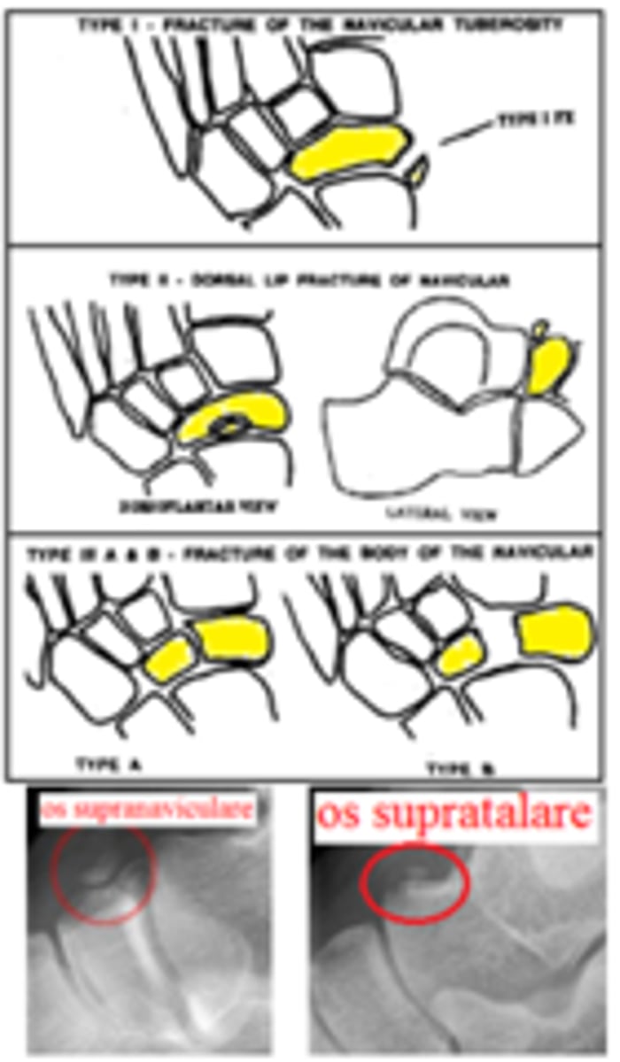
What is the Watson-Jones classification used for and its types? (Prism p50, Watkins p485)
Navicular Fractures
Type 1 - navicular tuberosity fracture
Type 2 - dorsal lip fracture
Type 3 - transverse body fracture
3A - fracture of body without displacement
3B - fracture of body with displacement
Type 4 - stress fracture
What is a Resnick type 3-B?
Puncture wound. Bone penetration w/o signs of infection
What is the Rowe Classification used for and its types? (Prism p52, Watkins p475)
Calcaneal fx
Type 1 -
1A - fracture of plantar tuberosity 1B - fracture of sustentaculum tali 1C - fracture of anterior process
Type 2
2A - ―beak fracture‖
2B - avulsion fracture of Achilles insertion
Type 3
3A - simple oblique fracture of body not involving STJ
3B - comminuted oblique fracture of body not involving STJ
Type 4 - intra-articular fracture involving STJ
Type 5 - intra-articular, comminuted, depression fracture with STJ involvement
Note: Rowe is primarily used for extra-articular fractures. Intra-articular fractures (Rowe 4 & 5)
are usually replaced by Essex & Lopresti.
What must ABX be if used in PMMA (polymethylmethacrylate)?
Heat-labile: Gentamycin, Tobramycin, Vancomycin, Ticarcillin, Cefazolin, Moxalactam, Cefotaxime
What is a Resnick type 4?
Puncture wound. Bone Penetration with osteomyelitis
What is a Patzakis type 1?
Wound location. Toe to met head. 50% incidence of osteomyelitis
What are the 2 layers of periosteum?
outer fibrous layer
inner cambium osteogenic layer (connected to cortex via Sharpey's fibers)
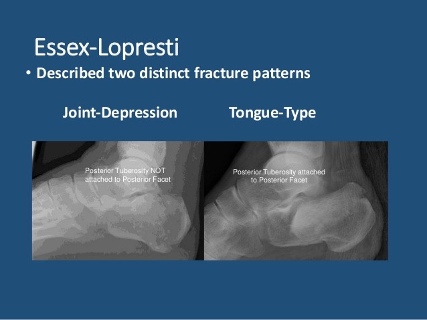
What is the Essex-Lopresti used for and its types? (Prism p52, Watkins p476)
ESSEX-LOPRESTI - Calcaneal fx
Type 1 - tongue fracture (vertical fracture line) without STJ involvement
Type 2 - joint depression fracture (horizontal fracture line) with STJ involvement
What is a Patzakis type 2?
Wound location=Midfoot. 17% incidence of osteomyelitis
What is the Degan Classification used for and its types?
DEGAN - Calcaneal Ant Process fx
Type 1 - non-displaced fracture of the anterior process
Type 2 - extra-articular, displaced fracture of the anterior process
Type 3 - intra-articular, displaced fracture of the anterior process involving C-C joint
Name of the venous plexus deep to the EHB and EDB muscle bellies
Ruch's Plexus
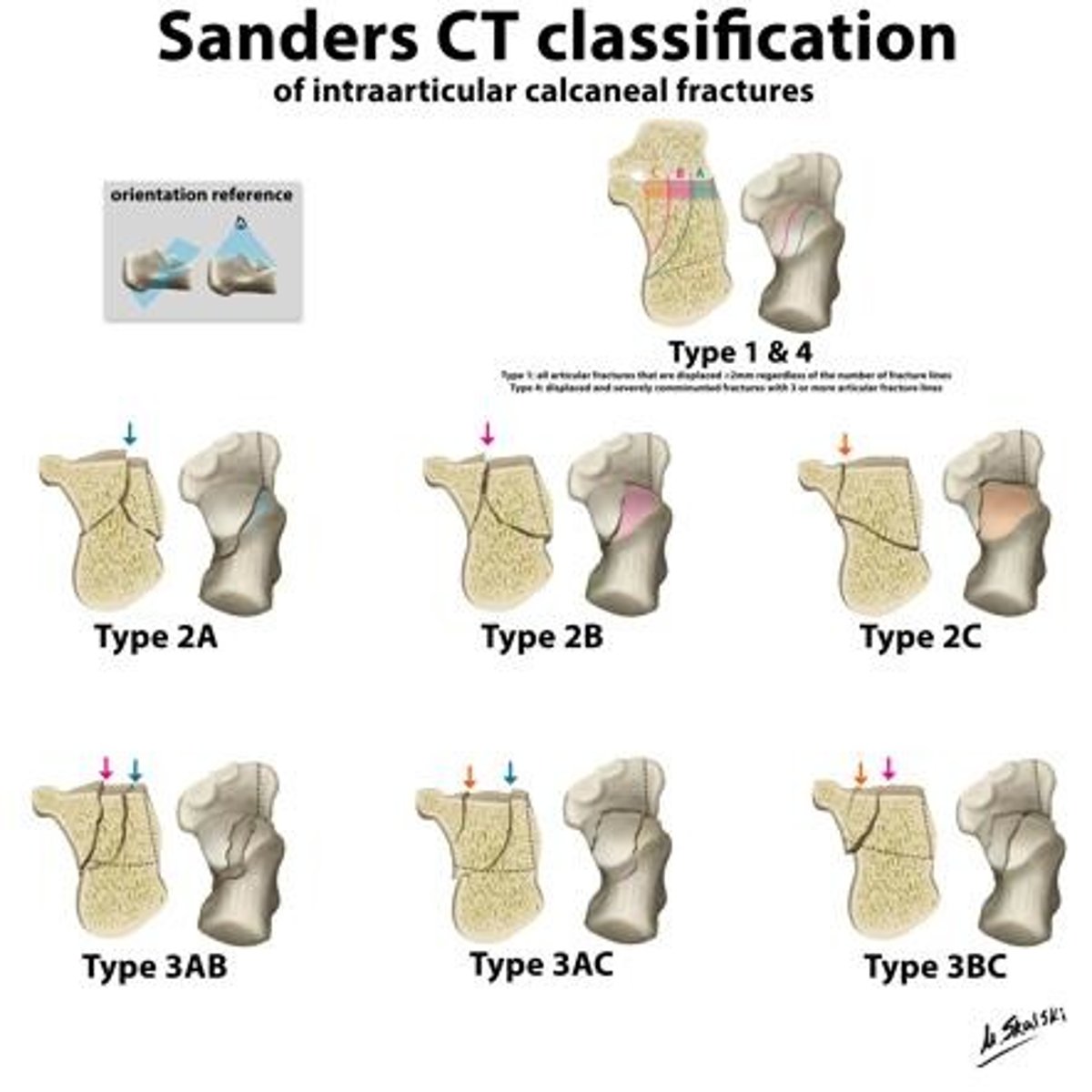
What is the Sanders classification used for and its types? (Prism p52, Watkins p477)
SANDERS - Calcaneal fx
Used for CT evaluation from coronal and axial views
Classified by number of pieces
Lines A and B divide the inferior portion of the posterior talar facet into 3 equal portions.
Line C separates the medial and posterior facets.
A - lateral
B - midline
C - medial (at sustentaculum tali)
1 - any number of fracture lines
- All non-displaced, extra-articular fractures
2 - one fracture line
Two-part fracture of posterior facet
Use one letter (2A, 2B, or 2C)
3 - two fracture lines
Three-part, intra-articular fracture of posterior facet with depressed central fragment
Use two letters (3AB, 3AC, or 3BC)
4 - three fracture lines
Four-part, intra-articular fracture of posterior facet and sustentaculum fragment with high degree of comminution
Incidences of peroneus tertius, plntaris, and peroneus quartus
PT 93%, PL 90%, PQ 7%
What is a Patzakis type 3?
Wound Location= Calcaneus. 33% incidence of osteomyelitis
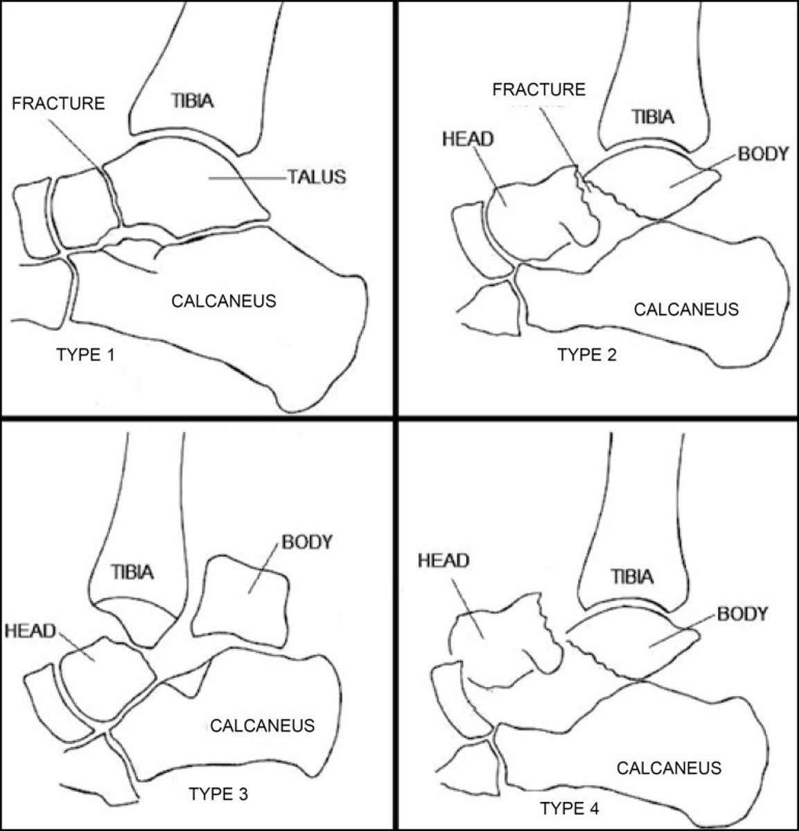
What is the Hawkins Classification used for and its Types? (Prism p51, Watkins p480)
HAWKINS - Talar neck fx
Type 1 - vertical fracture of talar neck that is nondisplaced
Type 2 - vertical fracture of talar neck with STJ dislocation/subluxation
Type 3 - vertical fracture of talar neck with STJ and ankle dislocation/subluxation
Type 4 - vertical fracture of talar neck with STJ, ankle, and T-N dislocation/subluxation
What is an Orr and Fainer stage one?
Frostbite. Erythema, Edema, White Plaque
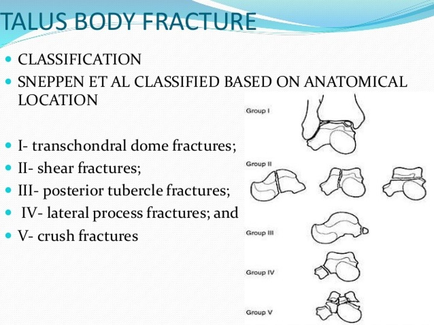
What is the Sneppen Classification used for and its types?
SNEPPEN - Talar Body Fractures
Type 1 - compressive fracture of the talar dome usually involving medial or lateral aspect Type 2 - shearing fracture of the talar body
2A - coronal shearing force 2B - sagittal shearing force 2C - horizontal shearing force
Type 3 - fracture of the posterior tubercle
Type 4 - fracture of the lateral process
Type 5 - crush fracture
Danger of rapid correction of hyponatremia?
demyelinating syndrome
What is an Orr and Fainer stage 2?
Frostbite. Blister or bleb formation
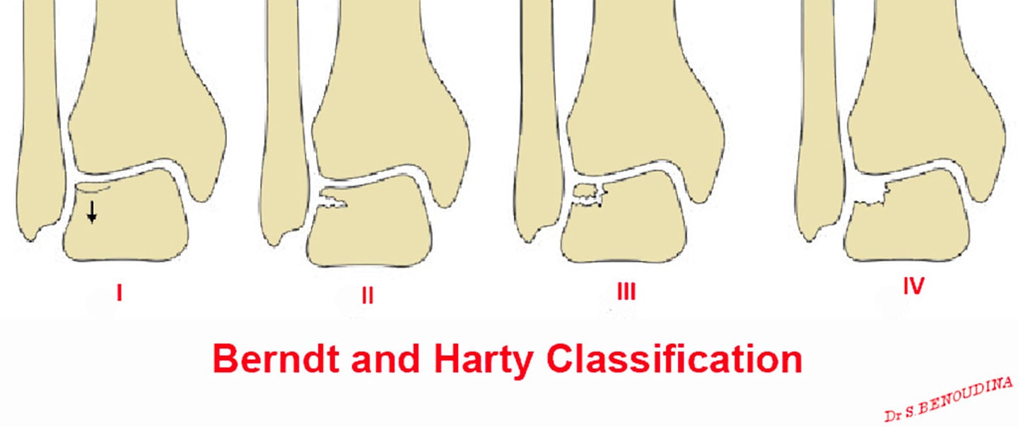
What is the Berndt & Hardy classification used for and its types? (Watkins p479)
BERNDT & HARDY - Talar dome fracture
Stage 1 - nondisplaced compression of talar dome
Stage 2 - partially detached osteochondral lesion
Stage 3 - completely detached, nondisplaced osteochondral lesion
Stage 4 - completely detached, displaced osteochondral lesion
How can a patient with a DVT develop a CVA if the occlusion is venous origin?
paradoxical embolus secondary to cardiac septal defect
What is an Orr and Fainer stage 3?
Frostbite. Full thickness w/ hemorrhagic blister + some necrosis
What is a Orr and Fainer stage 4?
Frostbite. Complete necrosis. Needs amputation
What is the Downey classification used for and its Types? (Prism p38)
DOWNEY - Tarsal Coalitions
Juvenile (Osseous Immature)
Type 1 - extra-articular coalition
A - no secondary arthritis
Tx: Badgley procedure
B - secondary arthritis
Tx: resection or triple
Type 2 - intra-articular
A - no secondary arthritis
Tx: resection, isolated arthrodesis, or triple B - secondary arthritis
Tx: triple
Adult (Osseous Mature)
Type 1 - extra-articular
A - no secondary arthritis
Tx: resection or triple
B - secondary arthritis
Tx: triple
Type 2 - intra-articular
A - no secondary arthritis
Tx: isolated or triple B - secondary arthritis
Tx: triple
Formula for calculating blood sugar from HBA1C?
6%= 110, then add 30 for each %
What is stages of Lauge-Hansen SAD?
Supination-Adduction (SAD) - No tib-fib diastasis
Stage l - rupture of lateral collaterals or * transverse fracture of lateral malleous below level of ankle joint (WEBER A)
Stage 2 - * vertical fracture of medial malleolus
What Abx is ineffective for pulmonary infections?
Daptomycin (inactivated by lung surfactant)
What is a Salter Harris type 1?
Epiphyseal Fx. Epiphysis seperated from the metaphysis w/o boney fracture
What are the stages of Lauge-Hansen PAB?
Pronation-Abduction (PAB)
Stage 1 - rupture or deltoid ligament or transverse avulsion fracture of medial malleolus
Stage 2 - rupture of anterior and/or posterior distal tib-fib ligaments
Stage 3 - * short fibular fracture (oblique on AP, trans, on lateral) at level of ankle joint
Critical lab to order for patient on Linezolid
CBC (risk of thrombocytopenia)
What is a Salter-Harris type 2?
Epiphyseal Fx. "Thurston Holland" fx through the epiphysis transversly going through the metaphysis. Most common.
What are the stages of Lauge-Hansen SER? (Prism p52)
Supination-Eversion (SER) - most common
Stage 1 - disruption of anterior tib-fib ligament with either a tibial avulsion (Tillaux-Chaput) or a fibular avulsion (Wagstaffe)
Stage 2 - * spiral oblique fracture of the fibula at level of ankle joint
Stage 3 - rupture of posterior tib-fib ligament or tibial avulsion (Volkmann)
Stage 4 - ruptured of deltoid or transverse fracture of medial malleolus (WEBER B)
Who first described the triple arthrodesis?
Ryerson 1928
What is a Salter-Harris type 3?
Epiphyseal Fx. Intraarticular fx from joint though the epiphysis, then extending along the plate to the periphery
What are the stages of Lauge-Hansen PER? (Prism p52)
Pronation-External Rotation (PER)
Stage 1 - rupture of deltoid ligament or transverse avulsion of med malleolus
Stage 2 - disruption of anterior tib-fib ligament with rupture of interosseous ligament and
Tillaux-Chaput or Wagstaffe avulsions
Stage 3 - * high fibular fracture above level of ankle joint (Maisonneuve fracture) - (WEBER C)
Stage 4 - Posterior tib-fib ligament or Volkmann fracture
Common complication of iliac bone graft harvesting?
meralgia paresthetica: lateral femoral cutaneous nerve entrapment
What is a Salter Harris type 4?
Epiphyseal Fx. Intraarticular fx from the joint through the epiphysis and physeal plate and through the metaphysis
What is a Salter Harris type 5?
Epiphyseal Fx. Compression of plate due to crushing injury
What is bone wax made of?
bees wax and paraffin
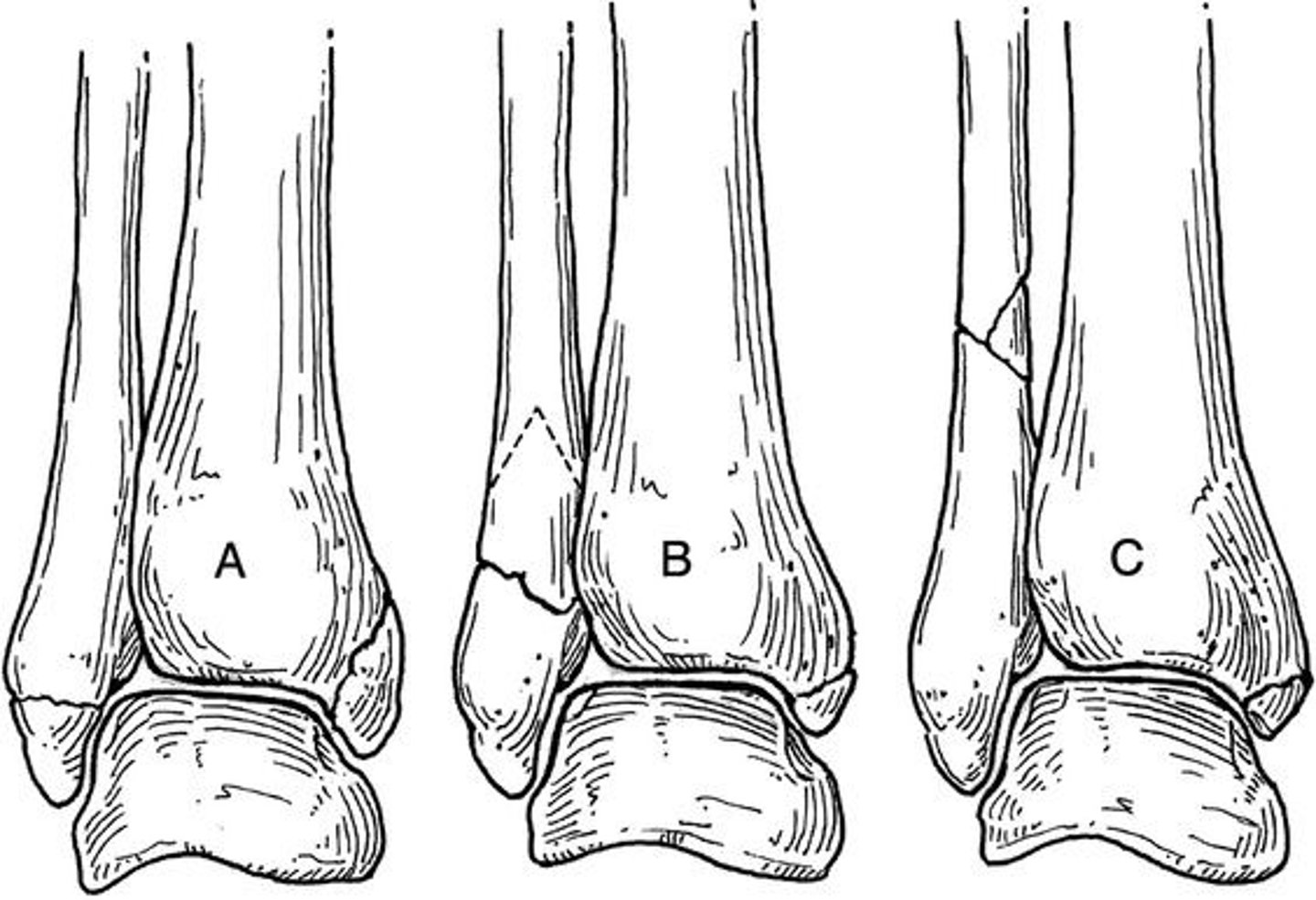
What is Danis Weber and which Lauge Hansen corresponds with which type? (Prism p52)
DANIS WEBER - Describes location of fibular fracture.
Type A - transverse avulsion fracture below the level of the ankle joint
(corresponds with Lauge-Hansen SAD)
Type B - spiral or oblique fracture at the level of the ankle joint
(corresponds with Lauge-Hansen SER and PAB)
Type C - fracture above the level of the ankle joint (Maisonneuve fracture)
(corresponds with Lauge-Hansen PER)
Why doesn't Apligraf come pre-fenestrated?
It is living tissue and will heal before application
What is a Salter Harris type 6?
Epiphyseal Fx. Peripheral bruise to Zone of Ranvier leading to angular deformity
What Ruedi & Allgower classification and its types? (Prism p55)
RUEDI & ALLGOWER - Pilon fractures - distal tibial metaphyseal fracture.
Type 1 - non-displaced tibial fragments
Type 2 - intra-articular tibial fracture without comminution
Type 3 - comminution and disruption of tibial articular surface
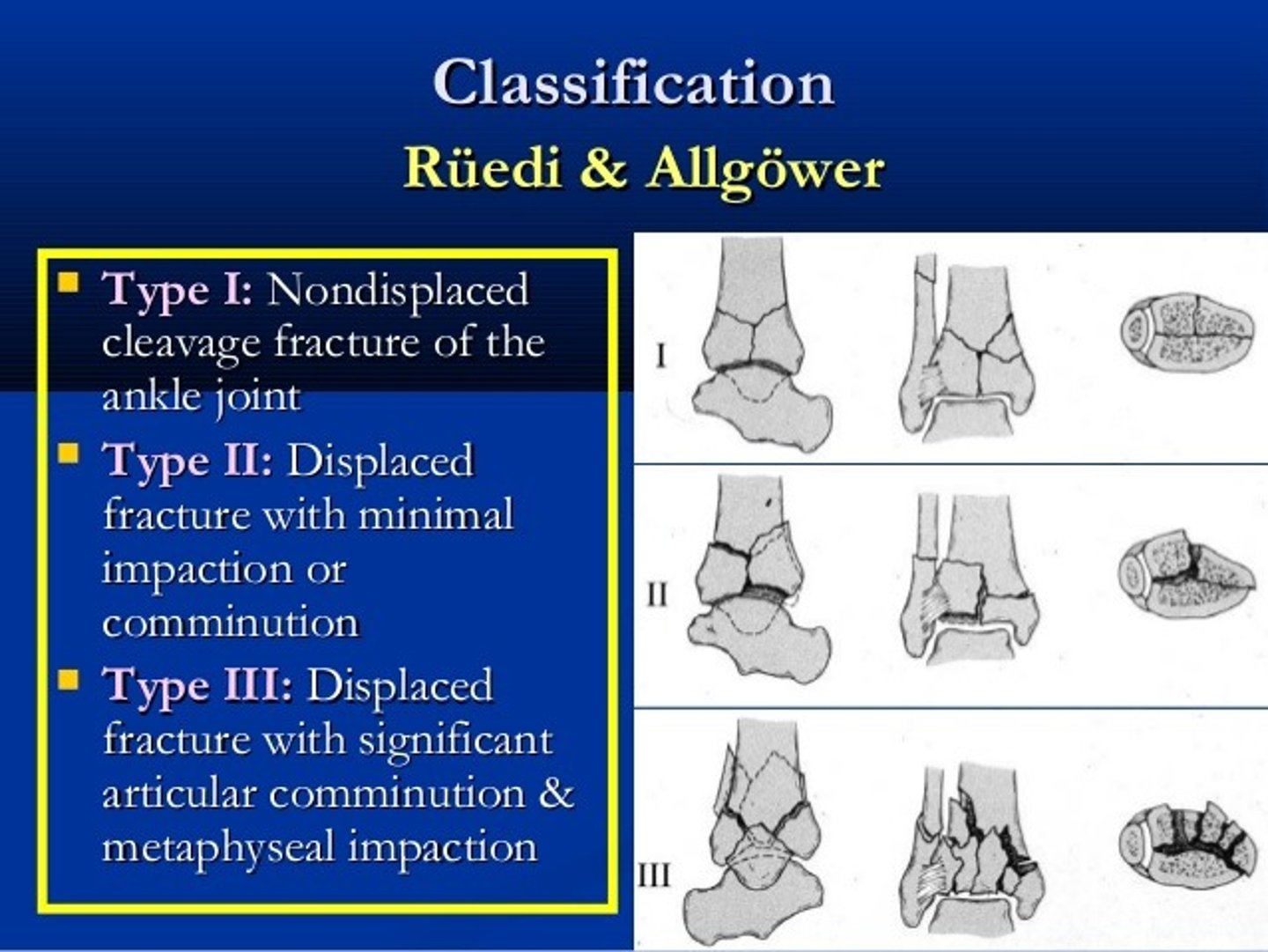
What is a Reudi-Allgower type 1?
Tibial Plafond Fx. Minimal displacement.
What are the components of Virchows triad?
Hypercoagulable State, Endothelial Damage, Stasis
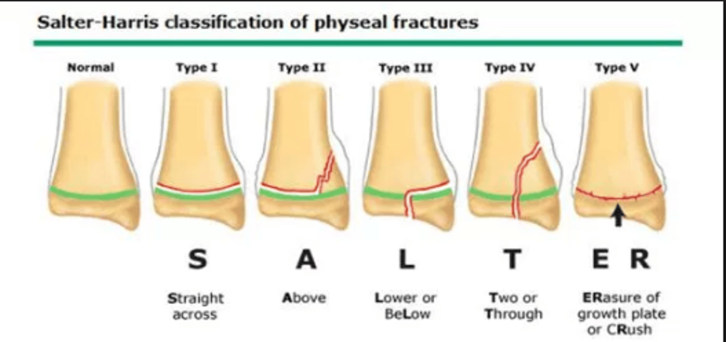
What is the Salter-Harris classification used for and its types? (Prism p61)
SALTER-HARRIS - Physeal Injuries
(SMACK - Same, Metaphysis, Articulation, Continuous, Krush) (SALTR - Same, Above, Lower, Through, Really bad)
1 - fracture through physis
2 - fracture through physis into metaphysis
3 - intra-articular fracture through physis into epiphysis
4 - intra-articular fracture through epiphysis, physis, and metaphysis 5 - crush injury
How can you tell if abx are in the pulse lavage?
bubbles/foams
What is a Reudi-Allgower type 2?
Tibial Plafound Fx. Significant Displacement
What is CONTI Classification used for and its stages?
CONTI - PTTD - MRI rapture
Stage 1 - 1-2 fine, longitudinal tears
Stage 2 - intramural degeneration, variable diameter
Stage 3 - diffuse swelling
What is a Reudi-Allgower type 3?
Tibial Plafound Fx. Impaction and comminution.
Incidence of vertebral fx with calc fx
10% L1
What is the Kuwada Classification for and its Types? (Prism p55)
KUWADA - Achilles Tendon Ruptures
Type 1 - partial tear <50%
Tx: cast with foot plantarflexed
Type 2 - complete tear with <3 cm defect after debridement
Tx: end-to-end attachment
Type 3 - complete tear with 3-6 cm defect after debridement
Tx: end-to-end attachment and tendon flap
Type 4 - complete tear with >6 cm defect after debridement
Tx: end-to-end attachment, recession, or graft
What bugs do quinolones not cover?
enterococcus
What is the AO-OTA classifications 4 elements?
Pilon Fx. Bone, Segment, Articular, Complexity
What is the Mueller classification used for an its types?
MUELLER - Post Tib Tendon Inury
1 - direct injury
2 - pathologic rupture (RA)
3 - idiopathic
4 - functional abnormality
What is Danis-Weber AO classification Type A?
Ankle Fx. Fibular fx below the level of the syndesmosis
How much STJ motion do you lose with a TN joint fusion?
91%
What is the Gustilo & Anderson Classification used for and its types? (Prism p47)
Open Fractures GUSTILLO & ANDERSON
Type 1 - wound
Type 3 - extensive skin, soft tissue, muscle, and neurovascular damage
- 3A - adequate tissue coverage, high energy trauma
- 3B - periosteal stripping, massive comminution
- 3C - arterial injury
What is Danis-Weber AO classification Type B?
Ankle Fx. Fx at level of syndesmosis
What is the Weber and Cech classification used for? (Prism p34)
WEBER & CECH- Non-Union of Fractures
Hypertrophic - hypervascular (90%)
Elephant foot
Horse hoof
Oligotrophic
Atrophic - avascular (10%)
Torsion wedge
Comminuted
Defect
Atrophic
Concerns with prone surgery?
retinal detachment and peri-orbital edema
What is Danis-Weber AO classification Type C?
Ankle Fx. Fibula Fx above the level of the syndesmosis
What is the Wagner Classification used for and its grades? (Prism p9)
Ulcers
Grade 0 - no open lesions but bony prominence and/or structural deformity present
Grade 1 - superficial ulcer without penetration to the deep layers
Grade 2 - deep ulcer penetrating to tendon, joint capsule, or bone
Grade 3 - Grade 2 depth with the presence of infection
Grade 4 - gangrene of the forefoot
Grade 5 - gangrene of the entire foot
First person to describe charcot?
Musgrave 1703- venereal disease
What is a SAD 1?
Ankle Fx. Transverse fx of the lateral mal or CFL
What is the UT Wound system used for an its grades? (Prism p9)
University of Texas at San Antonio - UTSA (J Foot Ankle Surg, 35:528-531, 1996)
Grade 0 - completely epithelialized pre- or post-ulcerative lesion
Grade 1 - superficial wound not involving tendon, capsule, or bone
Grade 2 - wound penetrating to tendon or capsule
Grade 3 - wound penetrating to bone or joint
Within each grade, there are 4 subtypes:
A - non-ischemic, clean wound
B - infected wound
C - ischemic wound
D - infected and ischemic wound
What is a SAD 2?
Ankle Fx. Type 1 + Vertical Fx of the medial mal/ Deltoid
First person to describe Charcot in diabetics?
Jordan in 1936
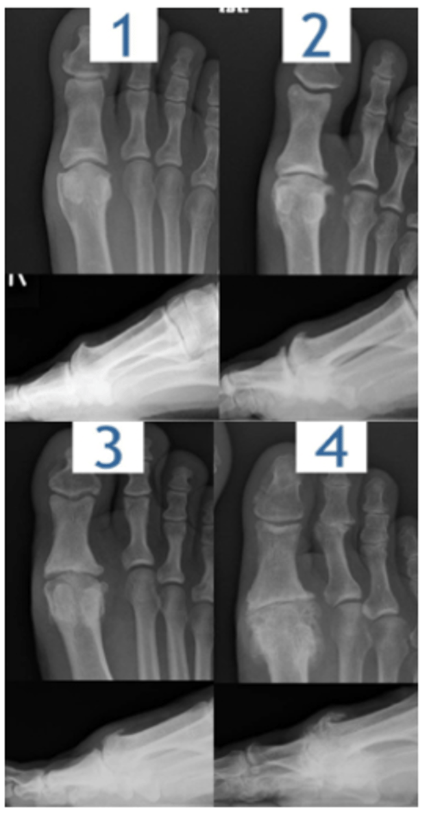
What is the Drago Oloff Jacobs classification used for an its stages?
MPJ Joint Degeneration
Stage I: "Functional limitus" - MPJ unable to fully dorsiflex
during gait. NO radiographic changes yet.
Stage II: "Joint Adaptation" - small dorsal exostosis and
met head flattening. Pain at end ROM.
Stage III: Asymmetric joint space narrowing, large dorsal
spurring, severe met head flattening. Pain and crepitus w
full ROM, OA well established now.
Stage IV: Anklyosis of joint. Severe osteophytes and joint
mice present. Pain usually from skin irritation due to
friction from rubbing over large osteophytes. Less pain
from ROM at this stage as joint has no motion.
What does V.A.C stand for?
vacuum assisted closure
What is a SER 1?
Ankle Fx. ATFL Rupture
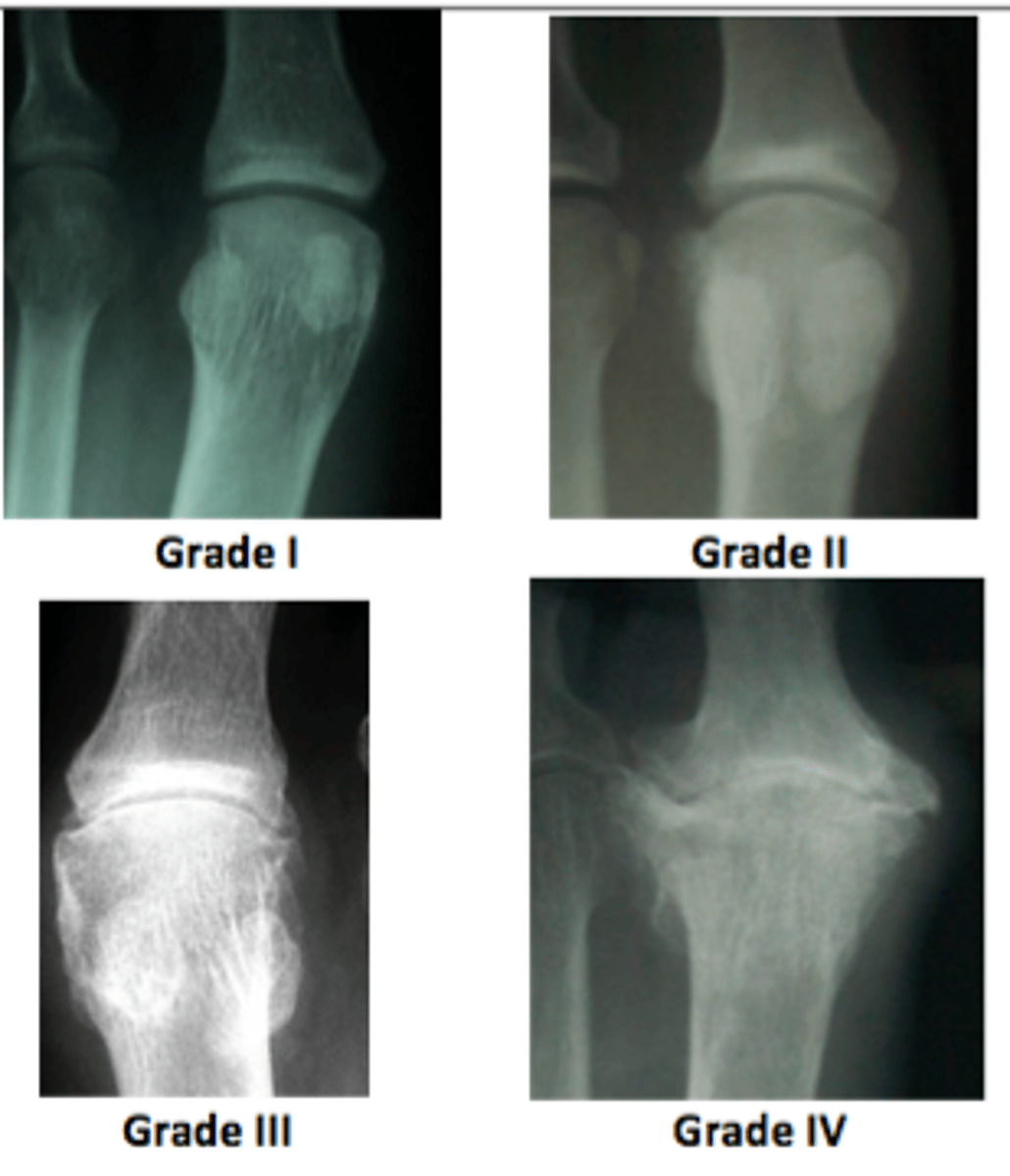
What is the Kellgren and Lawrence classification used for and its grades?
Classification for Osteoarthritis
Grade I: Doubtful narrowing of joint space with possible osteophytic lipping.
Grade II: Definite osteophytes with possible narrowing of joint space.
Grade III: Multiple osteophytes, definite jt. space narrowing, sclerosis, possible deformity of bone contour.
Grade IV: Large osteophytes, significant jt space narrowing, severe sclerosis, definite deformity of bone contour.
How does a wound V.A.C work?
creates a negative pressure environment which stimulates mitosis and angiogenesis of granulation tissue
What is an SER 2?
Ankle Fx. SER 1 + SO lateral mal Fx . Posterior Spike.
What is the Fallat & Buckholts Classification used for and its types? (Prism p31)
Classification for: Tailor's Bunionette
Type 1: enlarged lateral 5th metatarsal head (hypertrophy) Type 2: lateral bowing of 5th metatarsal (IM= normal, no hypertrophy)
Type 3: Increased IM angle (lateral deviation angle is normal)
Type 4: combo
What is an SER 3?
Ankle Fx. SER 2 + PITFl rupture or post mal FX