Main Body: Mediastinum and Pleural Cavity
 Call Kai
 Learn
Learn Practice Test
 Spaced Repetition
 Match
Match Flashcards
 Knowt Play
1/46

There's no tags or description
Looks like no tags are added yet.
Name | Mastery | Learn | Test | Matching | Spaced | Call with Kai | Chat |
|---|
No analytics yet
Send a link to your students to track their progress
47 Terms
transthoracic plane
divides teh superior mediastinum from the inferior mediastinum, through the sternal angle anteriorly and the body of T5 posteriorly- cuts the pleural cavity into 1/3 and 2/3 parts
main compartments in the thoracic cavity
2 pleural cavities, 1 mediastinum
structures at the transthoracic plane
ascending aorta becomes arch of aorta, which becomes the descending aorta, brachiocephalic veins become the superior vena cava, trachea bifurcates into two main bronchi (carina)
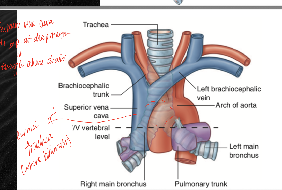
carina
name for the portion of the trachea that bifurcates into the two primary bronchi
nerves found within the superior mediastinum
vagus nerve and cardiac nerves from the sympathetic trunk
function of the heart
self-adjusting suction and pressure pump, has 4 chambers, atria are the receiving and ventricles are discharging chambers
apex of the heart
inferolateral part of the left ventricle, posterior to the left 5th intercostal space
base of the heart
hearts posterior aspect, mainly the left atrium
position of the heart
is tilted about 45 degrees to the left and the apex tilts about 45 degrees in the anterior-posterior plane, is also slightly centered to the left, which 2/3 of the heart left of the midsternal line
fibrous pericardium
outermost layer of the heart, function to envelop and protect the heart and aids in contractile efficiency of the heart by constricting how much the muscle can expand (like the deep fascia of the leg kinda)
serous pericardium
lines the inner surface of the fibrous pericardium, consists of the parietal pericardium and the visceral pericardium the directly lines the heart, in between the layers is the pericardial space in which serous fluid is found
serous fluid
found in the pericardial space and functions as lubrication between the pericardium of the heart and the heart itself
blood supply of the pericardium
80% of the blood supply is derived from the right and left internal thoracic arteries by way of their pericardiacophrenic branches (since blood from inside the heart itself moves too quickly to diffuse and supply it)
innervation of the pericardium
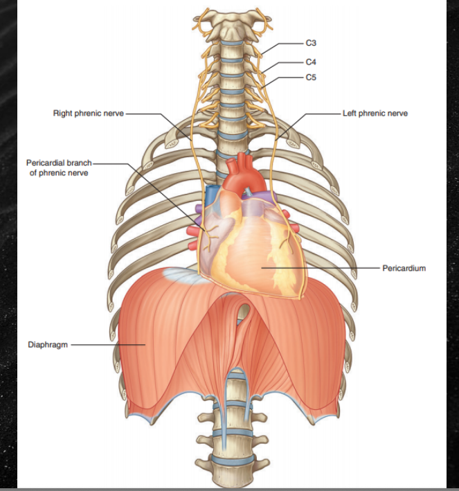
phrenic nerve provides sensory innervation from the parietal pericardium, and the general visceral afferent of the vagus nerves carries pain from the visceral pericardium
right atrium
sits posterior in situ, vessels entering are teh SVC, IVC, and the coronary sinus, also technically the foramen ovale (oval fossa) which is closed after first breath but in utero functioned as a pathway between the R and L atria
tricuspid valve
valve found between the right and left atria
right ventricle
ejects blood into the pulmonary trunk through the semilunar valves (3)
papillary muscles
myocardium that attach to cordinae tendinae and help to open and close the bicuspid and tricuspid valves
left atria
recieves blood from the 4 pulmonary veins, atria/ventricle separated by the bicuspid valve (mitral valve)
left ventricle
contains semilunar valves between it and the arch of the aorta, has the thickest muscular wall since ejects the blood into the aorta and rest of the body from here
valves open during diastole
during diastole, the heart fills and the tricuspid and bicuspid are open (aortic and pulmonary valves closed)
valves open during systole
during systole, the heart empties and the aortic and pulmonary semilunar valves are open while the tricuspid and bicuspid are closed
branches from the arch of the aorta
R brachiocephalic trunk, L common carotid, L subclavian
blood supply of the heart
supplied by branches from the left and right coronary arteries, which branch just distal to the start of the aorta
branches of the left coronary artery
is the dominant anterior artery, give rise to
circumflex branch which wraps under the L auricle
anterior interventricular branch that runs between the ventricle on the anterior surface
branches of the right coronary artery
wraps under the right auricle, gives rise to the right marginal artery and posterior interventricular artery
innervation of the heart
vagus nerve and many of its branches, as well as sympathetic nerves
veins of the heart
begins at the coronary sinus, see branches of the small cardiac vein, the great cardiac vein, and the middle cardiac vein
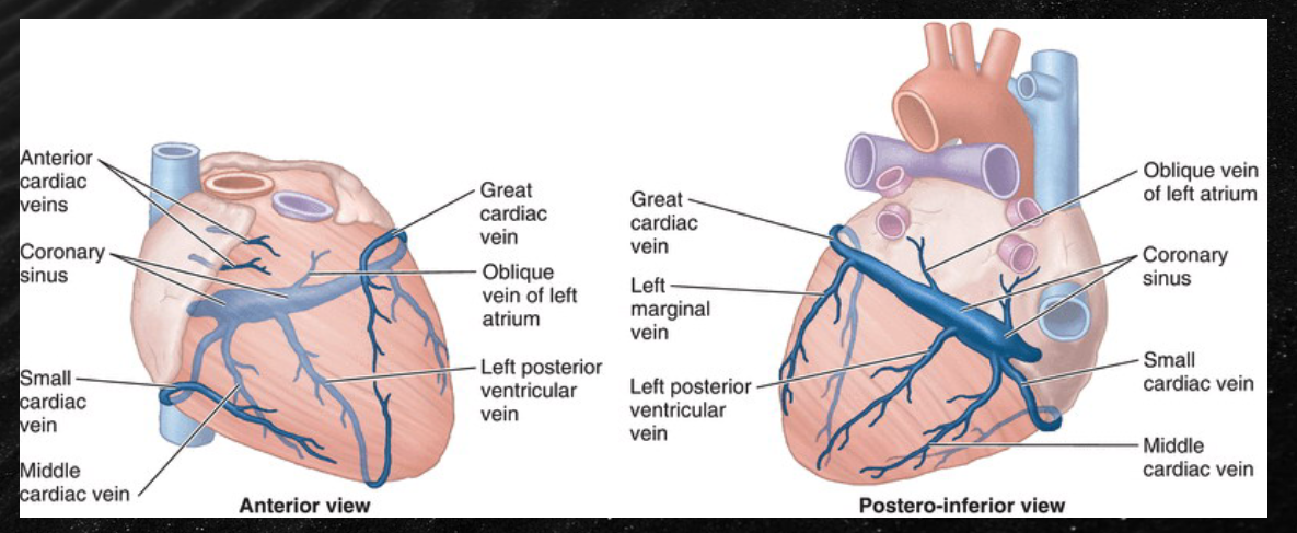
great anatomists must practice so much
great cardiac vein runs with anterior interventricular artery
middle cardiac vein runs with posterior interventricular artery
small cardiac vein runs with right marginal artery
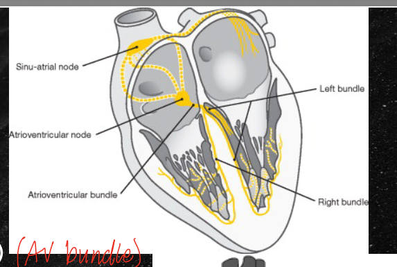
conduction of the heart
begins at the SA node, travels to the AV node, then to the AV bundle, then to the right and left bundles, then to the purkinje fibers (which are formed by the purkinje cells)
pleural cavity components
parietal pleura (outermost layer), visceral pleura (lines the lungs), and the parietal cavity between them (potential space filled with pleural fluid)
right lung
has three lobes, superior, middle, and inferior with a horizontal and oblique fissure
left lung
only has a superior and inferior lobe with a oblique fissure, also has a cardiac notch and the lingula
root of the lungs
is surrounded by the hilum, contains veins, arteries, and bronchus
right bronchi
is shorter and stouter, also more vertical which predisposes it to aspirated objects which get dumped into the middle lobe
apex of lungs
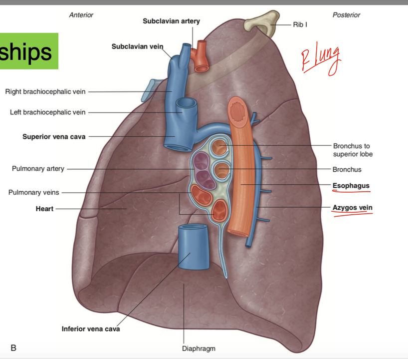
found just above the 1st rib right under the root of the subclavian artery and vein
pulmonary arteries and veins
unlike all other arteries and veins, pulmonary arteries carry deoxygenated blood from the heart and veins carry oxygenated blood to the heart
indentations seen in the R lung
see indentation from the superior vena cava, the esophagus, and the inferior vena cava
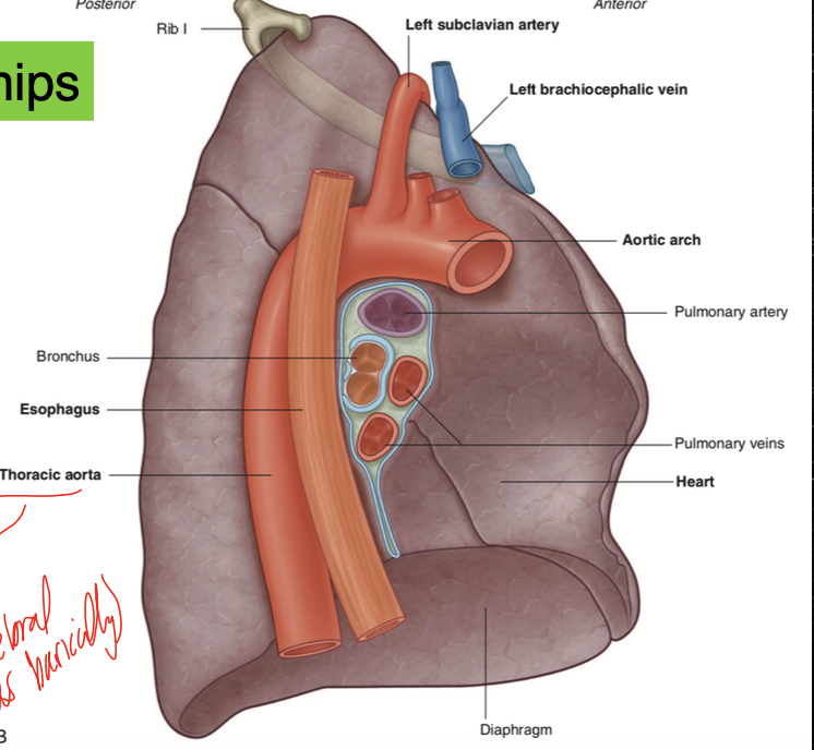
indentations in the left lung
see indentations from the thoracic aorta and aortic arch, and from the esophagus
flow of blood through lungs
pulmonary artery to the alveolar sac to the pulmonary vein a
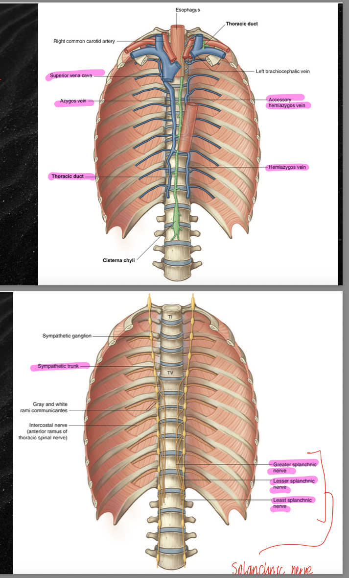
azygous system
consists of the azygous vein and the hemiazygous vein and accessory hemiazygous vein which crosses over to the L side, drains into the right and left bronchial veins
posterior mediastinum
consists of the aorta, hemiazygous vein, azygous vein, the accessory hemiazygous vein, the SVC, and the brachiocephalic vein, the thoracic duct, and the sympathetic chains and splanchnic nerves
diaphragm
innervated by the phrenic nerve (C3, C4, C5) and functions in inspiration. Has openings for the major structures passing between the abdomen and thorax, the anterior portion is found at the level of T9 and the posterior is found at the level of T12
IVC hiatus
found at the level of T8
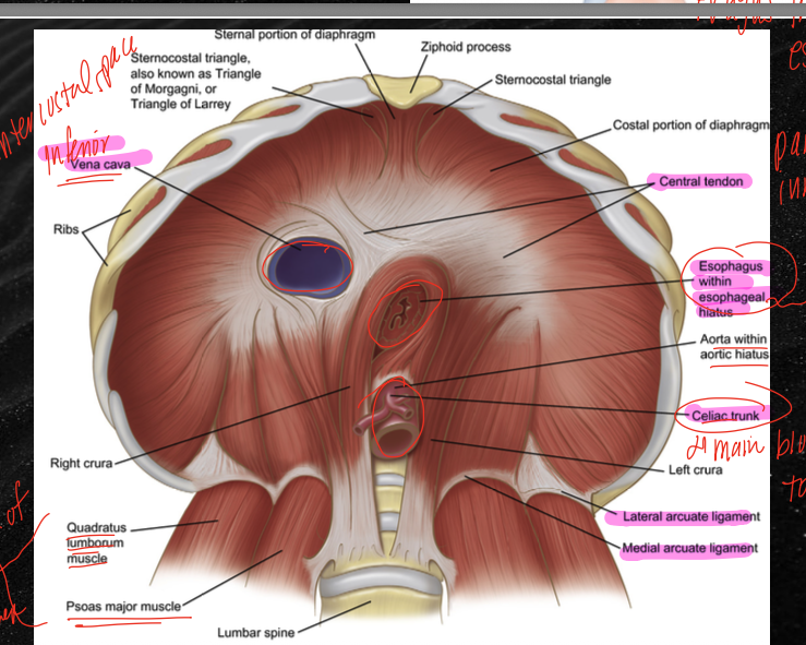
esophageal hiatus
found at the level of T10
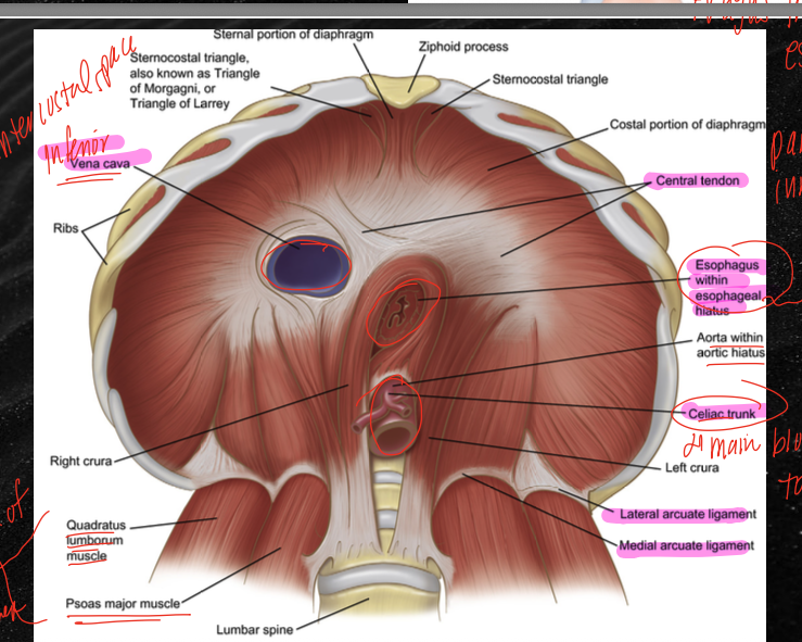
aortic hiatus
found at the level of T12
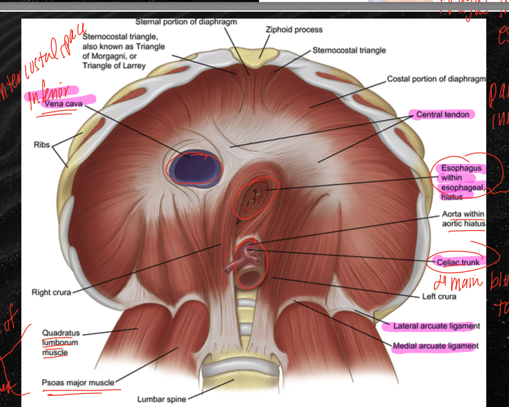
celiac trunk
branches off the aorta right underneath the diaphragm, is the main blood supply to teh viscera