Lecture 1: Innate Immune System
 Call Kai
 Learn
Learn Practice Test
 Spaced Repetition
 Match
Match Flashcards
 Knowt Play
1/8

There's no tags or description
Looks like no tags are added yet.
Name | Mastery | Learn | Test | Matching | Spaced | Call with Kai | Chat |
|---|
No analytics yet
Send a link to your students to track their progress
9 Terms
Innate Immunity
Early Recognition:
First line of host defense (skin, mucosal barrier, blood brain barrier, chemical barriers
Detects microbes and initiates mechanisms to remove threats
Distant characteristics:
Local effect
Nonspecific
Lacks memory
Works quickly
Pre-programmed
Protective Proteins: Keratin, Interferons, Complement
Protective cells: Neutrophils and Macrophages
Protective processes: Fever and Inflammation
External Barriers:
Acid mantle: Inhibit bacterial growth
Dermicidin: Antibacterial peptide in sweat
Defensins and Cathelicidins: Destroy bacteria, viruses and fungi. These are enhanced by Vitamin D
Mucous Membranes:
Chemical barriers and Physical barriers
Sticky consistency
Lysozyme (mucus, tears, saliva): Destroys bacterial cell walls
Hyaluronic acid: In a healthy state, large, intact hyaluronic acid molecules act like a tough structural glue in your mucus membranes. It physically traps bacteria and viruses so your body can flush them out, while sending an "all clear, anti-inflammatory" signal to keep your immune system calm
Hyaluronidase: The enzyme that chops up hyaluronic acid, and it plays a dual role depending on who is using it. Your own body releases hyaluronidase at an injury site to intentionally break down the hyaluronic shield. This creates the "distress flare" fragments needed to activate your immune defenses and allows healing immune cells to easily move through the tissue to reach the wound
Cells of the Innate Immune System
Granulocytes:
Neutrophiles (60-70% of leukocytes):
Granules contain hydrolytic enzymes or defensins (antimicrobials)
Pattern Recognition Receptors on surface
Phagocytic: “bacteria slayers”
Highly Regulated-to prevent unwanted damage to host cells
Degranulation: When bacteria are detected in immediate area, lysosomes migrate to the cell surface and discharge enzymes into the tissue fluid, and releases antimicrobials to kill bacteria
Phagocytosis: Engulfing microbes with pseudopods and destroying with lysosomal enzymes
Neutrophil Extracellular Traps (NETs): Release of chromatin to neutralize and kill bacteria, fungi, viruses and parasites and prevent dissemination of bacteria (too many?-can cause blood clots and tissue damage!)
Eosinophils (2-4% of leukocytes):
Red to crimson (acidophilic) coarse, lysosome-like granules
Stand guard against parasites, allergens and other foes
Secrete superoxide, H2O2 and neurotoxins
Kill parasites such as tapeworms and roundworms
Phagocytized and degrade antigen-antibody complexes, allergens and inflammatory chemicals
Found in the mucous membranes
Can degranulate
Basophils (<0.5% of leukocytes)
Large, purplish-black (basophilic) granules contain histamine
Secrete chemicals to aid in mobility and action of other leukocytes
Secrete histamine (vasodilator) - increased blood flow; heparin (anticoagulant) - mobility of WBC’s prevents clotting; leukotrienes - activate and attract neutrophils and eosinophils
Mast cells are very similar - found in tissues rather than in bloodstream
Eosinophils promote the action of basophils and mast cells
Agranulocytes:
Monocytes (3-8% of leukocytes):
Differentiate into macrophages (large phagocytic cells found in tissues)
Found in connective tissue (some specialized microglia-nervous system; alveolar macrophages-lungs; hepatic macrophages-liver)
Phagocytize pathogens, dead neutrophils and debris of dead cells
Present antigens to activate other cells of the immune system
Dendritic Cells:
Antigen presenting cells
Found in cells in contact with external antigens (gastrointestinal mucosa, skin epithelia)
Travel to lymph nodes
Lymphocytes (25-33% of leukocytes)
Increase during infections and immune responses
Broadly: destroy cancer cells, present antigens to activate other cells of the immune system, secrete antibodies, serve in memory
Three types of lymphocytes: Natural killer cells, T cells, B cells
All can travel in and out of tissues and the bloodstream
Natural Killer Cells (Lymphocyte)
Continually patrol the body for pathogens and diseased cells
Target cells infected with viruses, cells of transplanted organs and tissues, and cancer cells
NK cells bind, release perforins, create a hole in the membrane of the invading cell
Also secretes granzymes to destroy target cell’s enzymes and induce apoptosis

Antimicrobial Proteins
Can inhibit microbial reproduction and provide short-term, non-specific resistance to pathogenic bacteria and viruses (dermicidin, cathelicidin)
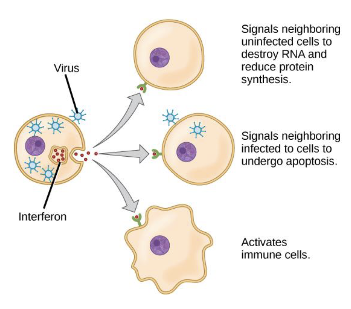
Interferons: Little benefit to cell that secretes them; messenger to alert neighboring cells of danger
Complement System: 30 or more globulins that contribute to both innate and adaptive immunity. Contributes to pathogen destruction through: inflammation, immune clearance, phagocytosis and cytolysis
Interferons
When certain cells are infected, they secrete interferons
Complement System
Group of >30 globulin proteins that contribute to immunity. Plasma proteins

Innate Immunity: Pattern Recognition Receptors
Activation of these receptors—triggers intracellular pathways that results in production of cytokines, chemokines and cell-surface molecules
Molecular security cameras—broad ability to detect
(cytokines—proteins secreted by cells that interact with specific receptors on other cells and induce a functional response)
Broadly what do they recognize?
Bacterial cell wall components (LPS, peptidoglycan), viral RNA/DNA, or fungal carbohydrates.
Specifically PRRs recognize:
Pathogen-Associated Molecular Patterns (PAMPs): Essential conserved microbial components
Damage-Associated Molecular Patterns (DAMPs): Markers of tissue damage or death on host cells
Major Classes of Pattern Recognition Receptors
Toll-like receptors (TLRs): Detect microbial lipids, proteins, nucleic acids (found on cell surfaces & endosomes).
NOD-like receptors (NLRs): Cytoplasmic sensors for bacterial products and cell stress; some form inflammasomes.
RIG-I-like receptors (RLRs): Detect viral RNA inside cells, triggering interferon production.
C-type lectin receptors (CLRs): Bind fungal and bacterial carbohydrates.
AIM2-like receptors (ALRs): Detect cytosolic DNA
Inflammation
Local defensive response to tissue injury—trauma or infection
Purposes:
Limit spread of pathogens
Remove debris of damaged tissue
Initiate tissue repair
4 Signs of Inflammation:
Redness
Swelling
Heat
Pain
Mobilization of defenses (Signal cytokine released by damaged tissues) —> Containment and Destruction of Pathogens (Fibrinogen) —> Tissue Cleanup and Repair (Monocytes—macrophages)
