1) Renal
 Call Kai
 Learn
Learn Practice Test
 Spaced Repetition
 Match
Match Flashcards
 Knowt Play
1/69

There's no tags or description
Looks like no tags are added yet.
Name | Mastery | Learn | Test | Matching | Spaced | Call with Kai | Chat |
|---|
No analytics yet
Send a link to your students to track their progress
70 Terms
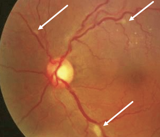
What are Cotton Wool Spots (CWS)?
→ White, yellow, fluffy retinal lesions usually indicative of vascular occlusion
can be the initial sign of SLE and subclinical glomerulonephritis
What are the 3 primary categories of renal function?
Excretory - removing waste (ammonia, urea nitrogen) in the form of urine
Regulatory -maintaining fluid/ion/acid-base balance
Endocrine - hormone production
Which 3 hormones are produced and released by the kidneys?
Renin - regulate BP
Erythropoietin/EPO - make RBC
Calcitriol - activate Vitamin D for Ca2+ absorption
How is it Erythropoietin produced?
→ made by erythropoietin producing cells found between PCT & peritubular capillaries
read O2 levels
if O2 drops → EPO is released into the blood
What role do the PCT play in Vitamin D activation?
Proximal convoluted tubular epithelial cells convert inactive Vitamin D (25-hydroxyvitamin D) → active 1,25-dihydroxyvitamin D (calcitriol) using 25(OH)D-1α-hydroxylase (1-hydroxylase)
Why is the right kidney typically lower than the left kidney?
During embryogenesis, the kidneys rise from the pelvis
right kidney "hits its head" on the liver sooner & stays lower
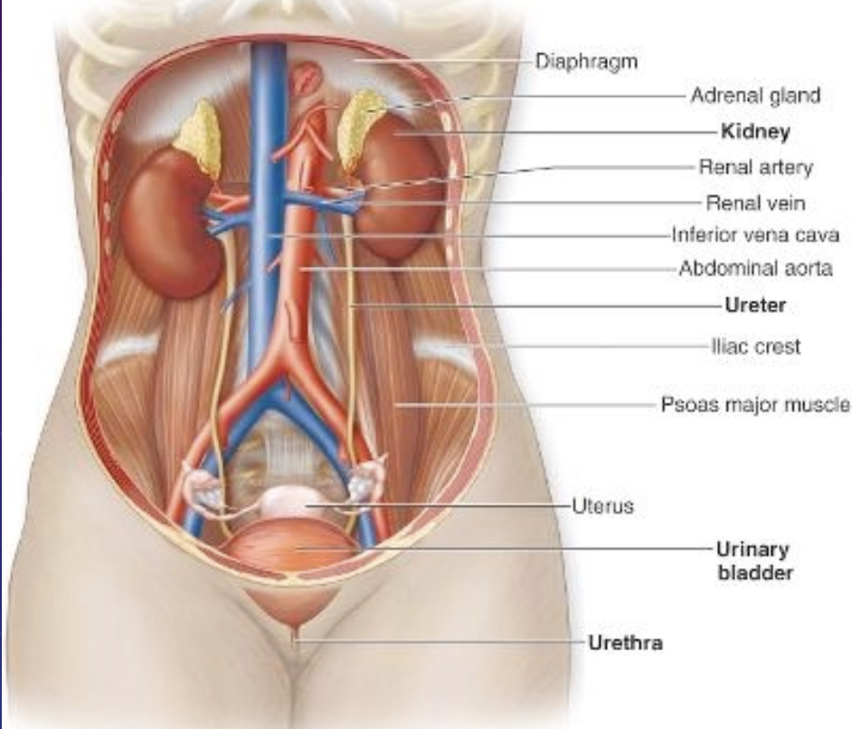
What does "retroperitoneal" mean for the kidneys?
→ located “behind” the peritoneum
b/c of this, kidney disease presents as flank pain (pain on the sides of the back)
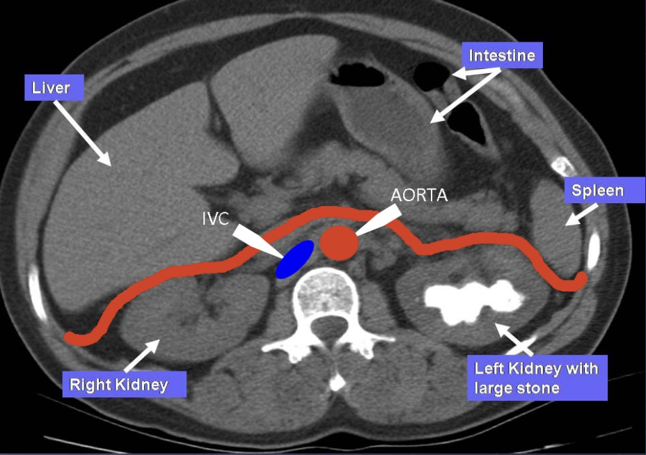
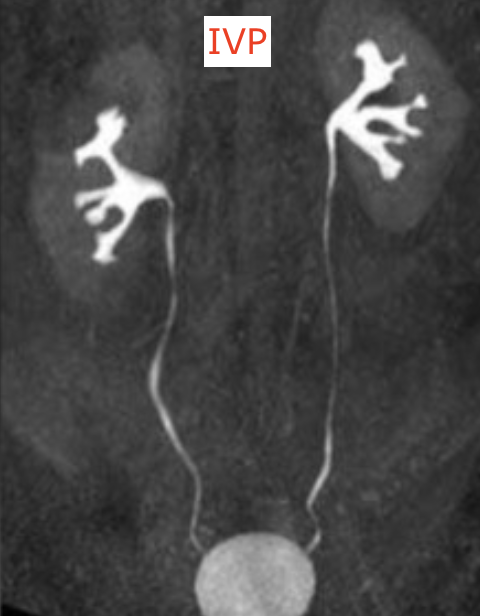
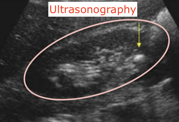
How do Intravenous Pyelograms (IVP) and Ultrasonography differ?
IVP - uses iodine-based dye to trace urine pathway
urine moved via peristalsis
Ultrasonography - used frequently to avoid dye reactions when looking for obstructions (e.g., stones)
Define the Uriniferous Tubule.
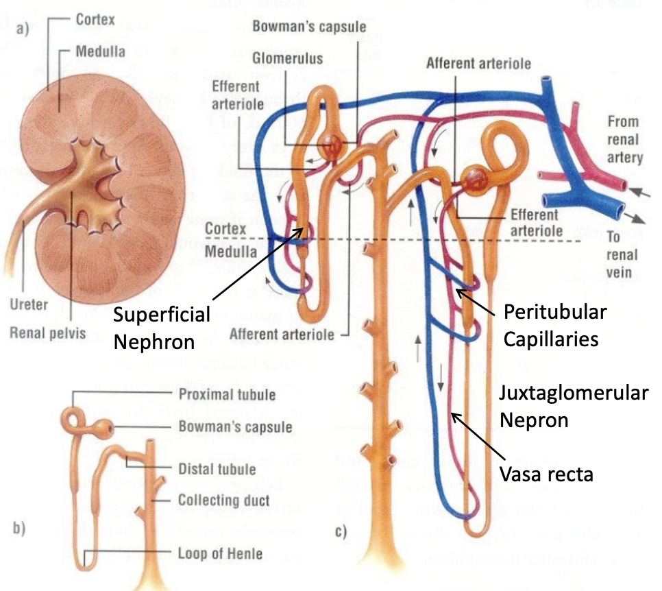
CT + ALL the nephrons that feed into it
Define the Nephron.
Renal corpuscle + its associated duct system (PT, LOH, DT)
Define the Renal Corpuscle.
Glomerulus (capillary tuft) + Bowman’s space + Bowman’s capsule
What percentage of cardiac output reaches the kidneys?
~25% (about 1.25 L/min or 1800L per day)
What is the difference between Renal Blood Flow and Renal Plasma Flow (RPF)?
RPF - accounts only for the plasma (~55% of blood volume)
kidney acts on plasma to filter 180L/day, reabsorbing 99% of initial filtrate
What is Total renal blood flow proportional to?
pressure difference b/w the renal artery & renal vein
What is Total renal blood flow inversely proportional to?
total resistance of renal vasculature
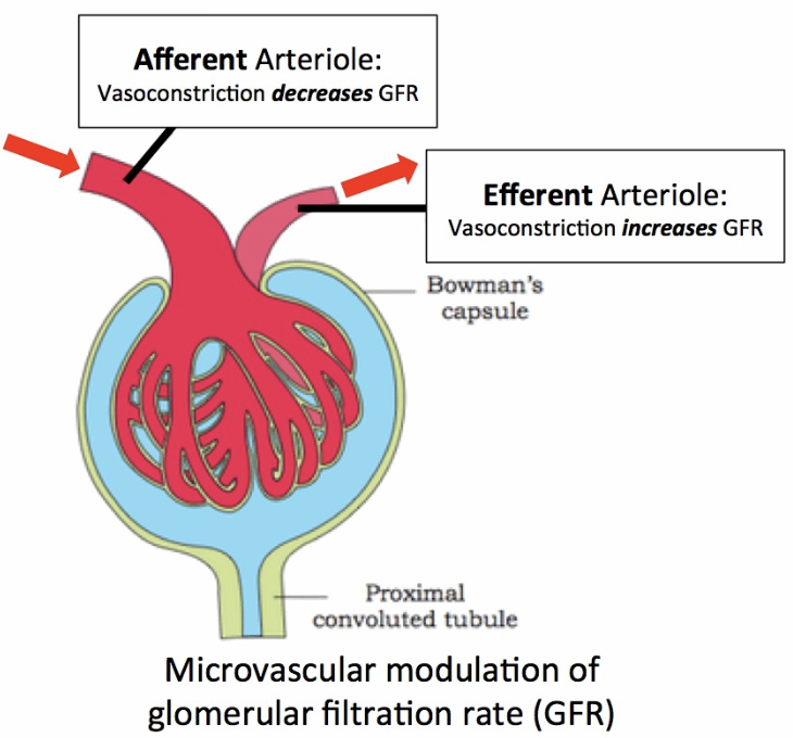
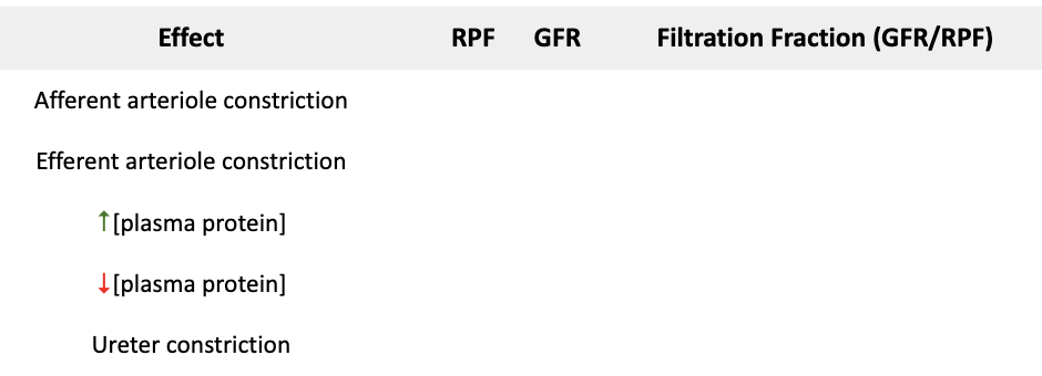
How does arteriolar constriction affect the Glomerular Filtration Rate (GFR)?
Afferent constriction = ↓ GFR
Efferent constriction = ↑ GFR
Why is the independent control of constriction & dilation of the afferent and efferent arterioles important? What does it imply?
→ helps control GFR
implies that there’s no drop in hydrostatic pressure when capillaries lead to venules
How do sympathetics regulate GFR?
↑ sympathetic tone (e.g., in blood loss) → net vasoconstriction (mediated by α1 receptors on afferent side) → ↓ GFR to retain fluid
Note: Both afferent and efferent arterioles receive SNS input, but there’s more afferent arterioles → ↓ GFR
What is the dose-dependent effect of Angiotensin II on GFR?
↓ Ang 2 constrict only efferent arteriole = ↑ GFR
↑ Ang 2 constrict both afferent & efferent arteriole = ↓ GFR →↑ BP
What is the role of Prostaglandins (PGE2, PGI2) in the kidney?
→ produced locally by macula densa cells in DCT to cause vasodilation of afferent & efferent arteriole
act as a “check” against excessive vasoconstriction to prevent renal failure
Why should NSAIDs be avoided in situations like hemorrhage?
↓ prostaglandin synthesis which can interfere with the compensatory vasodilation that occurs during hemorrhage
List some vasodilators.
NO
Bradykinin
Dopamine
What is Autoregulation in the kidney?
→ ability of renal vessels to constrict or dilate based on pressure differences on its wall without needing autonomic inputs
autoregulation ensures constant blood flow to kidneys during varying pressure
Name the 3 cell types of the Juxtaglomerular Apparatus (JGA). Describe them.
Juxtaglomerular cells
occupy the tunica media of the afferent arteriole
receive SNS innervation
contain renin
Macula densa cells
modified cells of DCT
sense Na+ in filtrate near the afferent & efferent arteriole junction
Mesangial cells
provide mechanical stability ensuring macula densa & afferent + efferent arteriole remain locked together as a unit
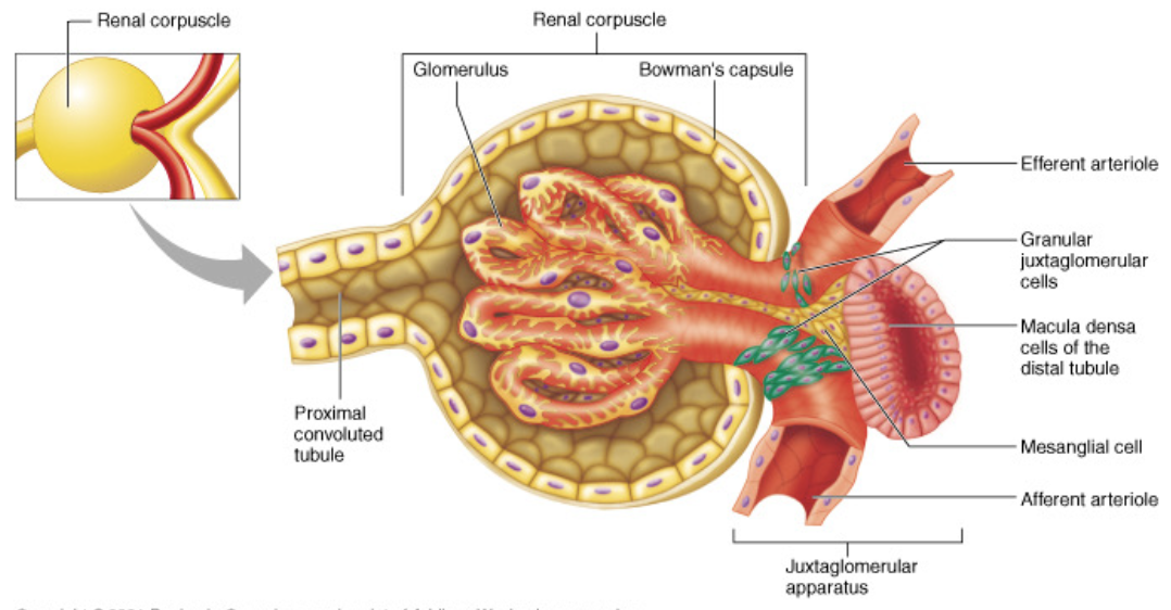
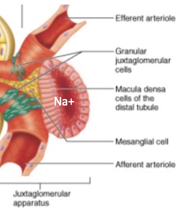
How do Macula Densa cells initiate the tubuloglomerular feedback loop?
→ Senses ↓ Na+ (indicating ↓ BP/filtration)
Afferent arteriole dilation → ↑ GFR
↑ Renin release from juxtaglomerular cells via paracrine stimulation → activates RAAS → ↑BP
What are the 3 triggers for Renin release?
↓ Na+ detected by macula densa
SNS input (Eph/NE binding to β1 receptors)
Direct response to ↓ renal perfusion pressure
Outline the RAAS pathway from Renin to Aldosterone.
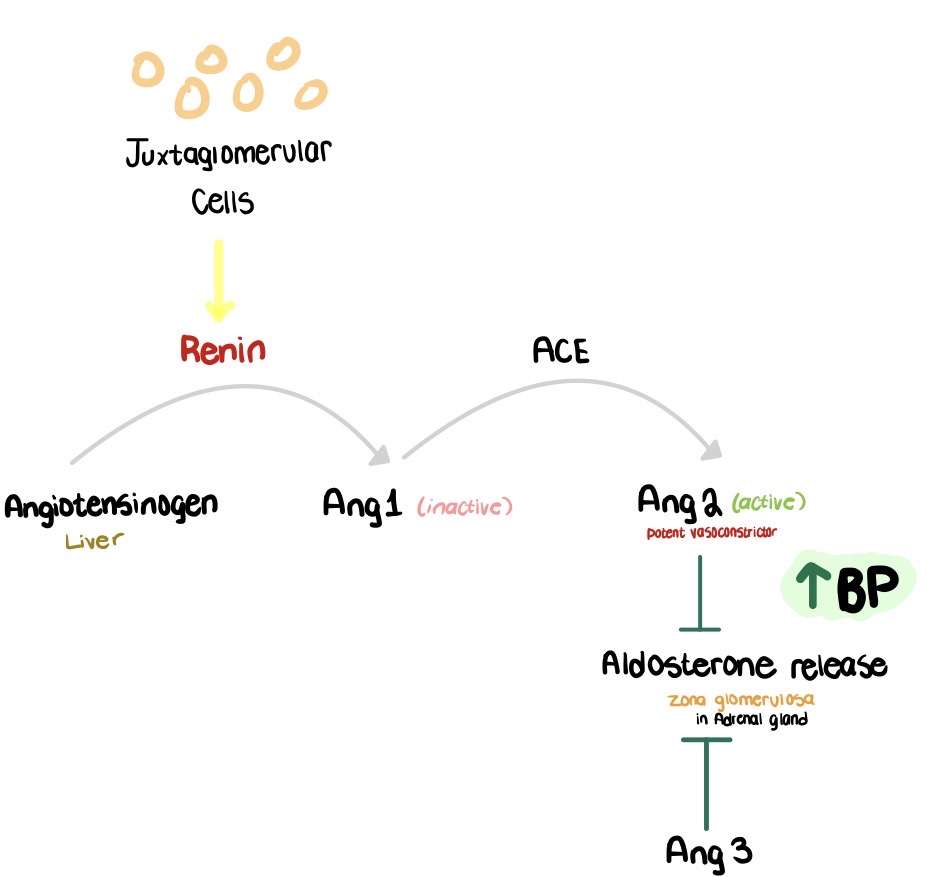
List the functions of Ang 2.
potent vasoconstrictor (↑ BP)
inhibits NO (vasodilator)
stimulates aldosterone release → Na+ and H2O retention → ↑ BP
What is Ang 3? Mention what it’s known as?
→ stimulates aldosterone release just like Ang 2, but doesn’t contribute to “constriction” as much
aka: des-ASP heptapeptide
True or False - The concentration of ACE in the bloodstream influences how much BP can increase.
False - ACE is just an enzyme, and it itself doesn’t raise blood pressure. If there is not excess Ang 1 around, the amount of ACE is in the blood doesn’t matter
What is the function of ACE2?
Cleaves Ang 2 → Angiotensin 1-7 = ↓ BP
countering the effects of ACE
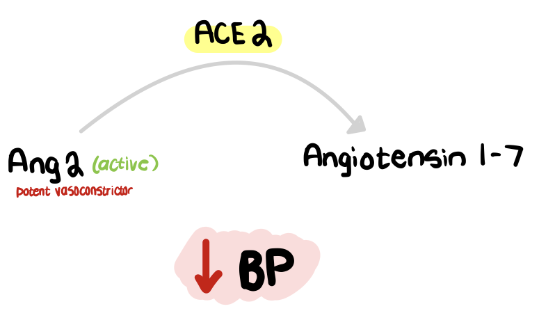
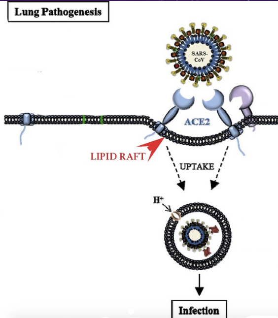
What is the clinical relevance of ACE2 and COVID-19?
ACE2 (bound to lung, heart, renal, intestinal cells) acts as the receptor used by COVID-19 to enter target cells
List the 6 major factors that control Renin secretion.
Renal vascular receptor responds to changes in tension in the afferent arteriolar wall
Macula densa detecting changes in Na+ in DT
Circulating angiotensin has (-) feedback on renin secretion
SNS stimulates renin secretion via the renal nerve
Endothelin
↓ ANP (↑ renin)
What is Endothelin?
→ powerful, vascular, endothelial vasoconstrictor
produces a salt sensitive ↑ BP
activates local RAAS
What is NO?
→ Endothelial derived relaxant factor
produced by arterial and venous endothelium → diffuses through the vessel wall into the smooth muscle causing vasodilation
What is The Atrial Natriuretic Peptide (ANP)?
→ hormone released by atria in response to ↑ blood volume that ↑ Na+ & water excretion (natural diuretic)
↓ ANP = fluid retention & HTN
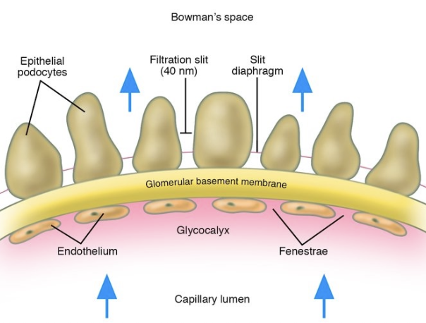
Describe the components of the Glomerular Capillary filtration barrier.
Large endothelial pores (70-100nm)
Thick basement membrane with small pores (allows plasma protein to pass)
Podocyte foot processes with filtration slits (25-60nm)
Glycocalyx
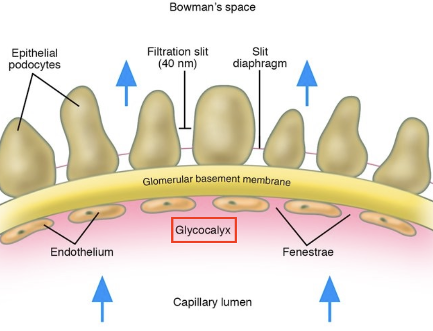
What is the significance of the Glomerular Glycocalyx?
→ highly (-) charged, repelling plasma proteins → ↓[protein] urine
disruption → proteinuria (protein in urine)
How do Starling Forces differ in the glomerulus compared to other capillaries?
theres’s no venous end only arterial on both ends
Hydrostatic pressure is dominant along the entire length → fluid never moves back into the capillary

PGC– PBS = pressure difference across the vessel wall
Ki = permeability of the filtering membrane
πGC = oncotic pressure created by [protein] difference inside vs outside in Bowman’s space
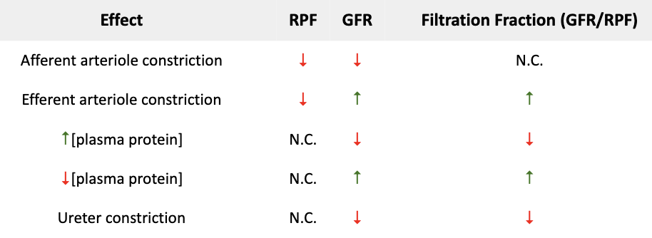
Why does the efferent arteriole blood promote fluid reabsorption in the proximal tubule?
Glomerulus filtration → ↑ [protein] in efferent arteriole → ↑ oncotic pressure in peritubular capillaries → ↑ fluid reabsorption from PT
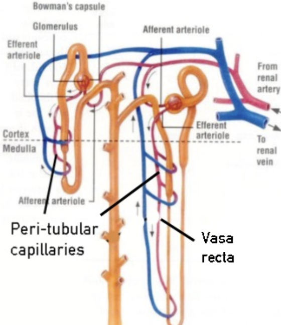
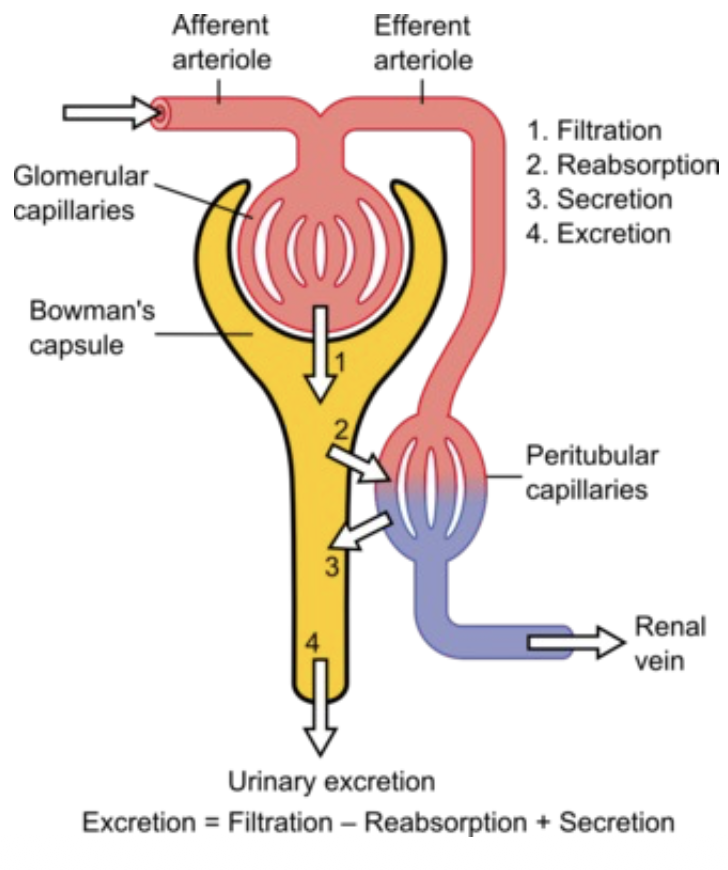
What’s the difference between secretion and excretion?
Secretion: Transferred from blood to filtrate
Excretion: Final urine composition
Excretion = Filter - Reabsorb + Secretion
Why is Inulin the ideal marker for GFR?
freely filtered
not reabsorbed or secreted
its clearance rate is a direct measure of GFR
Where is Creatinine produced? Where are its levels measured?
→ produced by the muscle at a constant rate
measured in blood and urine
Why is GFR calculated from Creatinine called "estimated" (eGFR)?
→ Creatinine is filtered but a small amount is also secreted
this can cause blood levels to slightly underestimate GFR & overestimate filtration
↓ GFR declines = ↑ creatinine levels in blood
What are Serum creatinine levels in men and women?
Men = 0.7-1.3 mg/dl
Women = 0.6-1.1 mg/dl
higher in men b/c they have more muscle
What is BUN (Blood Urea Nitrogen), and where is it reabsorbed?
→ amount of nitrogen in your blood that comes from the waste product urea
reabsorbed in the PT and CT (via ADH)
How does BUN/ Creatinine ratio change?
Dehydration, bleeding → retains BUN and creatinine → ↑BUN/creatinine ratio
What is the BUN/creatinine ratio a valuable indicator for?
→ Distinguishing whether ↓GFR is from extrarenal or renal causes
If ↓GFR is renal cause → both BUN & Creatine will reduce closer to 1:1
What is the Normal BUN/ Creatinine ratio?
10:1 to 20:1
What does a BUN/Creatinine ratio > 20:1 typically indicate? List the factors that can cause this.
→ Extrarenal cause of ↓GFR
Dehydration
Gut bleeding
Hyperthyroidism
Congestive heart failure
Kidney disease
Drugs (tetracycline, corticosteroids)
What does a BUN/Creatinine ratio < 10:1 typically indicate? List the factors that can cause this.
→ Renal causes of ↓GFR
Low protein intake
Advanced liver disease
Sickle cell anemia
Hypothyroidism
Rhabdomyolysis
Kidney damage & failure
Drug (acetazolamide)
What is a Kidney dialysis?
→ done to ↓ ammonia levels in blood
IV Na+ benzoate and phenylacetate promote nitrogen disposal
arginine is supplemented for NO production
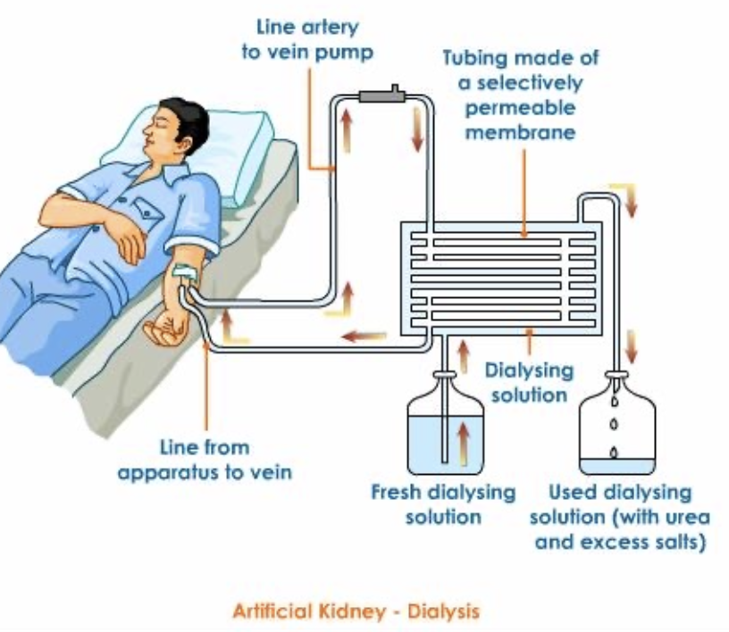
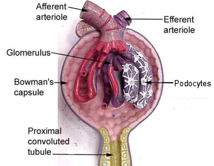
Describe the series of steps that occurs before Urine formation.
Plasma under pressure passes into the glomerulus (highly permeable)
Ultrafiltrate of plasma is “sieved” into the Bowman’s space
Ultrafiltrate passes into PCT
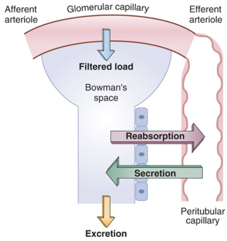
Once the filtrate is formed, what 3 processes occur?
Reabsorb from filtrate to blood via tubular epithelial cell
Secrete organic acids, bases & K+ from blood to filtrate
Excretion
Why is Na⁺ important for proximal tubule reabsorption?
Na⁺/K⁺ ATPase pump creates a Na⁺ gradient by pumping Na+ out to basal side, that allows Na⁺ to enter from the filtrate at the apical side
Na⁺ movement drives the reabsorption of glucose, amino acids etc, while water follows via osmosis
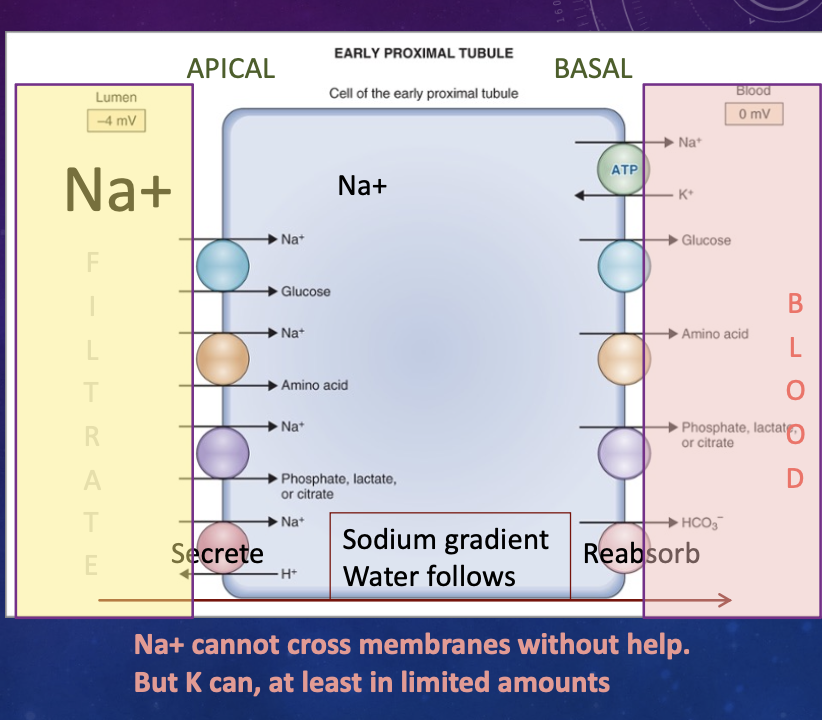
What happens in the Early Proximal Tubule?
Reabsorption into blood:
100% glucose &nd AA
85% HCO3-
67% Na+ & water
.
Secretion into filtrate:
H+ for pH balance
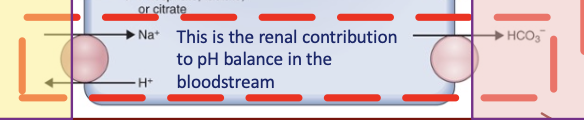
What is the role of CA in the renal contribution to pH balance in the bloodstream?
Carbonic anhydrase generates H+ + HCO3-
excreting H+ into the forming urine → ↑ pH in blood
HCO3- bind additional H+ to ↑ pH in blood more
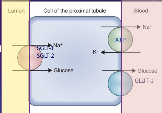
How is Glucose handled in the PCT?
Reabsorbed via:
SGLT-1 & SGLT-2 co-transporters (apical) - Na+ & glucose
GLUT-1 transporters (basal) - transports glucose using the Na+ gradient
normally ALL glucose reabsorbed before the LOH
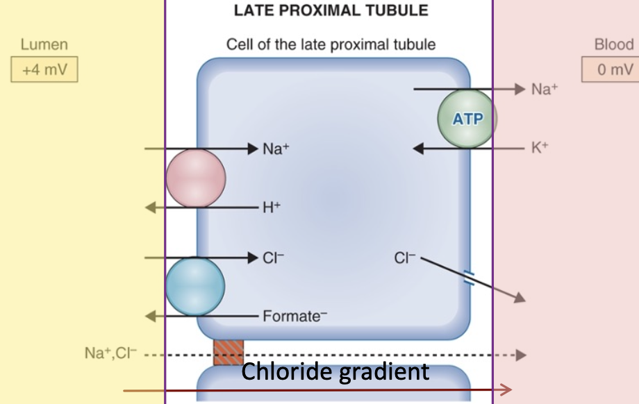
What occurs in the Late PCT?
Lots of Cl- in blood which enters via anionic antiporter based on formate → Cl- diffuses along its gradient to the blood
Na & Cl also pass paracellularly along their gradients
where Kidney converts inactive Vit D → active Vit D, releasing into blood
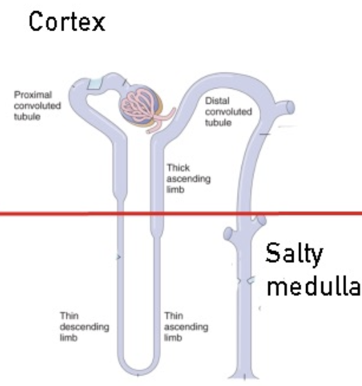
List the 3 parts of the LOH.
Thin descending
Thin ascending
Thick ascending
→ paralleled by Vasa recta
Describe the permeability of the LOH limbs.
Thin Descending limb: Permeable to water, NOT ions → water gushes out
Thin Ascending limb: Permeable to ions, NOT water → ions flow out
Thick ascending limb: impermeable to water due to tight junctions
can pumps ions using basal Na-K ATPase pump and 2er active transport on apical side via Na+, K+, 2Cl- co-transporter → hypertonic medulla
How do Loop Diuretics (e.g., Furosemide) work?
→ Block Na+-K+-2Cl- co-transporterin thick ascending limb
prevent water reabsorption → diuresis
How do Thiazide Diuretics work?
Block Na-Cl co-transporter in DCT
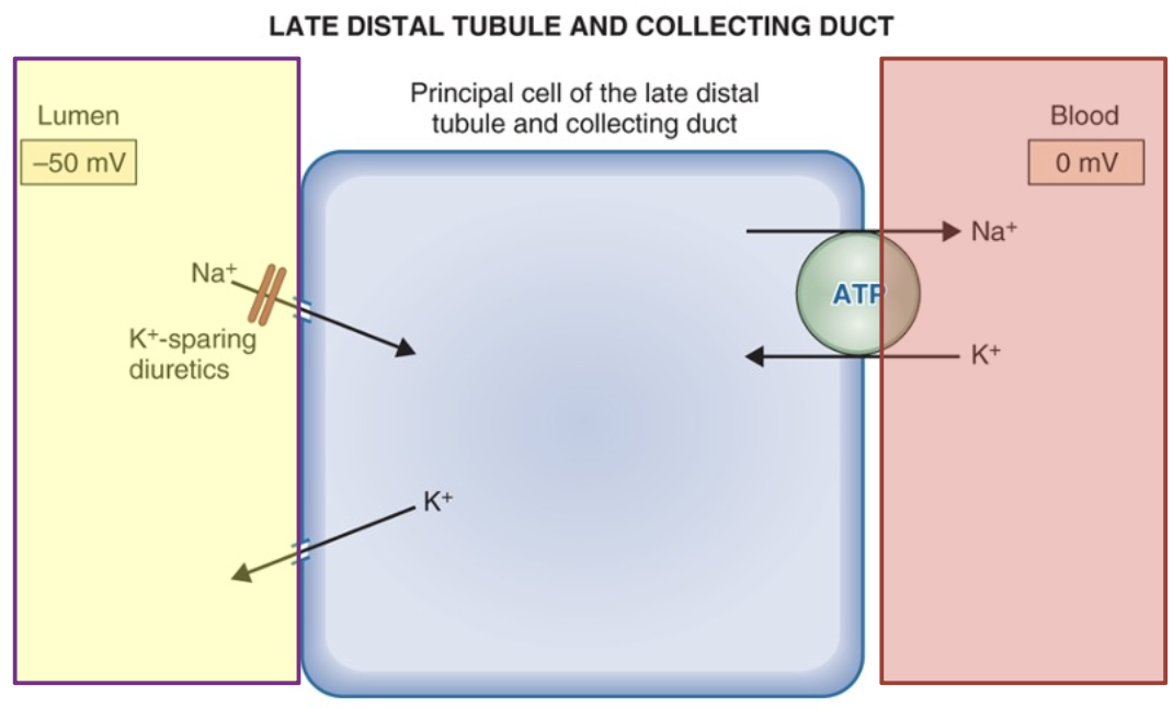
What are the roles of Aldosterone and ADH in the collecting ducts?
Aldosterone = ↑ Na+ & water reabsorption
ADH = ↑ water & urea permeability (concentrating the urine)
released by thirst
Why does the CT continue to reabsorb water as it passes through the medulla?
CT passes back through the hypertonic medulla as it travels toward the renal papilla → causes water to move out of the CT (additional water reabsorption)
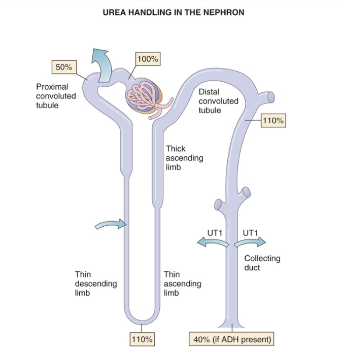
How is urea handled along different parts of the kidney?
PT → 50% urea reabsorbed
Thin loop → urea secreted back
Medullary CT + ADH → urea reabsorbed to maintain medullary osmolarity
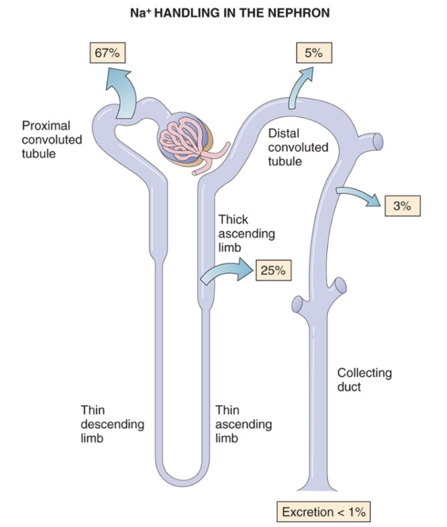
How is Na+ handled along different parts of the kidney?
PT → 66% Na⁺ reabsorption
Thick ascending limb LOH → additional Na⁺ reabsorption
Collecting ducts → final 3–4% of Na⁺ reabsorption (controlled by aldosterone)
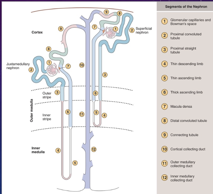
Compare Juxtamedullary vs. Superficial nephrons.
Juxtamedullary nephrons (20-30%) have loops that dive deeper into the salty medulla → more concentrated urine
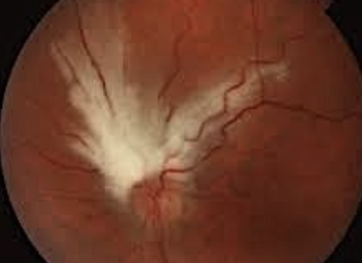
How does Myelinated Nerve Fibers present the eye?
Cause: Myelination fails to stop at the lamina cribrosa
Effect: Blocks light to photoreceptors → non-progressive VF defect