ANATOMY LAB 1
 Call Kai
 Learn
Learn Practice Test
 Spaced Repetition
 Match
Match Flashcards
 Knowt Play
1/59

There's no tags or description
Looks like no tags are added yet.
Name | Mastery | Learn | Test | Matching | Spaced | Call with Kai | Chat |
|---|
No analytics yet
Send a link to your students to track their progress
60 Terms
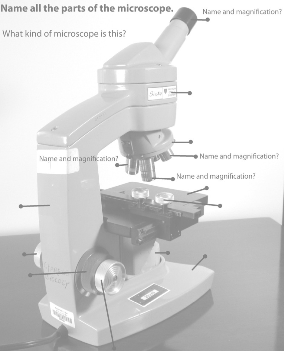
Name the parts of the microscope.

What is the ocular lens used for?
Lens you look through;
magnifies image about 10×

What are the objective lenses?
Lenses that have different magnifications
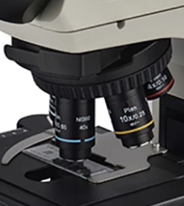
What is the scanning objective lens (SP)?
Scanning power (SP)
Lowest magnification 5X
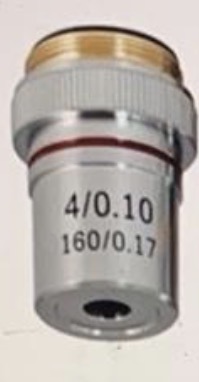
What is the lower power objective lens (LP)?
Used to view more detail 10X

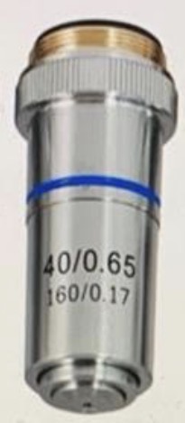
What are high power objective lens (HP)?
Used for greater detail 40-45X
What is the oil immersion objective lens (OI)?
Highest magnification; 100X
Only used with immersion oil

What is the body tube of a microscope?
Holds/connects the ocular and objective lenses

What is the rotating nose piece?
Holds the objective lenses and rotates to change magnification

What is the stage?
Supports the slide being viewed

What is the Iris diaphragm?
Controls the amount of light reaching the specimen
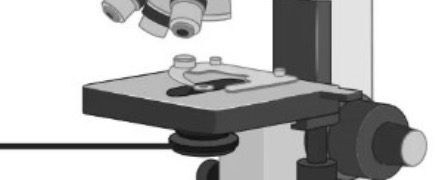
What is the rheostat?
control that adjusts the brightness of the light

What is the Illuminator?
light source for the microscope
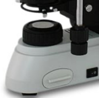
What are the stage clips?
Holds the slide in place
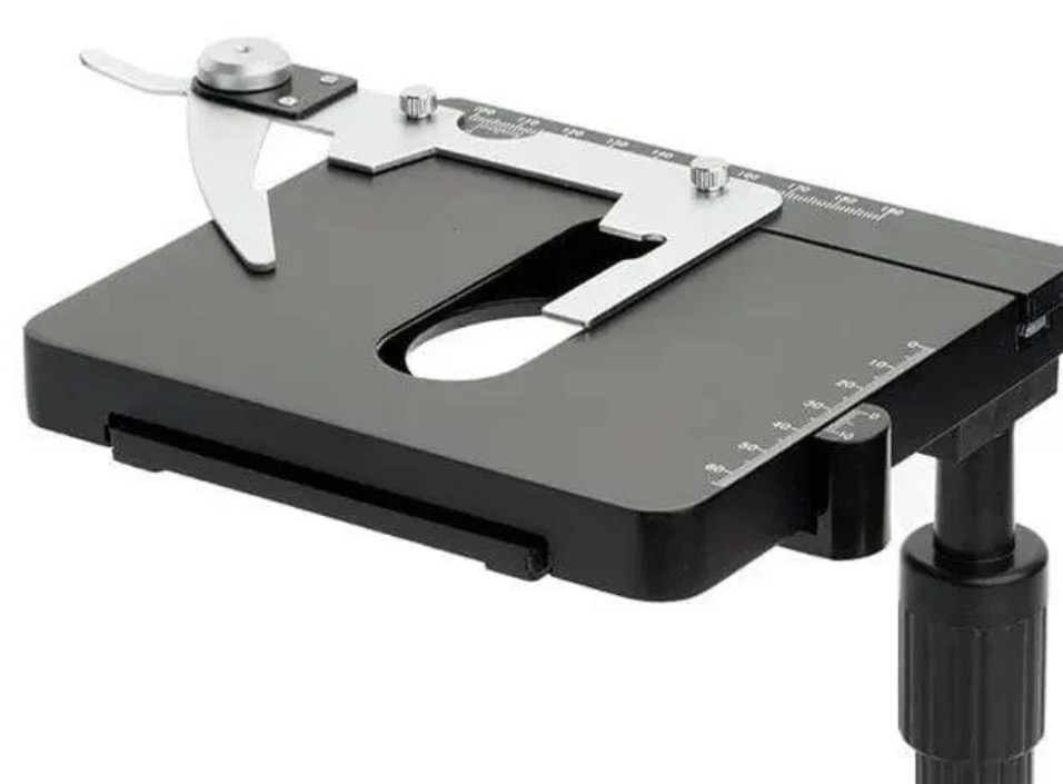
What is the manipulator in a microscope?
Moves the slide around
What is the base?
Supports the microscope
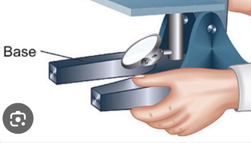
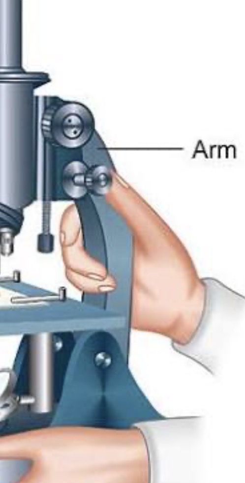
What is the arm?
Used to support the microscope when carried
What is the Coarse focus knob?
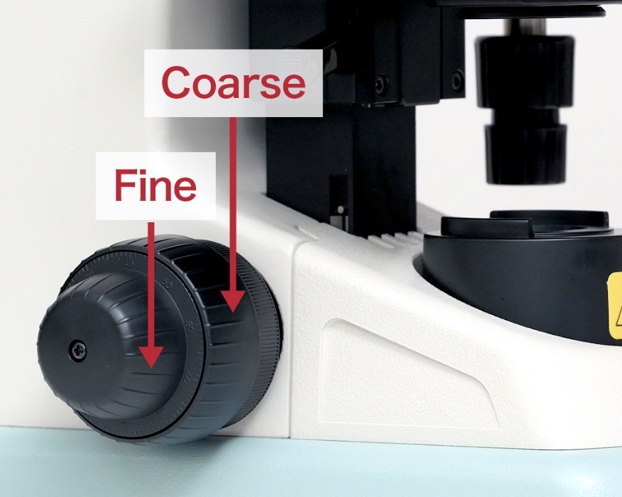
Moves the stage/objective quickly for initial focusing

What is the Fine focus knob?
Makes small adjustments to sharpen the image
What are the eight safety rules for the practicum?
1. Always store microscope without any specimen on stage
2. Always store microscope with SP or LP objective lens rotated into position and with maximum distance between lens and stage.
3. Load specimen onto stage only when in SP or LP is rotated into position.
4. Always begin viewing a specimen with the SP or LP objective lens, never the HP or Ol objective lens.
5. Only clean a microscope lens with official "lens paper."
6. Always carry a microscope with 2 hands. Strong hand on microscope arm and weak hand on microscope base.
7.Never use the course focus knob with the high power or oi mmersion objective lenses
8. Use the course focus knob only when using the SP or LP objective lenses.
What are the steps while using a microscope?
1. Plug electrical cord into a working electrical socket.
2. Switch on illuminator.
3. Be sure that all the objective lenses are securely screwed into the nosepiece.
4. Rotate nosepiece so that SP or LP lens is clicked into position. Always begin to locate a specimen by using the scanning or LP objective lens. Never start to locate a specimen by using the HP or Ol lens.
4. Place specimen on stage and center over the condenser lens.
5. Dim the light using the iris diaphragm or rheostat.
6. Lower the objective lens all the way down to the specimen using both the course and fine focus knobs, if necessary.
7. Now look into the ocular and begin to find the specimen using the fine focus knob to slowly raise the objective lens. Turn the fine focus knob about 1 turn. If you do not see specimen then go to # 8, otherwise go to # 9.
8. Now use the course focus knob to slowly raise the objective lens until the specimen comes into rough focus.
9. Use the fine focus knob and clearly focus on the specimen.
10. Use the iris diaphragm to adjust the light for best contrast.
11. If the specimen is thin you may now switch to the next higher power objective lens. Do not move ANY focus knob when switching from LP to HP objective lenses. You do not have to be unduly concerned about crashing the lens into the specimen as long as you do not touch the focus knobs.
12. When in HP, use ONLY the fine focus knob to clearly focus, and the iris diaphragm to adjust the light for best viewing sharpness and contrast.
What does the term parfocal mean?
when you change from one objective lens to another, the specimen stays mostly in focus
What is a cell?
The smallest basic unit of life
What is the nucleus?
the central core or control center of a cell, an atom, or group
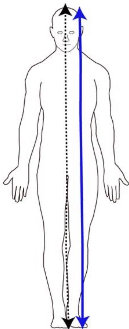
What is sagittal?
Divides body into RIGHT and LEFT
(Vertical)

What is midsagittal (median) plane?
divides the body into equal left and right halves
(Vertical)

What is a parasagittal plane?
divides the body into unequal left and right portions
(Vertical)
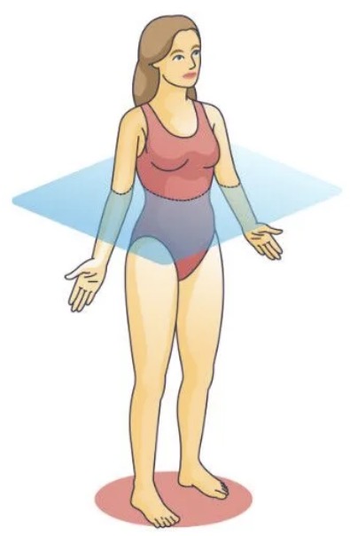
What is a transverse (horizontal or cross sections) plane?
divides the body into upper and lower portions as
What is a coronal (frontal) plane?
divides the body into front (anterior) and back (posterior) portions
(Vertical)
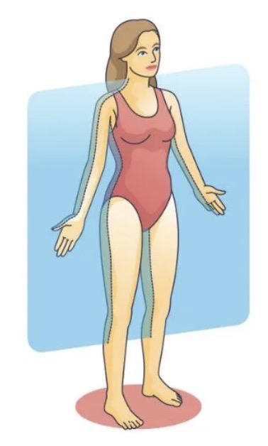
What is Anterior (ventral)?
toward the front of the body
Ventral=Belly
Face, chest, and abdomen
Your chest is anterior to your spine because your chest is in front of your spine.

What is Posterior (dorsal)?
toward the back of the body
Dorsal=Back
spine is posterior (dorsal) to your heart because the spine is behind the heart

What is superior?
toward the top of the body or above another body part
head is superior to your chest
chest is superior to your stomach
What is inferior?
toward the bottom of the body or below another body part
Your stomach is inferior to your heart
Your feet are inferior to your knees
What is cranial?
related to the skull (cranium) or the head
What is caudal?
toward the tail or lower end of the body
What is medial?
toward the middle of the body or closer to the body’s midline
Your nose is medial to your eyes
Your heart is medial to your arms
What is lateral?
away from the middle of the body
arms are lateral to your heart because they are farther away from the middle
What is proximal?
closer to where a body part starts/attaches
Shoulder is proximal to the elbow because it’s closer to the body
Elbow is proximal to the wrist
What is distal?
farther away from the body
The fingers are distal to the elbow
Because the fingers are farther away from the body than the elbow
What is the dorsal cavity?
HOLDS BOTH-
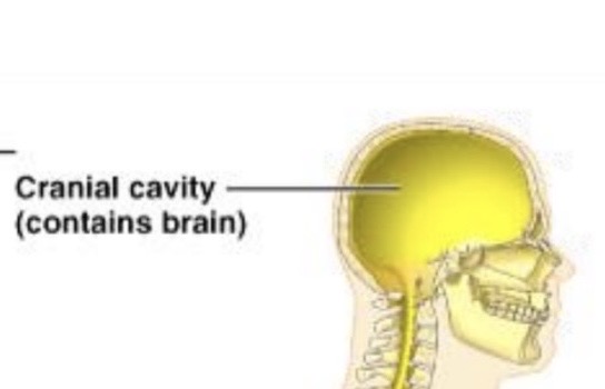
Cranial cavity → holds your brain
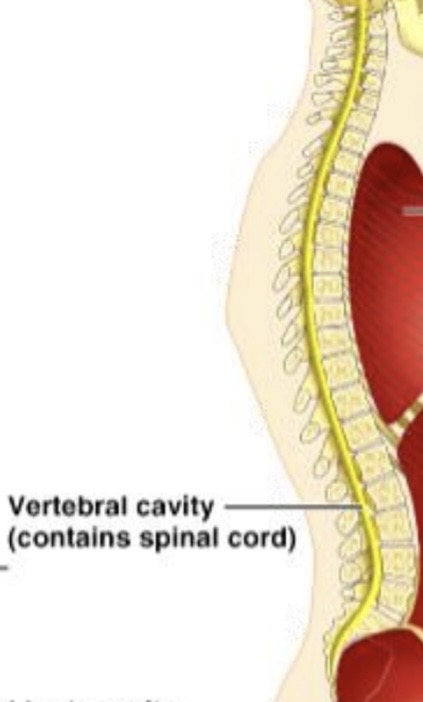
Vertebral (spinal) cavity → holds your spinal cord

What is the cranial cavity?
holds your brain
Inside dorsal body cavity
What is the vertebral cavity?
holds the spinal cord
Inside the dorsal body cavity
What is the ventral cavity?
FRONT
Contains:
↳ Thoracic cavity → chest
↳ Pleural → lungs
↳ Pericardial → heart
↳ Mediastinum → esophagus/trachea

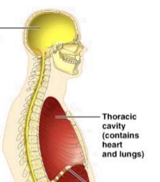
What is the thoracic cavity?
Inside ventral cavity
The cavity is inside your chest
Inside it are smaller spaces:
Pleural cavities → lungs
Pericardial cavity → heart
Mediastinum → trachea and esophagus
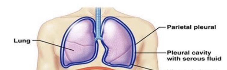
What is the pleural cavity?
space around each lung that contains pleural fluid
Inside thoracic cavity
What is the pericardial cavity?
space around the heart
Inside thoracic cavity

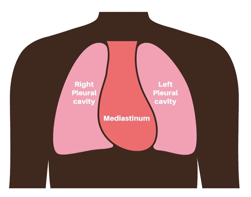
What is the mediastinum?
space in the middle of your thoracic (chest) cavity, between your two lungs.
Inside thoracic cavity
contains the esophagus and trachea
What is the abdominopelvic cavity?
large space below your chest that contains many of your organs
Contains abdominal and pelvic cavity
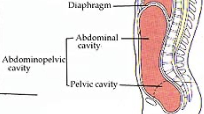
What is the abdominal cavity?
BELLY
space where my stomach and intestines are
Contains:
Stomach, Liver, small and large Intestines

What is the pelvic cavity?
space inside your pelvis, basically the lower belly area
Contains:
Bladder

What is the oral cavity?
space inside your mouth
What is the nasal cavity?
space inside your nose
What are the orbital cavities?
spaces in your skull that hold your eyes
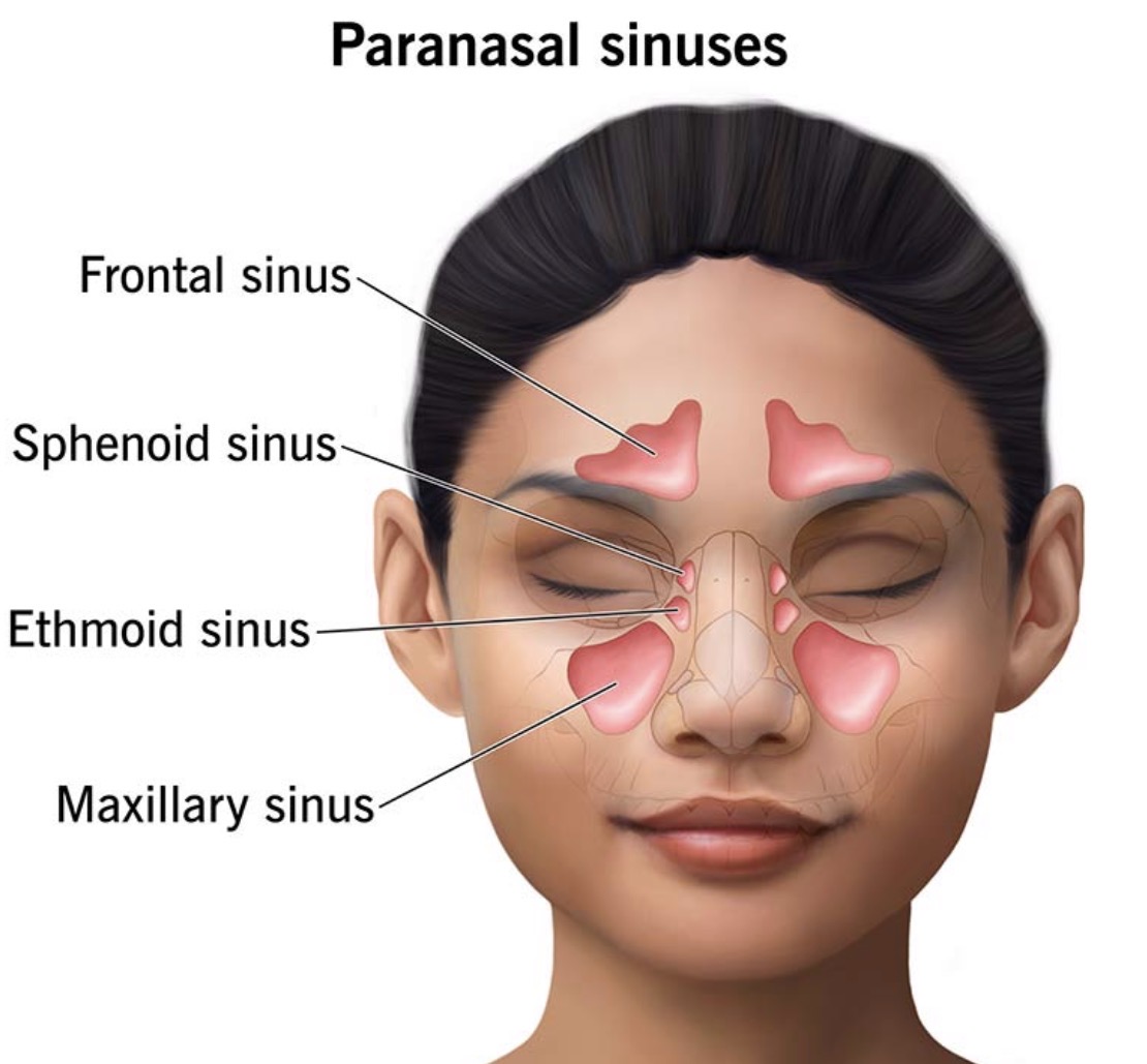
What are the paranasal sinuses/cavities?
air-filled spaces around your nose
What is mitosis?
phase of life cycle when cell divides
Chromosomes are condensed and duplicated
Consist of 2 duplicate strands called chromatids
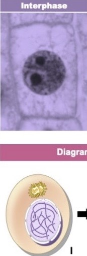
What is interphase?
cell is getting ready
“before the cell divides.”
cell grows
The cell does its normal job
The cell copies its DNA so it’s ready to make 2 cells
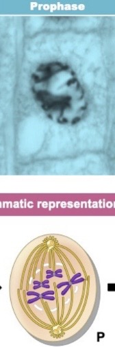
What is prophase?
chromosomes become visible distinct structures
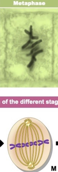
What is metaphase?
chromosomes line up single file along equator(middle)
– Positioned there by spindle fibers
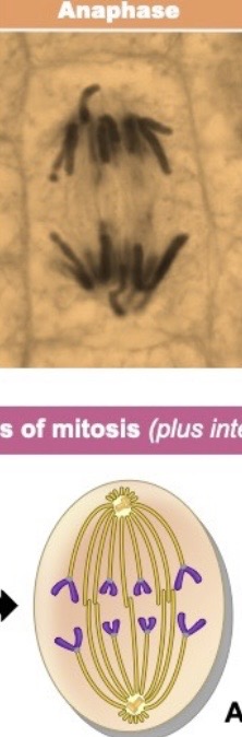
What is anaphase?
centromeres split
– Spindle fibers pull each chromatid to opposite poles
two groups of chromosomes, moving away from the middle
What is telophase?
cytoplasm is divided (cytokinesis), producing 2 daughter cells
