chapt. 24 PENN The Fetal Head and Brain
1/59
There's no tags or description
Looks like no tags are added yet.
Name | Mastery | Learn | Test | Matching | Spaced | Call with Kai | Chat |
|---|
No analytics yet
Send a link to your students to track their progress
60 Terms
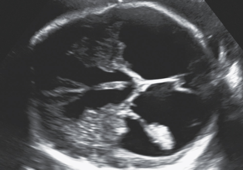
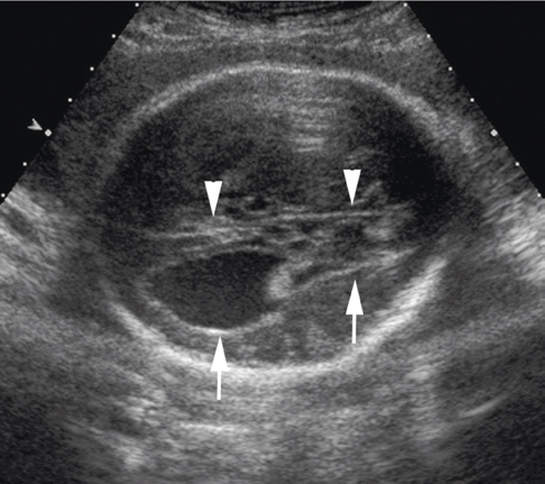
1. The image in Figure 24-31 is indicative of what abnormality?
Figure 24-31
a. Ventriculomegaly
b. Holoprosencephaly
c. Agenesis of the corpus callosum
d. Hydranencephaly
a. Ventriculomegaly

2. What is the sonographic sign in Figure 24-31?
Figure 24-31
a. Pancake sign
b. Monoventricle
c. Dangling choroid
d. Bilateral choroid plexus cysts
c. Dangling choroid
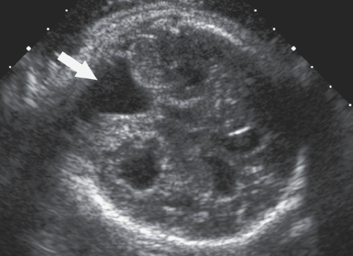
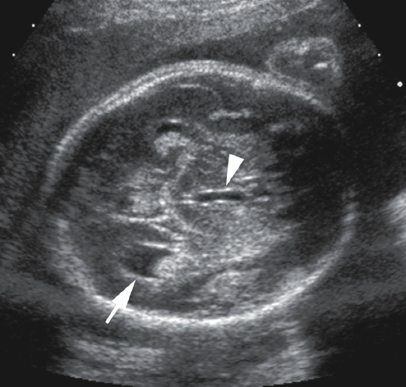
3. What is the abnormality noted by the arrow in Figure 24-32?
Figure 24-32
a. Subarachnoid hemorrhage
b. Porencephaly
c. DWM
d. Hydranencephaly
c. DWM
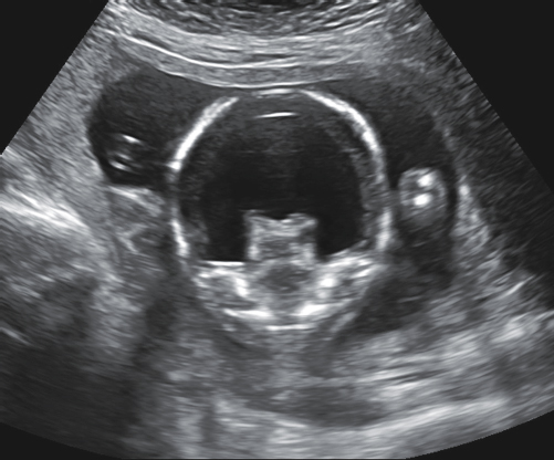
4. Which of the following is the most likely diagnosis for the fetus in Figure 24-33?
Figure 24-33
a. Mega cisterna magna
b. Holoprosencephaly
c. Apert syndrome
d. Anencephaly
b. Holoprosencephaly

5. What sonographic finding is present in Figure 24-33?
Figure 24-33
a. Absent cranial vault
b. Choroid plexus cysts
c. Colpocephaly
d. Fused thalami
d. Fused thalami

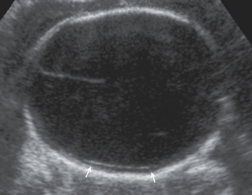
6. What do the arrows in Figure 24-34 indicate?
Figure 24-34
a. Temporal thickening
b. Ethmoidal enlargement
c. Scalloped frontal bones
d. Occipital flattening
c. Scalloped frontal bones
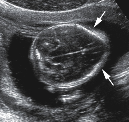
7. What is the sonographic sign indicated by the arrows in Figure 24-34?
Figure 24-34
a. Lemon sign
b. Banana sign
c. Dangling choroid sign
d. Pancake sign
a. Lemon sign

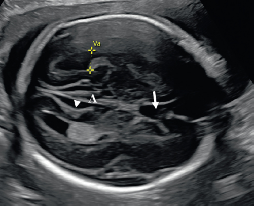
8. The arrowheads in Figure 24-35 indicate the:
Figure 24-35
a. corpus callosum.
b. CSP.
c. falx cerebri.
d. cerebellar vermis.
c. falx cerebri.
9. The arrows in Figure 24-35 are indicating what sonographic finding?
Figure 24-35
a. Colpocephaly
b. Cebocephaly
c. Schizencephaly
d. Dangling choroid
a. Colpocephaly
10. Which of the following is not part of Apert syndrome?
a. Craniosynostosis
b. Aqueductal stenosis
c. Midline facial hypoplasia
d. Syndactyly
b. Aqueductal stenosis

11. What does the arrowhead in Figure 24-36 indicate?
Figure 24-36
a. Cerebral aqueduct
b. Lateral ventricle
c. Third ventricle
d. Fourth ventricle
c. Third ventricle
12. What part of the lateral ventricle is the arrow indicating in Figure 24-36?
Figure 24-36
a. Temporal horn
b. Body
c. Anterior horn
d. Occipital horn
d. Occipital horn
13. Which of the following would be among the list of differential diagnoses for Figure 24-37?
Figure 24-37
a. Dandy–Walker variant
b. Severe ventriculomegaly
c. Ventriculitis
d. Mega cisterna magna
b. Severe ventriculomegaly
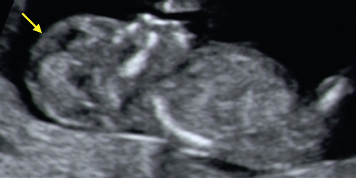
14. What abnormality is indicated by the arrow in Figure 24-38?
Figure 24-38
a. Holoprosencephaly
b. Arnold–Chiari II malformation
c. Acrania
d. Trisomy 18
c. Acrania
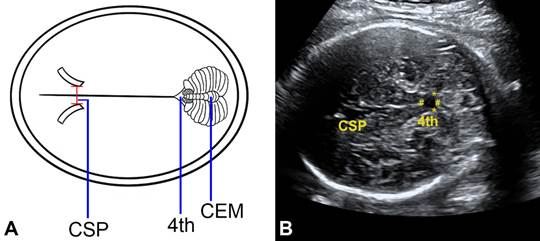
15. What does the arrow in Figure 24-39 indicate?
Figure 24-39
a. Third ventricle
b. Fourth ventricle
c. CSP
d. Cerebral aqueduct
c. CSP
16. What part of the lateral ventricle is routinely measured within the fetal cranium?
a. Atrium
b. Frontal horn
c. Temporal horn
d. Occipital horn
a. Atrium
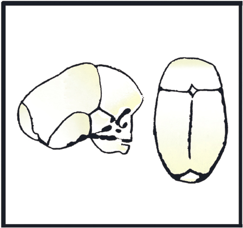
17. What does the drawing in Figure 24-40 represent?
Figure 24-40
a. Dolichocephaly
b. Cloverleaf skull
c. Brachycephaly
d. Microcephaly
a. Dolichocephaly
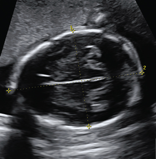
18. What measurement is being obtained with caliper #2 in Figure 24-41?
Figure 24-41
a. BPD
b. HC
c. TCD
d. OFD
d. OFD
19. Which of the following is thought to be caused by bilateral occlusion of the fetal internal carotid arteries?
a. Hydrocephalus
b. Holoprosencephaly
c. DWM
d. Hydranencephaly
d. Hydranencephaly

20. Which of the following is typically not seen at the level demonstrated in Figure 24-41?
Figure 24-41
a. Thalamus
b. Fourth ventricle
c. Area of the third ventricle
d. CSP
b. Fourth ventricle
21. With what structure does the posterior fossa cyst associated with DWM communicate?
a. Fourth ventricle
b. Third ventricle
c. Cerebellar vermis
d. Cerebral aqueduct
a. Fourth ventricle
22. The choroid plexus cyst could be associated with an increased risk of:
a. trisomy 13.
b. trisomy 4.
c. Arnold–Chiari II malformation.
d. trisomy 18.
d. trisomy 18.
23. All of the following are sonographic findings of Arnold–Chiari II malformation except:
a. enlarged massa intermedia.
b. hydrocephalus.
c. obliteration of the cisterna magna.
d. strawberry sign.
d. strawberry sign.
24. Which of the following is located on both sides of the midline?
a. Interhemispheric fissures
b. Third and fourth ventricles
c. Lateral ventricles
d. Third ventricle and cerebral aqueduct
c. Lateral ventricles
25. Which of the following will also typically be absent with agenesis of the corpus callosum?
a. Cerebellar vermis
b. CSP
c. Third ventricle
d. Fourth ventricle
b. CSP
26. The double fold of dura mater that divides the cerebral hemispheres is the:
a. cerebellum.
b. CSP.
c. corpus callosum.
d. falx cerebri.
d. falx cerebri.
27. The development of fluid-filled cleft within the cerebrum is consistent with:
a. holoprosencephaly.
b. lissencephaly.
c. schizencephaly.
d. hydranencephaly.
c. schizencephaly.
28. The anechoic midline brain structure located between the frontal horns of the lateral ventricles is the:
a. CSP.
b. cavum vergae.
c. corpus callosum.
d. fourth ventricle.
a. CSP.
29. The “sunburst” of the cerebral sulci is a sonographic finding of:
a. DWM.
b. agenesis of the corpus callosum.
c. colpocephaly.
d. hydranencephaly.
b. agenesis of the corpus callosum.
30. Enlargement of the occipital horns and narrowing of the frontal horns is termed:
a. holoprosencephaly.
b. DWM.
c. colpocephaly.
d. Apert syndrome
c. colpocephaly.
31. The interthalamic adhesion (massa intermedia) passes through the:
a. third ventricle.
b. fourth ventricle.
c. cisterna magna.
d. CSP.
a. third ventricle.
32. The most severe form of holoprosencephaly is:
a. lobar.
b. alobar.
c. semilobar.
d. lobular.
b. alobar.
33. Which of the following is a genetic disorder that includes craniosynostosis, midline facial hypoplasia, and syndactyly?
a. Lobar holoprosencephaly
b. Beckwith–Wiedemann syndrome
c. Arnold–Chiari II malformation
d. Apert syndrome
d. Apert syndrome
34. The third ventricle is located:
a. anterior to the thalamus.
b. anterior to the cerebellar vermis.
c. between the two lobes of the thalamus.
d. superior to the corpus callosum.
c. between the two lobes of the thalamus.
35. What chromosomal aberration is most often associated with holoprosencephaly?
a. Anophthalmia
b. Trisomy 21
c. Trisomy 13
d. Trisomy 18
c. Trisomy 13
36. Dangling choroid sign is associated with:
a. ventriculomegaly.
b. hydranencephaly.
c. lissencephaly.
d. Meckel–Gruber syndrome.
a. ventriculomegaly.
37. The third ventricle communicates with the fourth ventricle at the:
a. foramen of Magendie.
b. foramen of Luschka.
c. foramen of Monro.
d. aqueduct of Sylvius.
d. aqueduct of Sylvius.
****38. The fourth ventricle is located:
a. posterior to the CSP.
b. between the frontal horns of the lateral ventricles.
c. anterior to the cerebellar vermis.
d. medial to the third ventricle.
c. anterior to the cerebellar vermis.
39. The structure located between the two lobes of the cerebellum is the:
a. cerebellar vermis.
b. cerebellar tonsils.
c. falx cerebri.
d. corpus callosum.
a. cerebellar vermis.
40. A normal shaped skull is termed:
a. dolichocephaly.
b. brachycephaly.
c. mesocephaly.
d. scaphocephaly.
c. mesocephaly.
41. What is the most accurate head measurement for estimating gestational age in the second trimester?
a. BPD.
b. HC.
c. transcerebellar measurement.
d. lateral ventricle.
b. HC.
42. The cisterna magna should not exceed ____ in the transcerebellar plane.
a. 4 mm
b. 2 mm
c. 8 mm
d. 10 mm
d. 10 mm
43. A strawberry-shaped skull is commonly associated with:
a. trisomy 21.
b. trisomy 15.
c. trisomy 18.
d. trisomy 13.
c. trisomy 18.
44. Which of the following would be the most likely fetal cranial findings with TORCH infections?
a. Intracranial calcifications
b. Cerebral atrophy
c. Porencephaly
d. Scaphocephaly
a. Intracranial calcifications
45. The band of tissue that allows communication between the right and left cerebral hemispheres is the:
a. falx cerebri.
b. corpus callosum.
c. cerebellar vermis.
d. CSP.
b. corpus callosum.
46. A cloverleaf-shaped skull is related to:
a. trisomy 18.
b. Meckel–Gruber syndrome.
c. trisomy 13.
d. thanatophoric dysplasia.
d. thanatophoric dysplasia.
47. A lemon-shaped skull is related to:
a. trisomy 2.
b. Arnold–Chiari II malformation.
c. thanatophoric dysplasia.
d. Beckwith–Wiedemann syndrome.
b. Arnold–Chiari II malformation.
48. All of the following are sonographic features of alobar holoprosencephaly except:
a. cyclopia.
b. monoventricle.
c. dorsal cyst.
d. fused thalamus.
c. dorsal cyst.
49. What cerebral abnormality are atypical facial features most commonly associated with?
a. DWM
b. Schizencephaly
c. Lissencephaly
d. Holoprosencephaly
d. Holoprosencephaly
50. The absence of the skull is:
a. hydranencephaly.
b. schizencephaly.
c. acrania.
d. ventriculomegaly.
c. acrania.
51. What fetal suture is located within the frontal bone along the midline of the forehead?
a. Squamosal suture
b. Sagittal suture
c. Lambdoidal suture
d. Metopic suture
d. Metopic suture
52. The most common cause of hydrocephalus in utero is:
a. cerebral hemorrhage.
b. holoprosencephaly.
c. brain tumors.
d. aqueductal stenosis.
d. aqueductal stenosis.
53. The sonographic finding of a fluid-filled cranium with the absence of cerebral tissue is consistent with:
a. hydrocephalus.
b. hydranencephaly.
c. holoprosencephaly.
d. schizencephaly.
b. hydranencephaly.
54. The lack of sulci within the fetal cerebrum is a reliable indicator of:
a. agenesis of the corpus callosum.
b. lissencephaly.
c. schizencephaly.
d. porencephaly.
b. lissencephaly.
55. A cisterna magna that measures 15 mm and a normal-appearing cerebellum is most likely:
a. Arnold–Chiari II malformation.
b. schizencephaly.
c. mega cisterna magna.
d. DWM.
c. mega cisterna magna.
56. What cerebral malformation is as a result of agenesis or hypoplasia of the cerebellar vermis?
a. Arnold–Chiari II malformation
b. Schizencephaly
c. Mega cisterna magna
d. DWM
d. DWM
57. Which of the following would not be normally located within the midline of the fetal brain?
a. CSP
b. Lobes of the thalamus
c. Third ventricle
d. Falx cerebri
b. Lobes of the thalamus
58. What structures located along the falx cerebri reabsorb CSF into the venous circulation?
a. Choroid plexus cells
b. Choroid villi
c. Arachnoid granulations
d. Meninges
c. Arachnoid granulations
59. Following an intracranial hemorrhage, a cyst is noted within the cerebrum that communicates with the lateral ventricle. This is referred to as:
a. schizencephaly.
b. lissencephaly.
c. holoprosencephaly.
d. porencephaly.
d. porencephaly.
60. Which of the following should not be included in the correct level for an HC measurement?
a. Falx cerebri
b. Fourth ventricle
c. Thalamus
d. CSP
b. Fourth ventricle