A and P 2 Unit Exam 1
 Call Kai
 Learn
Learn Practice Test
 Spaced Repetition
 Match
Match Flashcards
 Knowt Play
1/205

There's no tags or description
Looks like no tags are added yet.
Name | Mastery | Learn | Test | Matching | Spaced | Call with Kai | Chat |
|---|
No analytics yet
Send a link to your students to track their progress
206 Terms
The 3 main components of Cardiovascular system
1. Heart
2. Blood Vessels
3. Blood
Blood
- Connective tissue transported by cardiovascular system
- Different from other connective tissue because its matrix/ground substance is
- fluid
- Lacking fibers
- not produced by the tissue's cells.
- Average adult has 4-6 liters
Hematology
The study of blood
Blood plasma
- Fluid ground substance in blood
- Produced from tissue fluid (directly outside of body cells)
Blood composition
- Formed elements 45%
- Plasma 55%
Formed Elements
Red blood cells(erythrocytes)
White blood cells(leukocytes)
Platelets(thrombocytes)
Hematopoiesis
- The production of blood cells
- In red bone marrow from hematopoietic stem cells called hemocytoblasts
Types of hematopoiesis
Erythropoiesis
Leukopoiesis
Thrombopoiesis
Centrifuging
Testing commonly used to separate components of a blood sample
Plasma composition
- Mostly water 92%
- Proteins (plasma proteins) 7%
- Other solutes 1%
Plasma proteins
- Albumins
- Globulins
- Fibrinogen
Albumin
(Most abundant)
- Help maintain osmotic pressure
- Maintain solute concentration of blood and therefore control whether blood is gonna have water move in or out of it by osmosis
Globulins
- Antibodies: plasma proteins that help protect from infection, stick to pathogens and mark them for destruction by wbcs
- Some Transport substances in the body like lipids
Fibrinogen
(Least abundant)
- Protein that can be converted into insoluble fibers that get tangled together at a wound which serves as a scaffolding where a blood clot forms(Coagulation)
Solutes in plasma proteins
Electrolytes: sodium, potassium, calcium
Organic nutrients: glucose, monosaccharides, amino acids
Organic wastes: organic molecules produced from metabolic reactions; kidneys will filter out
Coagulation
Blood clot formation
Red blood cells
Erythrocytes
- ⅓ hemoglobin by volume
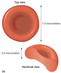
- Biconcave disk shape
- No nucleus at maturity (amitotic)
- 5.2 million/mm3
Hemoglobin
- Protein contained in cytoplasm of rbcs
- The protein that binds to oxygen gas and transports it inside the rbc
Biconcave shape
- High surface area to volume ratio
- All cytoplasm is near the plasma membrane
- Smooth and flexible
- Able to form stacks that slide through small capillaries
Anemia
Any condition in which the ability of the blood to transport oxygen gas is diminished
Anemia chart

Reticulocyte
Cells that become red blood cells
Sickle cell anemia
Hemoglobin is abnormal, which causes the red blood cells to become hard and sticky and look C-shaped
Oxyhemoglobin
- When hemoglobin binds to oxygen gas
- Bright red
- Happens with cuts because blood is exposed to air
Deoxyhemoglobin
When hemoglobin released and is not carrying oxygen gas
Darker red
Carbon monoxide
- Produced when you burn organic fuel
- Sticks to hemoglobin better than oxygen
Structure of hemoglobin
- Quaternary structure
- 4 polypeptides (chains of amino acids)
- 2 alpha chains
- 2 beta chains
Heme group(Blue) & Iron atom(red)
- The spot where oxygen gas binds, binds to iron atoms in heme groups
- Found in the center of each of the 4 polypeptides

Red blood cell life cycle
- Production of rbc is influenced by blood's ability to transport oxygen gas
- If blood oxygen gas level gets low it stimulates liver to secrete erythropoietin
- RBCs lifespan is about 120 days
- Spleen/liver get rid of damaged rbcs
erythropoietin
Hormone that stimulates red blood cell production
Macrophages (WBC)
- (big eater)
- Engulf rbcs when they are removed from circulation by spleen/liver
- Highly phagocytic
- Engulf and debris/remnants of broken cells and digest in cytoplasm
Bilirubin/Biliverdin
- What's left of the heme group when separated from iron
- A pigment molecule, orange
- Excreted from liver into small intestine
- Released from body in feces
Too much bilirubin
Causes jaundice (yellow eyes or skin)
Fate of hemoglobin following RBC destruction and phagocytosis by a macrophage
- Polypeptides are hydrolyzed and amino acids are recycled or metabolized
- Iron is removed from each heme and transported to red bone marrow to be recycled
- The remainder of each heme becomes biliverdin (green) or bilirubin (orange) which are transported to the liver for excretion
Phagocytosis
The process of removing debris and pathogens by engulfing and digesting them
RBC Surface Antigens
Genetically determined cell surface recognition molecules that can cause an immune response in individuals lacking those antigens
Antibodies
Plasma proteins (immunoglobins) from B-cells that bind to foreign antigens
Agglutination
Rbc clump together because antibodies bind to their antigens
ABO blood typing
Tests for:
- Antigens A and B
- Antibodies anti-A and anti-B

Rh blood typing
Tests for:
- Antigen D
- Antibody anti-D

What happens to antibodies bind to antigens
- Causes agglutination
- The blood cells that bare those antigens to clump
- Insoluble clump of antibodies bound to antigens on RBC surfaces
- Involves rbcs because antibodies have bond to their antigens
Antigen/Antibodies chart

Blood transfusion chart
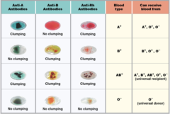
Why can't a person receive certain cells?
A person cannot receive cells that have antigens to which his/her antibodies can bind.
What happens if antibodies and antigen bind
It can cause clumping and hemolysis
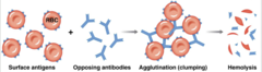
Hemolysis
Breaking of blood cells
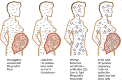
Rh blood system chart
Rh+ can receive + or -
Rh- can only receive -

Rh sensitization
White Blood Cells
Leukocytes
- Have a nucleus at maturity(nucleate)
- protect the body against infection
- Live for many years
- Function primarily outside of the cardiovascular system
- 7000/mm3
2 Categories of white blood cells
1. Granulocyte
2. Agranulocytes
What are Granulocytes
Cells that in cytoplasm have tiny granules, so cytoplasm looks grainy
Granulocyte white blood cells
- Neutrophils
- Eosinophils
- Basophils
What are Agranulocytes
cells that don't have granules
Agranulocyte white blood cells
- Monocytes
- Lymphocytes
Margination
White blood cell is stuck to the lining of the wall of a capillary/blood vessel wall
Emigration/Diapedesis
White blood cell crosses wall of capillary through the cells of wall(simple squamous)
Positive Chemotaxis
Movement in particular direction because of chemical, towards the chemical
5 types of White Blood Cells
Neutrophils
Lymphocyte
Monocyte
Eosinophil
Basophil
Neutrophil
- Most abundant type of white blood cells
- Round, nucleus has lobes(multilobe), cytoplasm has large pale inclusions
- Phagocytic- engulf pathogens or debris in injured/infected tissues
- Can release enzymes/chemicals that are toxic to infecting cells like bacteria
- Bigger than red blood cell
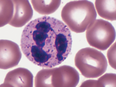
Eosinophil
- 4th most abundant white blood cell
- round, nucleus has 2 lobes, large granules, bright red
- Phagocytic- engulf antibody labeled materials
- Increase in abundance during allergic reaction

Basophil
- Rarest of white blood cells
- Round cell, nucleus can't be seen, dark purple staining
- Enter damaged tissues and release a chemical histamine

Lymphocyte
- 2nd in abundance
- Smallest, not much bigger than rbc
- Nucleus takes up almost whole interior, very little cytoplasm
- Cells of lymphatic system
- Provide immunity (protection against specific diseases)
- T cells and B cells

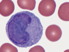
Monocytes
- 3rd in abundance
- Biggest white blood cell, 4-5x size of rbc, large cell, nulcuea shaped like kidney
- Enter tissue and become macrophages
- When monocytes are stimulated, they change into macrophages
Histamine
Chemical that promotes inflammation
Platelets
Thrombocytes
- Cell fragments of megakaryocytes
- Live 10 days on average
- 350,000/mm3
Platelets blood prevention
- By releasing chemicals that promote coagulation and vasoconstriction
- Can stimulate blood vessel to constrict so less blood goes through it
- Become sticky and will stick to collagen fibers in blood vessel wall and form a platelet plug
Vasoconstriction
the narrowing of blood vessels
Megakaryocyte
(cell with the big nucleus)
- Cell breaks up into platelets

Blood Cell Chart

Hemostasis
- The prevention of blood loss
- Maintaining a constant internal blood supply
- Uses hemostatic mechanisms to prevent blood loss
Hemostatic mechanisms
- Vascular phase
- Platelet phase
- Coagulation phase
Vascular phase
- If blood vessel is cut, stimulates vascular spasm, in blood vessel wall smooth muscle tissue will contract constricting blood vessels so lumen gets smaller/tighter so blood can't escape
- Following injury to a blood vessel, endothelial cells lining the blood vessel wall release chemicals that stimulate smooth muscle contraction.
- The endothelial cells also become sticky
- Cut ends of vessels can stick back together
- Opposite sides of capillary wall can stick together

Platelet phase
- Formation of a temporary platelet plug
- Platelets get sticky and stick to blood vessel walls, especially collagen fibers, and to each other and form platelet plug until clot forms

Coagulation phase
2 pathways for starting coagulation
Intrinsic
Extrinsic

Intrinsic pathway (right side of diagram)
Caused by blood contact with damaged or foreign surface (e.g. coagulation within a test tube)
- Starts with chemical that are normal components of the blood
- Chemicals that are part of blood come into contact in the body with an irregular surface
- Or outside of the body if the chemicals come into contact with a foreign surface
- Often occurs when blood comes into contact with a lining of blood vessel thast been damaged or has accumulation of plaque
Extrinsic pathway (left side)
Caused by chemicals released by broken blood vessels or damaged tissues
- Promoted by chemicals that are not part of the blood, that are released from damaged tissues
- Chemicals from another source
Common pathway for coagulation phase
- Both chemicals stimulate a chemical called factor x
- Activates molecule called prothrombin activator
- Changes a chemical prothrombin into thrombin, which
- changes plasma protein called fibrinogen into fibrin
- Fibrinogen is plasma protein that dissolves in blood plasma that when with thrombin it will precipitate forming solid protein threads in blood called fibrin, which gets tangled at wound site and form the netting that traps blood cells and forms clot
Heart Structure
- Fist-sized organ in the mediastinum of the thoracic cavity
- Wall has three layers
Endocardium
Myocardium
Epicardium
- Surrounded by a pericardial sac
Endocardium
Inner layer
- Consists of 2 tissue
1. Endothelium:
- Tissue that comes into contact with blood inside heart chamber, sits on top of areolar
- Simple squamous epithelium=endothelium
2. Areolar tissue: Yellow section of endocardium
- Connective tissue(glue) bind other tissues together
Myocardium
Middle layer
- Thick layer of cardiac muscle tissue
- Can contract in order to pump blood
Epicardium
Outer layer (Visceral Pericardium)
- 2 tissues
1. Areolar tissue
2. Simple squamous epithelium/Mesothelium: thin layer
Pericardial sac
Containing a serous fluid-filled pericardial cavity
- Visceral pericardium = epicardium
- Parietal pericardium
- Serous layer
- Fibrous layer
Base of heart
- Top of heart
- Blood vessels attached to it
Apex of heart
Bottom of heart
Sulcus
Shallow groove between heart structures
Coronary sulcus
- Sulcus dividing atria and ventricles
- Filled with fat and blood vessels
Anterior/posterior interventricular sulcus
Sulcus that divides the two ventricles
Septum
Wall between heart chambers
Interventricular septum
Wall between two ventricles
Interatrial septum
Wall between atria
Auracle
Ear-like flaps externally at the edge of the atria
Pectinate muscle
Muscle tissue that consists of interconnecting bands/ lining of atria
trabeculae carneae
Branchy muscle that lines ventricles
Papillary muscles
The muscle chordae tendineae connect too
Chordae tendineae
(heart strings) the string like fibrous connections between valves and papillary muscles
Blood Vessels of the heart
Aorta
Vena Cava
Pulmonary artery
Pulmonary veins
Heart Chambers
- Atria
- Ventricles
Right atrium (with auricle)
- Receives deoxygenated blood from the superior and inferior venae cavae
- Pumps blood to the right ventricle
Left atrium (with auricle)
- Receives oxygenated blood from the pulmonary veins
- Pumps blood to the left ventricle
Right ventricle
- Receives blood from the right atrium
- Pumps blood to the pulmonary trunk and pulmonary arteries