MRI Lumbar Spine
1/23
There's no tags or description
Looks like no tags are added yet.
Name | Mastery | Learn | Test | Matching | Spaced |
|---|
No study sessions yet.
24 Terms
routine projections L spine
Sagittal, Axial and Coronal Localizer
Sag T1
Sag T2
Sag STIR
Axial PD
what plane do you plan l spine sagittal slices on
coronal
angle of sagittal L spine
parallel to spinal cord
on axial it is parallel to spinous process
sat band L spine
over aorta to reduce artifact
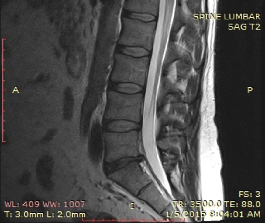
Sagittal T2 Lumbar
Sagittal T2 Lumbar what is bright
CSF
Sagittal plane L spine best demonstrates
degree of spinal stenosis
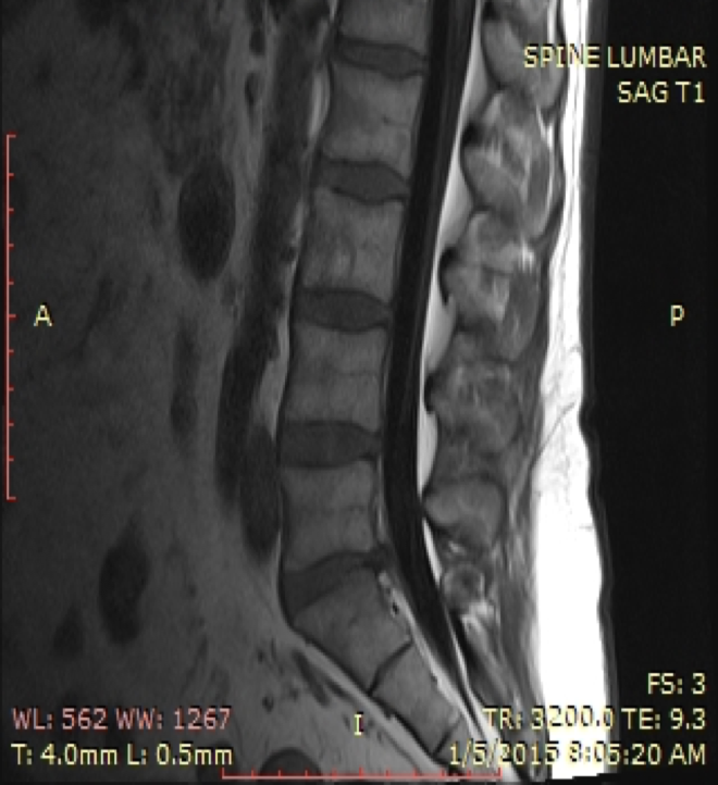
Sagittal T1 Lumbar
Dark on Sagittal T1 Lumbar
CSF
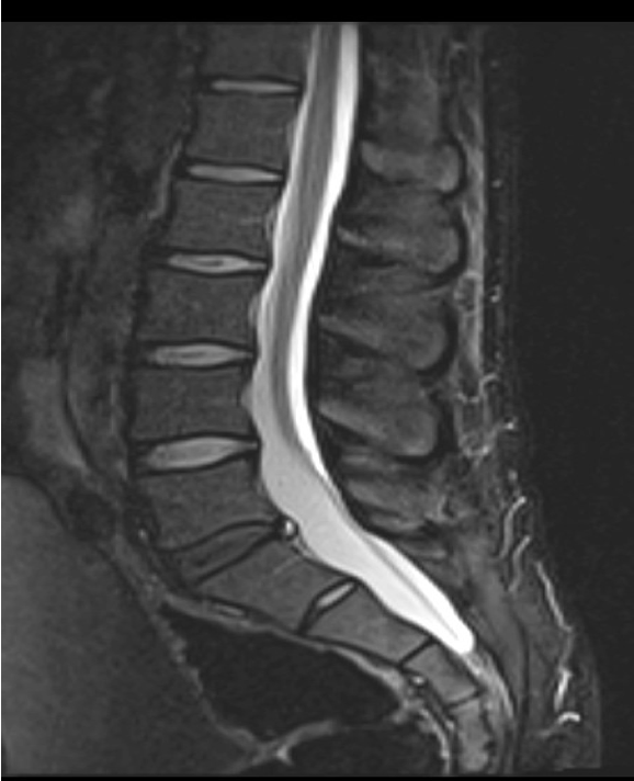
STIR lumbar
STIR
Short Tau Inversion Recovery
Inversion recovery sequence with specific timing to suppress fat based on differences in the T1 of tissues.
STIR Lumbar spine for
diagnosis of bone marrow diseases.
spine injury by interpreting whether vertebral compression fractures are acute, chronic or old.
Excellent detection of occult fx.
Valuable in diagnosing osteoporic fx.
dark on STIR Lumbar
Fat
Bright on Lumbar STIR
CSF
chronic fractures of vertebral bodies look
hyperintense on the STIR
acute/ subacute fracture on L spine
Hypointense T1 signal, hyperintensity on T2 and STIR,
axial slices for L spine plotted on
Sagittal Plane
how to plot axial slices L spine
Angle the first position block parallel to the L4-L5 intervertebral disc.
Continue through additional intervertebral disc spaces
how to plot disc axials L spine
After Sagittal T2 sequence is complete, axial slices are plotted through each disc space.
Additional slices may be added to cover pathology
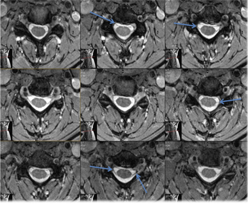
axial plane L spine best visualizes
both dorsal and ventral nerve roots. (blue arrows)
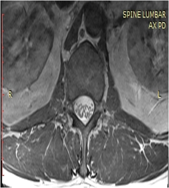
Axial PD L spine
Axial PD L spine
minimize effects of T1 and T2.
results in image weighting dependent primarily on the proton density of the image volume
Axial PD looks like
Higher proton density = higher image signal. (Bright)
Lower proton density = lower image signal. (Dark

Mri lumbar scoliosis how to plot
Coronal imaging is performed to visualize the spinal cord to accurately plot sagittal images.
After coronal T2 is completed, find the best midline cord slice and plot parallel with cord.
Sagittal may have to be done as upper and lower to compensate for curvature.
axials plotted off the coronal and sagittals