2 Clinical Manifestations of and Diagnostic Tests for Joint Disorders
 Call Kai
 Learn
Learn Practice Test
 Spaced Repetition
 Match
Match Flashcards
 Knowt Play
1/31

There's no tags or description
Looks like no tags are added yet.
Name | Mastery | Learn | Test | Matching | Spaced | Call with Kai | Chat |
|---|
No analytics yet
Send a link to your students to track their progress
32 Terms
Lameness
Gait Abnormality
Animals with joint disease commonly present with what history?
One joint/limb = traumatic or developmental
Multiple joints/limbs = degenerative or inflammatory
If the lameness is traumatic or developmental VS degenerative or inflammatory, how many joints/limbs are involved?
Inflammatory
Infectious (Borrelia burgdoferi)
Non-infectious
Erosive (rheumatoid arthritis)
Nonerosive (IMPA, SLE)
Non-inflammatory
Developmenal (UAP)
Degenerative (hip dysplasia)
Neoplastic
Traumatic processes
What are the two types of disorders affecting joints?
FALSE: this is true for inflammatory diease
TRUE OR FALSE: Noninflammatory diseases typically show signs of systemic disease with multiple joints affected.
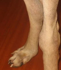
Effusion (fluid accumulation within a joint space or body cavity)
What is happening here?
Trauma
Panosteitis (age dependent/inflammation of long bones)
HOD: Hypertrophic Osteodystrophy (age dependent)
Osteomyelitis (infection of bone)
Bone neoplasia
You do a physical exam and there is pain upon palpation of the bones. What are some differentials?
Myositis
Strain/Sprain
You do a physical exam and there is pain upon palpation of the muscles. Give some differentials
Spinal cord or vertebral abnormalities
IVDD
intracranial disease (wide range of conditions that affect skull and surrounding structures)
meningitis
polyarthritis
You do a physical exam and there is pain upon palpation of the neck. Give some differentials.
Spinal cord or vertebral abnormalities
IVDD
discospondylitis (infection of intervertebral discs and endspace)
intervertebral facet joint inflammation
You do a physical exam and there is pain upon palpation of the spine. Give some differentials
*RADIOGRAPH*, maybe also CT or ultrasound
Also used when joint palpation reveals crepitation, instability or a restricted range of motion
Name some diagnostic imaging tools that can be used when only ONE joint is affected
Arthrocentesis and synovial fluid analysis
Name some diagnostic imaging tools that can be used when at least THREE TO FOUR joints are affected
If polyarthritis is suspected
When should synovial fluid should be analyzed from at least 3-4 joints?
FALSE: while arthrocentesis IS great for its minimal risk and high diagnostic yield, you ALWAYS aseptically prepare and wear gloves
TRUE OR FALSE: arthrocentesis is a great procedure for multiple joints because there is minimal risk with high diagnostic yield AND minimal aseptic requirements.
Thick, viscous fluid, clear to slightly yellow, high content of hyaluronic acid and lubricin
Describe normal synovial fluid
Normal synovial fluid
Which structure's primary function is to lubricate articular surfaces and facilitate movement within the joint
Normal
Describe this joint fluid
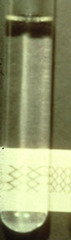
Noninflammatory
Describe this joint fluid
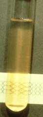
Inflammatory
Describe this joint fluid
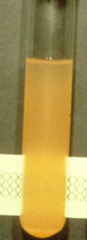
Septic
Describe this joint fluid

Hemorrhage
Describe this joint fluid
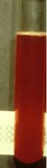
1. LEAST noninflammatory
2. inflammatory
3. septic
4. MOST hemorrhagic
List the following joint fluid conditions in order of increasing protein and cell count:
noninflammatory
hemorrhagic
septic
inflammatory
NORMAL = string effect
Is this a normal characteristic of joint fluid? What is it called when this happens?
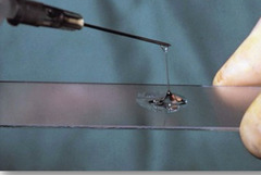
No more than 3 WBC per 40x field
Less than 10% neutrophils
Mix of large and small mononuclear cells
TP less than 2.5
(if 1 neutrophil/500 RBCs = blood contamination)
What should NORMAL joint or synovial fluid look like under a microscope?
DJD and acute trauma
If you see joint fluid with less than 3.5^2 TP and total nucleated cell counts less than 5.0 what should you be expecting?
Inflammatory joint disease (like immune mediated or infectious)
If you see joint fluid with more than 2.5^2 TP and total nucleated cell counts more than 5.0 what should you be expecting?
Neutrophils
Which cells increases in number as the inflammation in the joint becomes more severe?
1. monocytes, macrophages (remember monocytes have "holes eating thru cytoplasm", and are normally a pink/purple color)
2. noninflammatory (because of monocytes)
3. degenerative = arthritis or cruciate rupture
This is synovial fluid from a canine.
1. what cells are you seeing
2. inflammatory or noninflammatory?
3. name a common joint disease associated with this cytology? differentials?
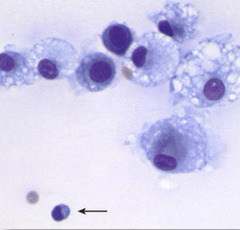
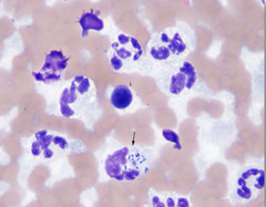
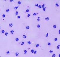
1. these are neutrophils
2. because excessive neutrophils are present = inflammatory
3. infectious (ehrlicia, anaplasma, RMSF) and immune mediated (polyarthritis)
This synovial fluid was taken from the carpus of a painful adult dog with a fever.
1. what are these cells
2. inflammatory or noninflammatory?
3. differentials?
Carpus, tarsus, stifle, elbow
Where do you take joint taps from?
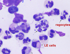
1. inflammatory
(this is actually polyarthritis, lupus erythmatosis)
This is joint fluid taken from an older cat. Describe what you see
1. inflammatory or noninflammatory?
Note: ragocytes are neutrophils in synovial fluid that contains cytoplasmic inclusions
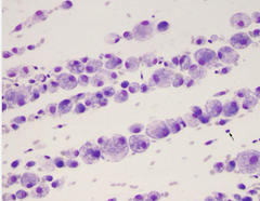
1. monocytes (fluffy with some vaculoes in them) = macrophages
2. noninflammatory based on the fact there are macrophages present
This is from a dog joint with chronic forelimb lameness
1. what kinds of cells do you see?
2. inflammatory or noninflammatory?
1. septic joint (because you see the bacteria being phagocytized by neutrophils) = inflammatory joint disease
2. RBC (maybe some contamination) and neutrophils plus one lymphocyte
This is from a horse joint. Describe what you see
1. inflammatory or noninflammatory?
2. cells?