OMFPR 7 - Principles of Radiographic Interpretation using Descriptive Terminology (Dr. Campos)
 Call Kai
 Learn
Learn Practice Test
 Spaced Repetition
 Match
Match Flashcards
 Knowt Play
1/82

There's no tags or description
Looks like no tags are added yet.
Name | Mastery | Learn | Test | Matching | Spaced | Call with Kai | Chat |
|---|
No analytics yet
Send a link to your students to track their progress
83 Terms
- Diagnostic images
- Analysis of abnormal findings (Use radiographic descriptive terminology- Consistency, reliability)
- Formulation of a disease category based on the description of the abnormality
- Develop a differential diagnosis (DDX) for the noted abnormality (Either a definitive interpretation or a short list of disease conditions - in the order of likelihood)
What does it take to interpret radiographs?
Differential diagnosis
Systematic radiographic analysis and description --> formulates ____________ through radiographic interpretation
- Location
- Extent
- Periphery/borders
- Shape/size
- Internal architecture
- Effect on adjacent structures
What are considered the six description categories of radiographic findings?
- Periphery/borders
- Internal architecture
- Effect on adjacent structures
of the 6 types of description categories of radiographic findings, what 3 are most important in guiding you towards a more definitive diagnosis?
Location
Define the descriptive terminology for abnormal findings:
- Localized vs generalized
- Periapical vs pericoronal vs interradicular vs other
- Extent - Focal, Multi-focal, Extensive
Periphery/Borders
Define the descriptive terminology for abnormal findings:
- Defined (corticated/non-corticated)
- Ill-defined, Infiltrative
- Size, Shape and Number
Corticated border
Define the following:
Radiopaque line along the periphery of a well-defined lesion
Internal architecture
Define the descriptive terminology for abnormal findings:
- Radiodensity
- Presence of septae
- Calcifications
Effect on Adjacent Structure
Define the descriptive terminology for abnormal findings:
- Displacement of teeth
- Root resorption
- Displacement of cortical boundaries or vital structures
- Osseous expansion
- Erosion/destruction
- Extension into adjacent soft tissue
Generalized
Define the following:
If an abnormal appearance affects all osseous structures of the maxillofacial region (usually in systemic or metabolic disease)
- Localized
- Periapical to #24
- Single lesion
Describe the abnormal findings about the location of the lesion: (3)

- Localized
- Pericoronal to impacted #17
- Single lesion
Describe the abnormal findings about the location of the lesion: (3)

Pericoronal
Define the following:
Lesion surrounding the crown
- Generalized
- Radiopaque
Describe the abnormal findings about the location of the lesion: (2)

- Multi-focal (Generalized)
- Periapical
- Confluent
Describe the abnormal findings of location of the lesion: (3)

- Unilocular
- Radiolucent
- Well-defined
- Corticated Borders
Describe the periphery/borders of the lesion: (4)

- Unilocular
- Radiolucent
- Well-defined
- Partially-Corticated Borders
Describe the periphery/borders of the lesion: (4)
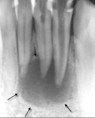
- Unilocular
- Radiolucent
- Well-defined
- Non-Corticated Borders
Describe the periphery/borders of the lesion: (4)

- Well defined
- Radiopacity soft tissue capsule with a radiolucent rim
- Non-Corticated Borders
- Attached to root (no PDL gap)
Describe the periphery/borders of the lesion: (4)
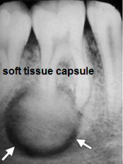
- Well defined
- Sclerotic border
- Not attached to root (PDL visible)
Describe the periphery/borders of the lesion: (3)

- Ill-defined invasive border (infiltrative)
- Radiolucent
Describe the periphery/borders of the lesion: (2)

- Ill-defined invasive border
- Radiolucent
(IAN is affected)
Describe the periphery/borders of the lesion: (2)

Ill-defined blending (fusiform)
(Note the gradual blending of the periphery of lesion with adjacent normal bone)
Describe the periphery/borders of the lesion:
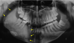
- Benign
- Developmental
- Inflammatory
well defined corticated lesions are usually found to be... (3)
- Inflammatory
- Benign
well defined non-corticated lesions are usually found to be... (2)
- Inflammatory
- Malignant
- Systemic
ill-defined lesions are usually found to be... (3)
- Inflammatory
- Malignant
ill-defined AND infiltrative lesions are usually found to be... (2)
fibro-osseous
Fusiform lesions are usually found to be...
Scalloped
(well defined, septa= multilocular)
Describe the periphery/borders of the lesion:

circular
(focal, well-defined, radiolucent, corticated)
Describe the periphery/borders of the lesion:
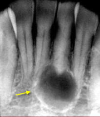
- Radiodensity
- Internal septation (unilocular/multilocular)
- Internal calcifications (for radiolucent lesions)
- Abnormal trabecular patterns (orange peel, ground glass, cotton wool, decreased trabeculations)
What are we analyzing when determining the internal architecture of the lesion?
Radiodensity
Define the following:
Lesions may be described as: radiolucent, radiopaque, or mixed
- Radiolucent
- Corticated border
- Well-defined
- Round shape
Describe the internal architecture of the lesion: (4)

Unilocular lesions
Define the following:
No septations hence lesions have a single chamber
Multilocular lesions
Define the following:
Presence of septae divide the internal aspect of the lesion into multiple chambers
- Unilocular
- Radiolucent
- Well-defined
- Corticated Borders
(Location: apical of 23, 24, 25, 26)
Describe the internal architecture of the lesion: (4)
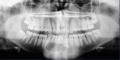
- Unilocular
- Radiolucent
- Non-Corticated Borders
- Sclerotic bone
Describe the internal architecture of the lesion: (4)

- Multilocular (multiple septae)
- Radiolucent
- Corticated
Describe the internal architecture of the lesion: (3)
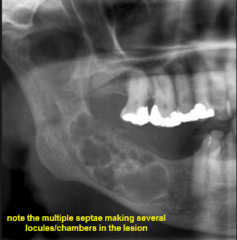
- Multilocular (multiple septae)
- Radiolucent
- Corticated
Describe the internal architecture of the lesion: (3)

- Periapical at #18
- Unilocular
- Radiolucent
- Well-defined
- Corticated
Describe the characteristics of this lesion: (5)
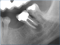
- Inter-radicular between #26 and #27
- Unilocular
- Radiolucent
- Well-defined
- Corticated
Describe the characteristics of this lesion: (4)

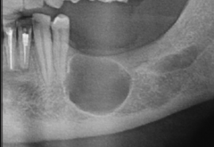
- In the edentulous zone (of 19-20)
- Unilocular
- Corticated
- Radiolucent
Describe the characteristics of this lesion: (4)
- Pericoronal of #17 (extends from coronoid process and sigmoid notch to apical region of 19)
- Unilocular
- Well-defined
- Corticated
- Radiolucent
- Oval shape
Describe the characteristics of this lesion: (6)

- Pericoronal of #17
- Unilocular
- Well-defined
- Radiolucent
- Corticated
Describe the characteristics of this lesion: (5)

- Pericoronal (Mesial CEJ to Distal CEJ of 17)
- Unilocular
- Well-defined
- Radiolucent
- Corticated
Describe the characteristics of this lesion: (5)
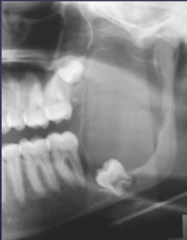
- Focal Opacity
- Target Lesion
- Multifocal Confluent
- Irregular, Ill-defined
- Ground Glass
- Soft Tissue Opacity
What are six types of appearances of radiopaque lesions?
focal radiopacity
(well-defined, round, not attached - PDL intact)
describe the appearance of the lesion:
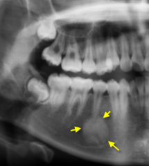
multifocal radiopacity
(apical to 30 and between to 28 and 29, well-defined, irregular shape)
describe the appearance of the lesion:

Multifocal Confluent Radiopacity
(confluence = connecting)
describe the appearance of the lesion:
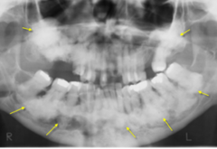
target lesion
Define the following:
Central opacity surrounded by a radiolucent zone with a sclerotic or radiopaque border
target lesion
describe the appearance of the lesion:

target lesion
describe the appearance of the lesion:
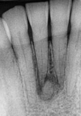
Trabecular pattern
Define the following:
Irregular and ill-defined radiopaque pattern
Trabecular pattern
(Extends beyond borders - most likely a malignancy)
describe the appearance of the lesion:

Ground glass radiopacity
Define the following:
Characterized by a diffuse, homogeneous radiopaque area with hazy pattern, resembling texture of finely ground glass
Ground glass radiopacity
(well-defined, expansion of IAN canal - bone is affected)
describe the appearance of the lesion:

Ground glass radiopacity
describe the appearance of the lesion:
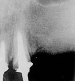
Ground glass radiopacity
describe the appearance of the lesions:
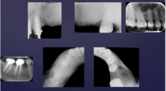
well-defined corticated borders with internal mixed density (mixed radiolucent-radiopaque) appearance, associated with impacted tooth
describe the appearance of the lesion:

Mixed (Lucent-Opaque) density lesion
(central radiopacity surrounded by a radiolucent area)
describe the appearance of the lesion:
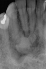
displacement of mandibular anterior teeth
(well-defined, non-corticated, apical to 20-27)
describe the effect of the lesion:
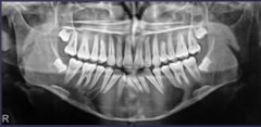
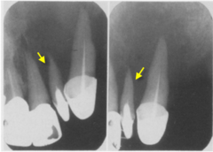
Root resorption - blunting of roots (slow process)
describe the effect of the lesion:
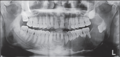
Root resorption - spiking (fast process)
("floating teeth", ill-defined radiolucency)
describe the effect of the lesion:
benign
blunting of roots is typically associated with ________ lesions

malignant
spiking of roots is typically associated with ________ lesions

Floating teeth
describe the effect of the lesion:
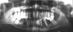
'Floating teeth'
The loss of bone support surrounding teeth due to a lesion is known as...
- Displacement of cortical boundaries
- Osseous Expansion and thinning of inferior mandibular cortex
- Inferior displacement of the left inferior alveolar nerve canal (IAN)
describe the effects of the lesion:
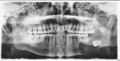
True
T/F: The lesion in the left posterior mandible can be described as well-defined, radiolucent, corticated, unilocular lesion causing displacement of tooth 18 distally and inferiorly
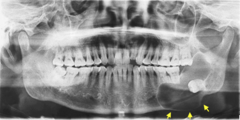
False (It is well-defined!)
T/F: The lesion in the left posterior mandible can be described as ill-defined, radiolucent, corticated, lesion causing displacement of the IAN canal inferiorly

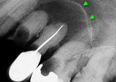
displacement of the floor of maxillary sinus
(lesion located in apical region of 12 and 13, well-defined, round, corticated, displacing floor of maxillary left sinus superiorly)
describe the effect of the lesion:
onion skin effect
The periosteal reaction, new bone formation parallel to the cortex of inferior mandibular border, is known as the ___________
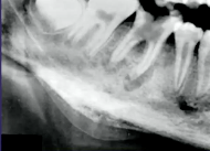
Outer cortical bone and periosteal reaction - 'onion skin effect'
(Area of 28-32, ill-defined, radiolucent)
describe the effect of the lesion:
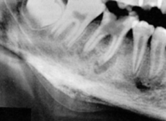
sunburst periosteal reaction
periosteal reaction, new bone formation perpendicular to the cortex of inferior mandibular border, is known as a ___________
Outer cortical bone and periosteal reaction - Sunburst reaction
(ill-defined, radiolucent lesion, invasive, IAN canal destroyed)
describe the effect of the lesion:
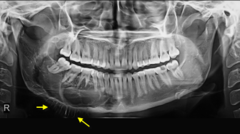
Outer cortical bone and periosteal reaction - Sunburst reaction
describe the effect of the lesion:

No reaction - destroyed inferior mandible, IAN canal, external oblique ridge of mandible, and PDL of 19
describe the effect of the lesion:

Ill defined radiolucent lesion with erosion/destruction of inferior mandibular border
Describe the characteristics of the lesion:
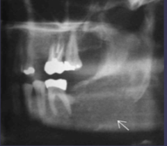
- Erosion of cortical bone boundary
- Ill defined radiolucent lesion with erosion/destruction of maxillary sinus floor
describe the effect of the lesion:

- Erosion of cortical bone boundary
- Soft tissue entity in the sinus perforating/eroding the floor of the sinus ( Right image)
describe the effect of the lesion:

- Left aspect of mandible; apical from 17 to 22 and to the ramus
- Multilocular
- Radiolucency
- #17 displaced distally
- Mandible is thin and expanded
- Resorption of root
- Expansion of external oblique ridge
Describe the characteristics of the lesion: (7)

- Right posterior mandible at #32
- Well-defined
- Corticated
- Radiopaque surrounded by radiolucent rim
- Displaced #32 distally/posteriorly
- External oblique branch expanded
- IAN canal displaced inferior
Describe the characteristics of the lesion: (7)

- Attached to #19 root
- Well-defined
- Oval shape
- Radiopaque with a radiolucent rim
- Non-corticated
- IAN canal displaced inferiorly
- Expansion of inferior border of mandible
- Sclerosis of bone
Describe the characteristics of the lesion: (7)
