Cell structure- OCR A Level Biology
1/64
There's no tags or description
Looks like no tags are added yet.
Name | Mastery | Learn | Test | Matching | Spaced | Call with Kai |
|---|
No analytics yet
Send a link to your students to track their progress
65 Terms
Cillia
Structure - Hair-like projections out of cells.
Function - Can be mobile or stationary. Mobile cilia help move
substances in a sweeping motion. Stationary cilia are important in
sensory organs, such as the nose
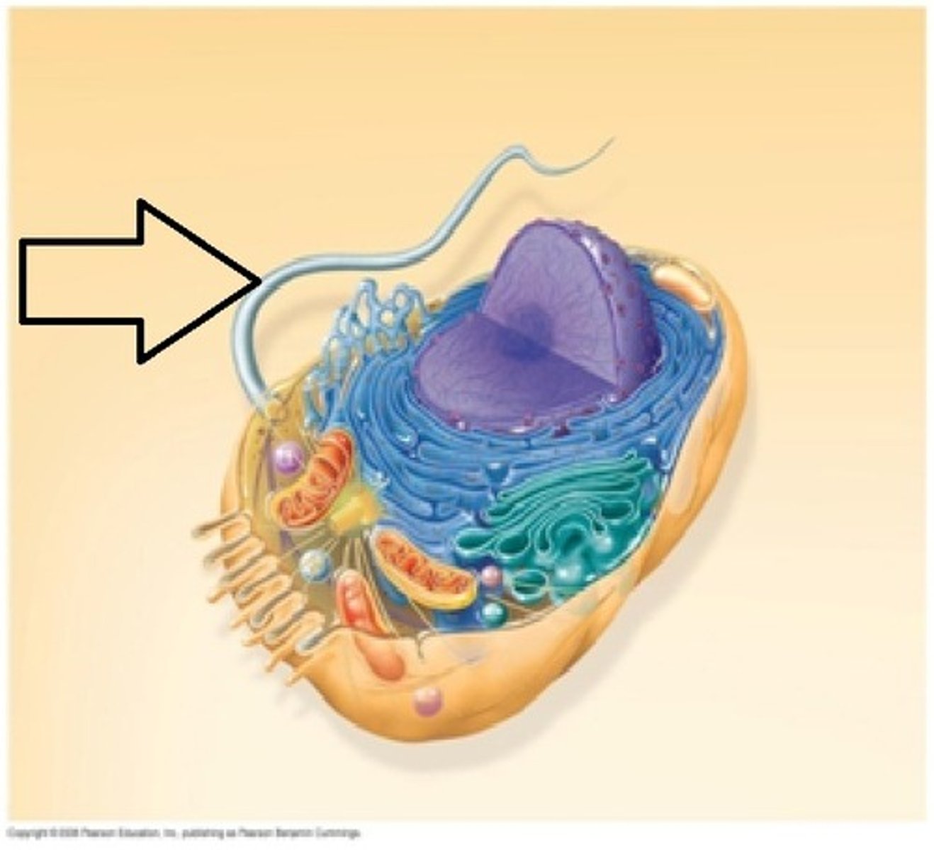
Flagella
Structure - Whip-like structure.
Function - For mobility, and sometimes as a sensory organelle
for chemical stimuli.
Centrioles
Structure - Made of microtubules. Occur in
pairs to form a centrosome.
Function -Involved in the production of
spindle fibre and organisation of chromosomes in cell
division.
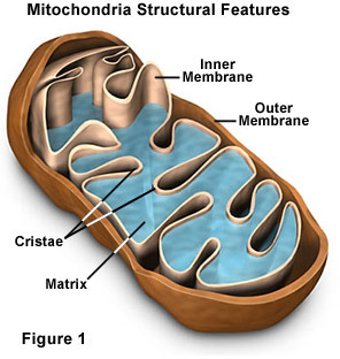
Mitochondria
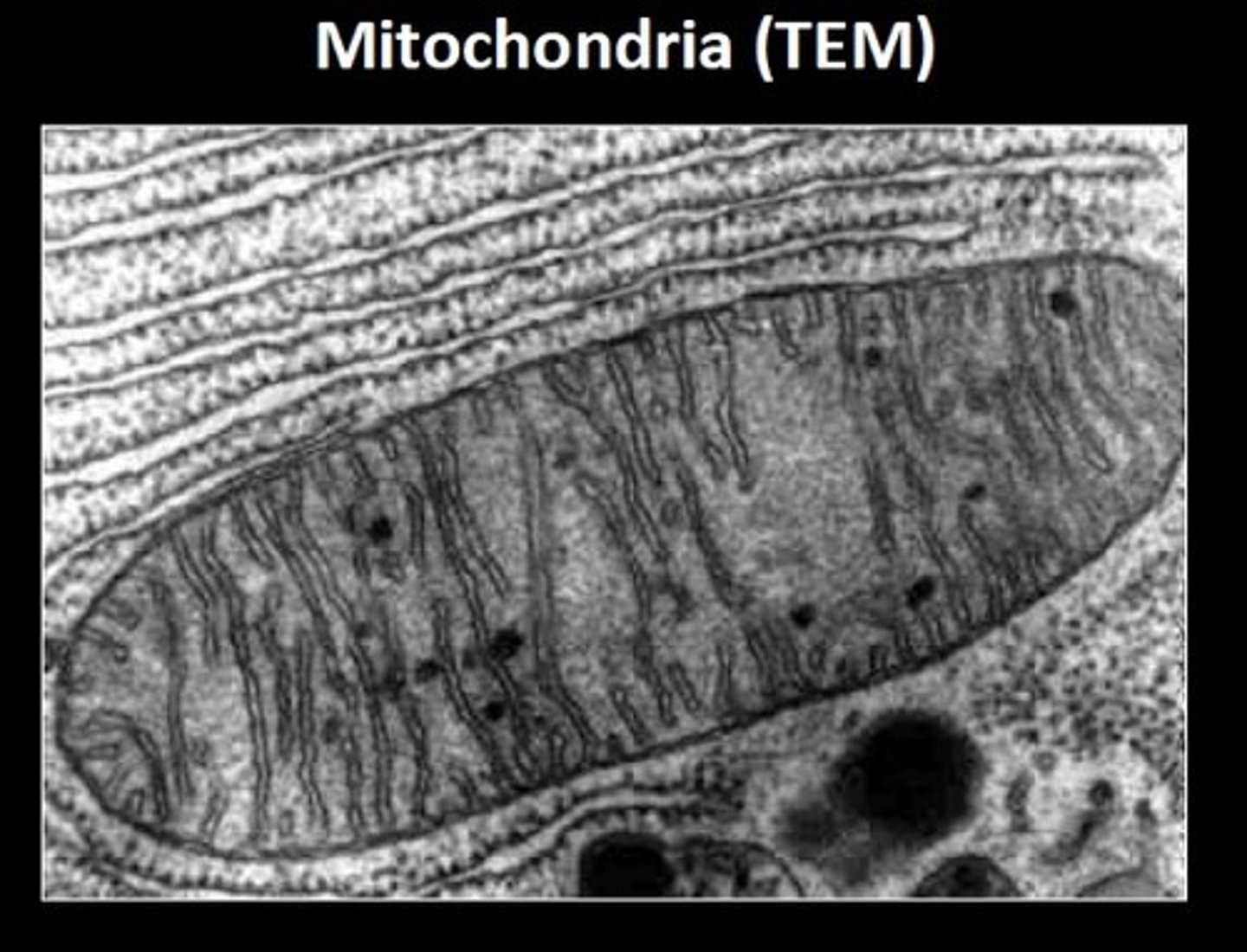
-The mitochondria are adapted for efficient aerobic respiration.
-The mitochondrial DNA contains the genes that code for the enzymes involved in aerobic respiration.
-The inner mitochondrial membrane (cristae) is folded to provide a large surface area to increase the number of electron carriers and ATP synthase that are embedded within the membrane (involved in oxidative phosphorylation).
-The outer membrane controls what can enter and exit the organelle.
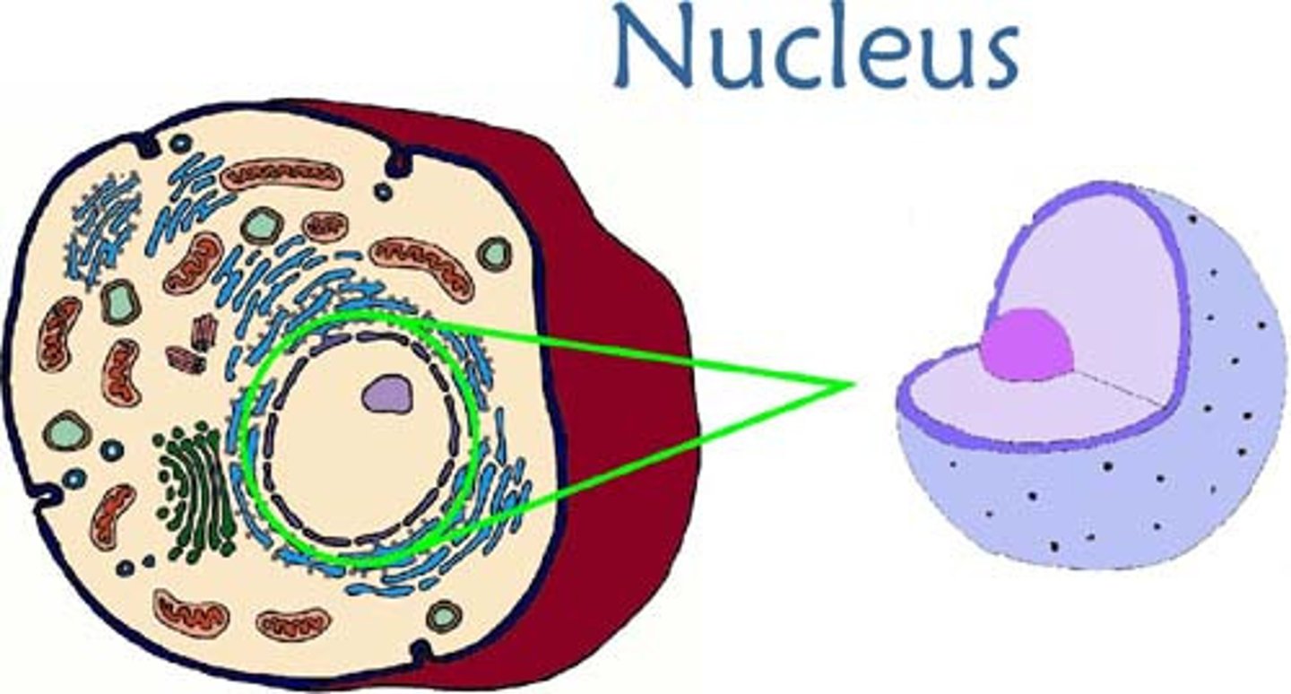
Nucleus
- Contains genetic material and chromosomes
- Controls what can enter and leave the cell
- Controls DNA transcription
- Controls gene expression, protein synthesis and storing DNA
Nucleolus
Site of ribosome production
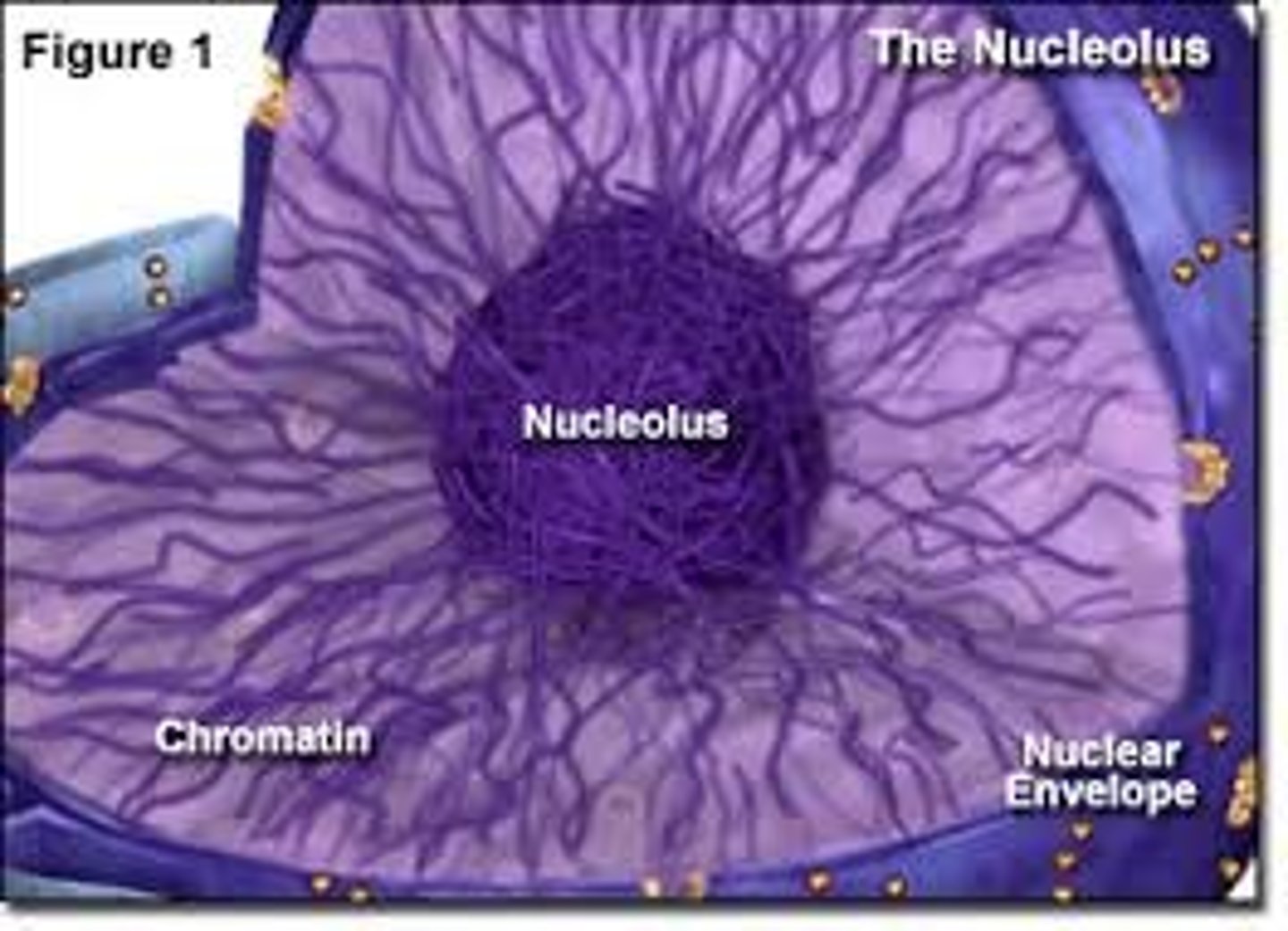
Nuclear envelope and pores
Seperates nucleus from cytoplasm. Spaces in the envelope form pores which allow movement of RNA out of the nucleus. There are ribosomes on the outer surface
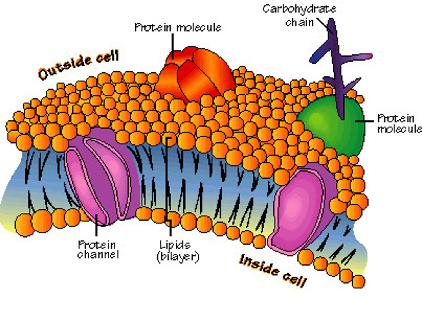
Cell membrane / plasma membrane
Structure - Consists of a phospholipid bilayer with proteins and cholesterol embedded within it.
Glycolipids and glycoproteins are located on the surface.
Function - The fluid-mosaic model of the membrane refers to the fluidity and range of molecules in the
membrane. Cholesterol provides strength and reduces fluidity; proteins are for transport and the
glycoproteins and glycolipids are for cell recognition and act as receptors.
Cytoplasm
A complex network of proteins that make up the cytoskeleton
Ribosome
- synthesises proteins from mRNA during translation
- fold amino acid chains into proteins
- consists of ribosomal proteins and ribosomal RNA
- made of large and small subunit, not surrounded by a membrane
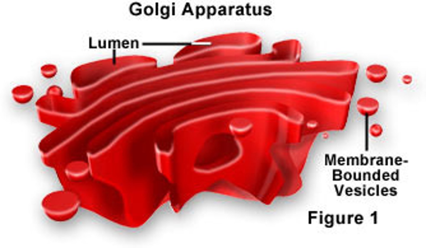
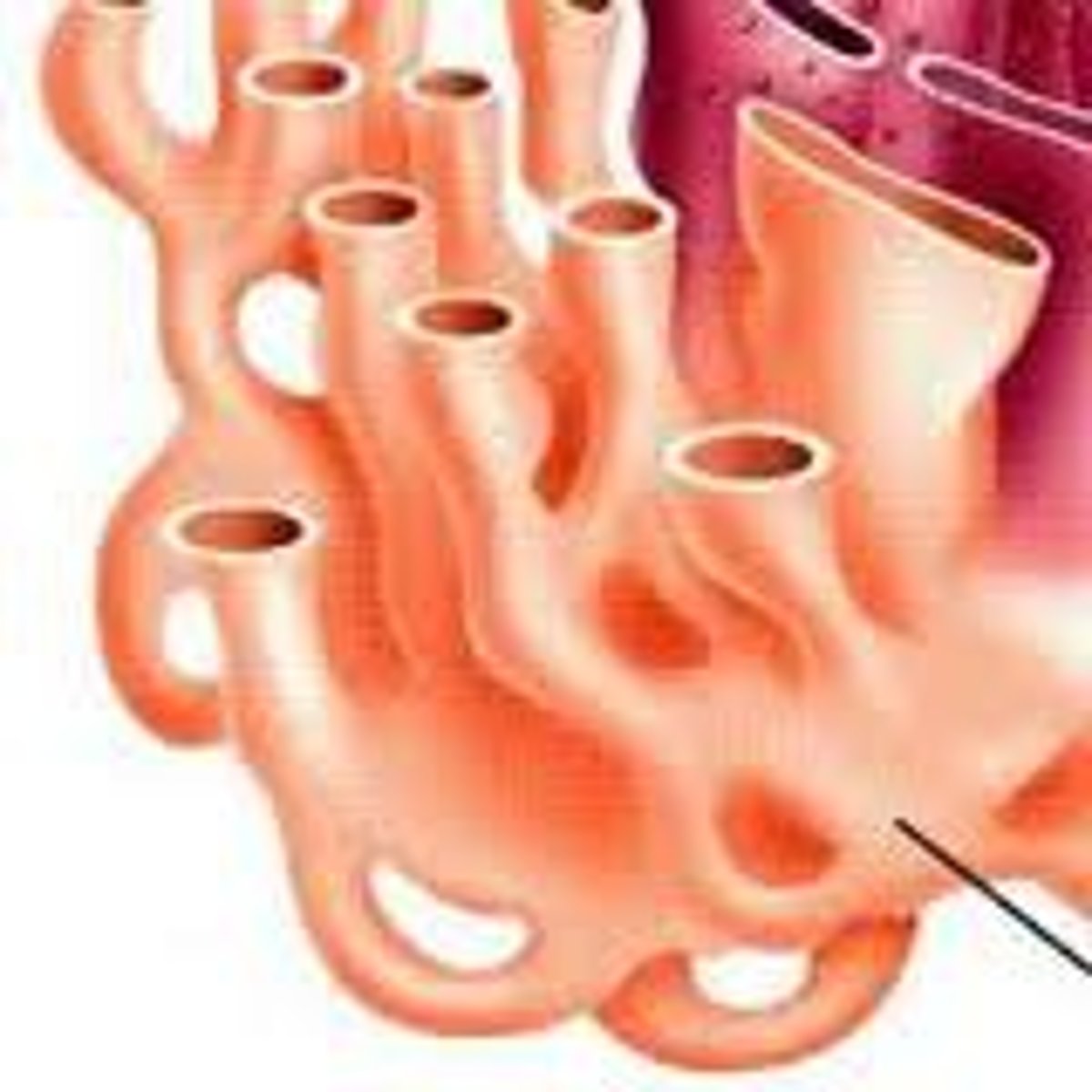
Golgi apparatus
Structure - Stacks of membranes creating flattened sacs called
cisternae, surrounded by small, round and hollow vesicles.
Function - Proteins and lipids and modified here. Carbohydrates
can be added to proteins to form glycoproteins. Finished products
are transported in the Golgi
vesicles
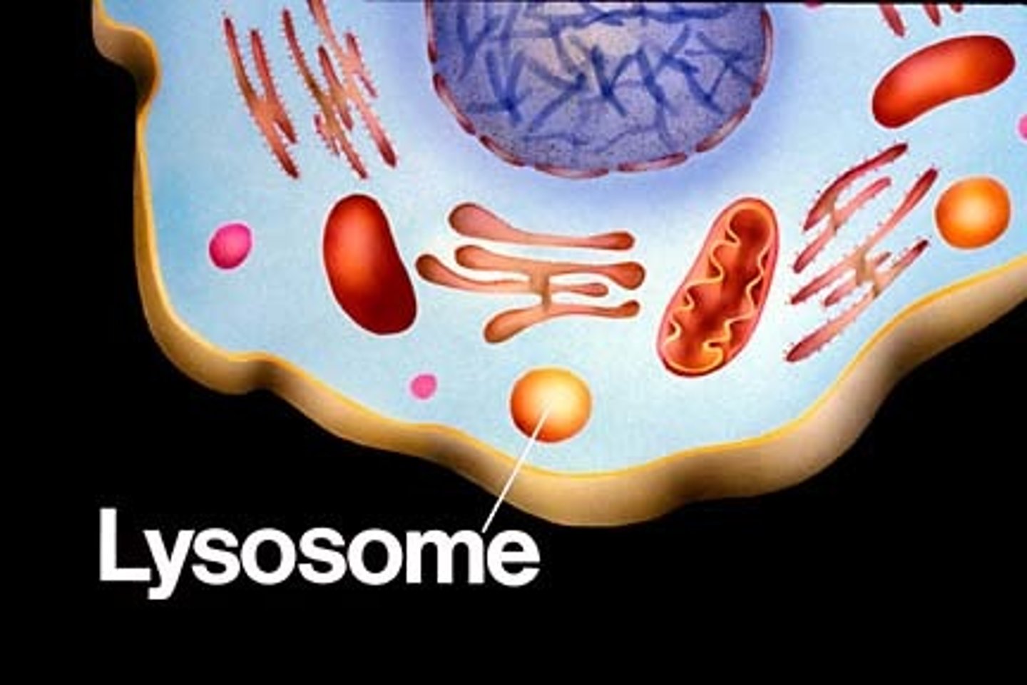
Lyosome
Structure - Formed when the Golgi
apparatus contains hydrolytic enzymes.
Function -A type of Golgi vesicle that
releases lysozymes.
- membrane prevents lysozymes from digesting the cytoplasm
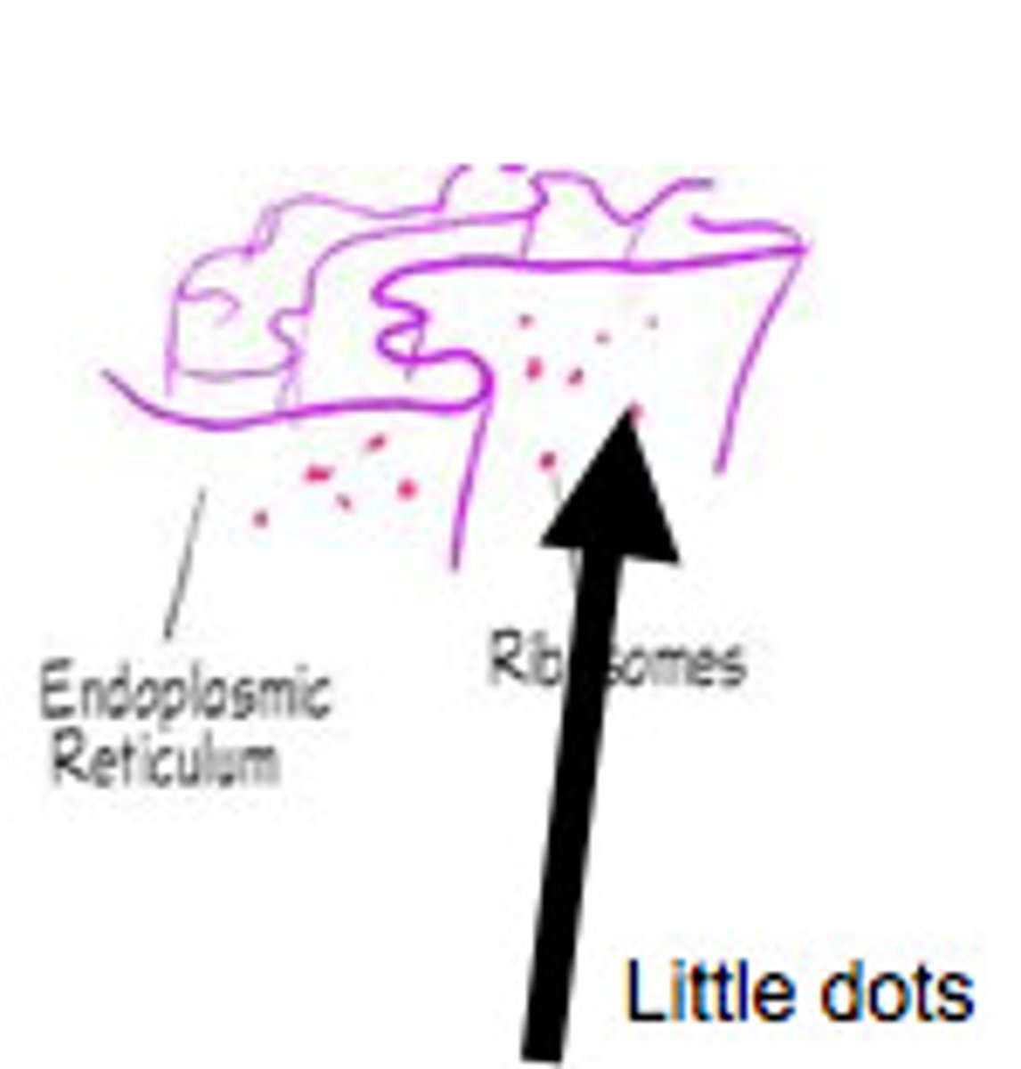
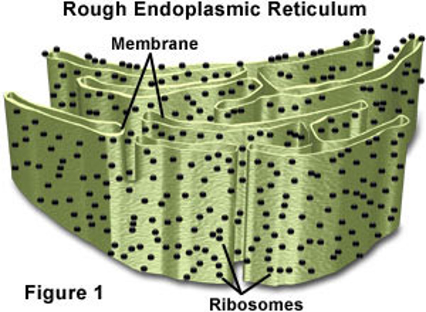
Rough Endoplasmic Reticulum (RER)
- processes and produces 3D structure of proteins
- site of glycoproteins synthesis
- Large SA for protein synthesis
- Have ribosomes along outer surface
- formed from flattened sacs called cisternae
Smooth Endoplasmic Reticulum (SER)
- no ribosomes of the surface
- stores, processes and synthesises phospholipids, cholesterol and lipids
- typically attached to RER and linked to nuclear membrane
- Large SA to increase rate of synthesis
- formed from flattened sacs called cisternae
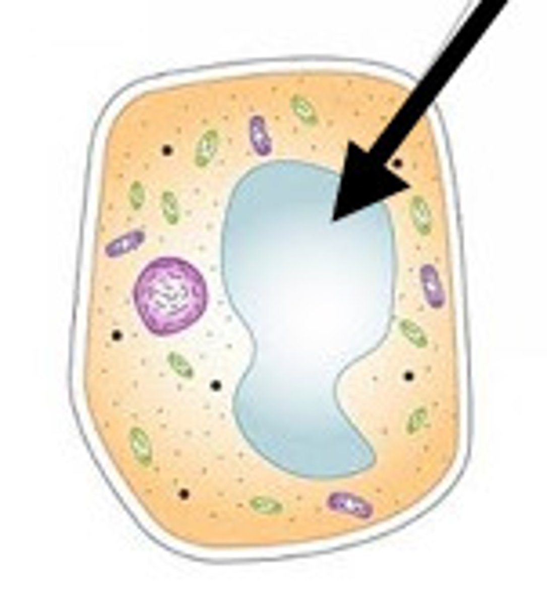
Permanent cell vacuole'
- plant cells only
- permanent pockets of cell sap, water and a solution of sugars
- surrounded by a membrane called tonoplast
- maintains osmotic pressure inside the cell
- ensures plant cells remain turgid which stops the plant from wilting
- stores unwanted chemicals that are discarded by the cell
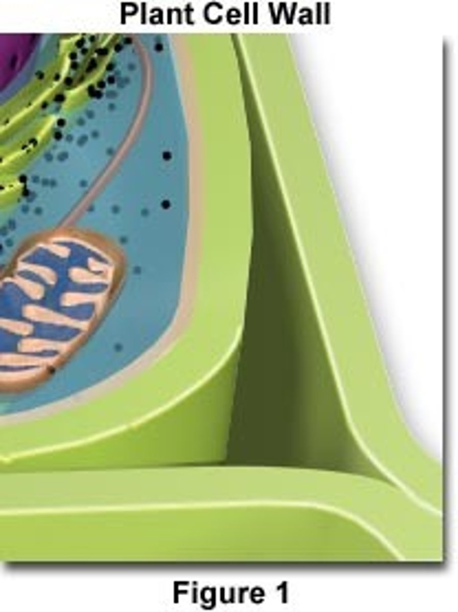
Cell wall
- Found in plant cells only
- Rigid structure surrounding cell membrane
- Maintains cell shape and structual support
- Protection from invading pathogens
- Made of cellulose (plants) and chitin (fungi)
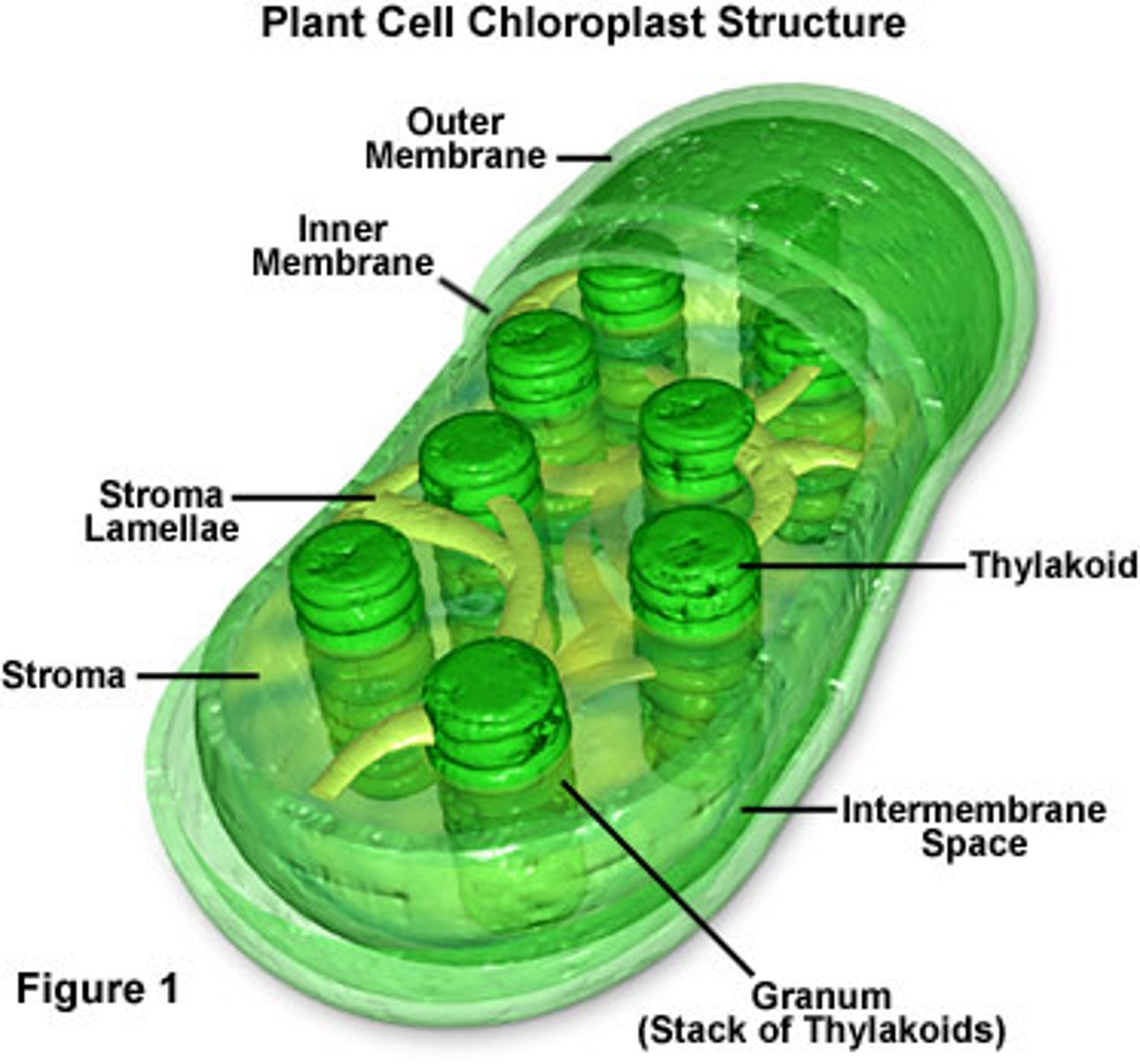
Chloroplast
-Thylakoid membranes: Folded membranes (grana) which contain photosynthetic proteins (chlorophyll) and embedded, transmembrane electron carrier proteins which are both involved in light-dependent stage.
-Stroma: Fluid centre which contains enzymes involved in the light-independent stage.
-Inner and outer membrane: Controls what can enter and leave the organelle (think plasma membranes).
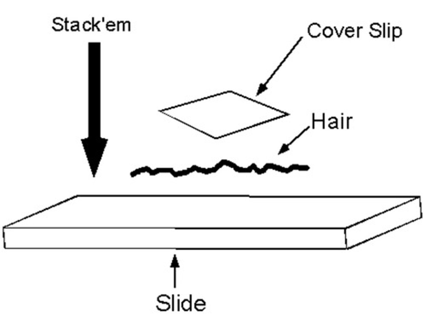
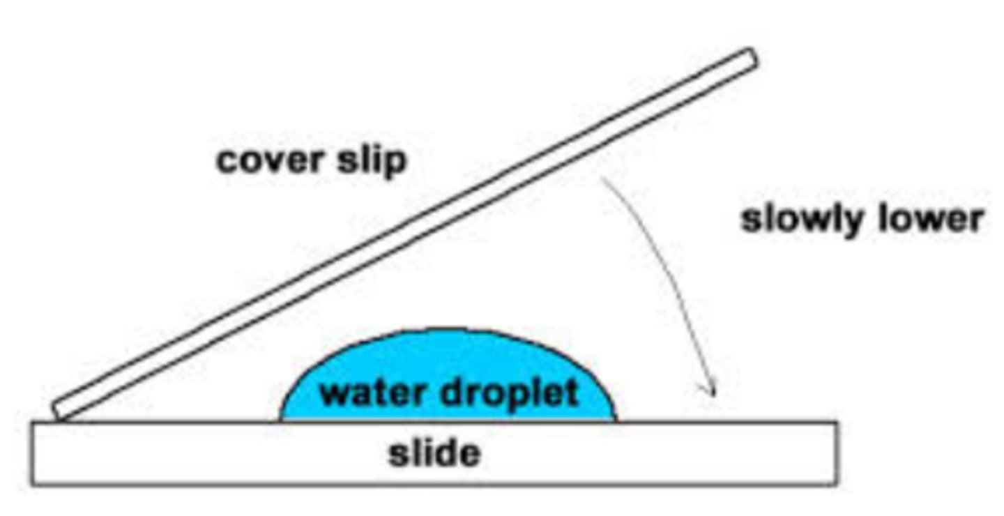
Dry mount slide
- Cut very thin sample of specimen, put specimen flat on slide, add a stain, place cover slip on with mounting needle
- Sample must be thin because light rays do not penetrate far through tissue
Wet mount slide
- Similar to dry mount but specimen is to be submerged in oil or water to prevent the tissue from drying out
Squash slide
- similar to dry mount but you press down between the cover slip and slide to squash the specimen
- not possible to see original arrangement of cells
- this method ensures that the specimen is thin enough to be seen through the microscope
Smear slides
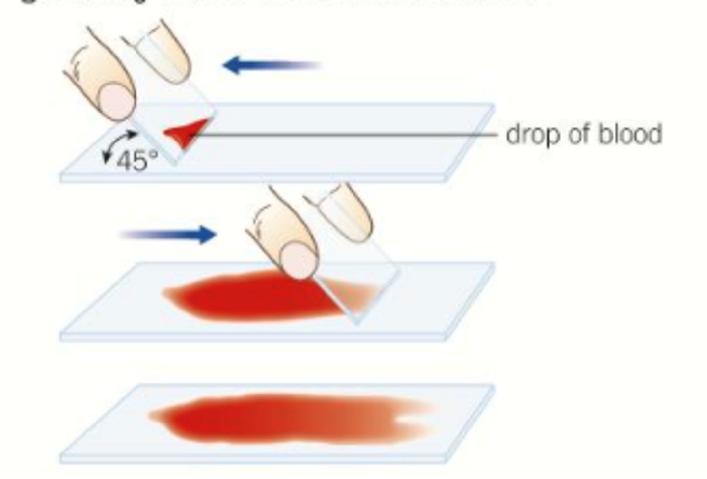
the edge of a slide is used to smear the sample, creating a thin, even coating on another slide, over which a cover slip is placed, e.g. blood.
Sectioning in the preparation of cells
This is done to obtain very thin sections of specimen from a thicker tissue
- freeze specimen in liquid nitrogen or carbon dioxide
- use cryostat to cut into thin slices
- put on slide, add stain, put on cover slip, remove air bubbles
Differential staining
- Many chemical stains used to distinguish different organelles or to provide contrast to cells Cell stain different colours depending on its property
- emphasises difference in structure of organelles and therefore differences in function
- Cells and cellular structures are usually transparent so dyes are used for staining
- eg. gram staining
Magnification equation
magnification = image size/actual size
Equation for total magnification
total magnification = eyepiece lens magnification x objective lens magnification
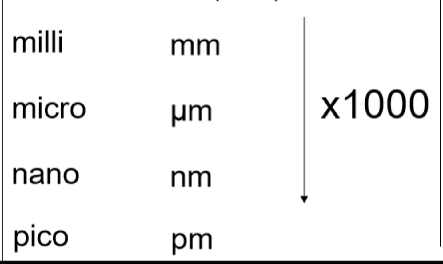
millimetres to micrometers
x1000

micrometers to nanometres
x1000
how to calibrate an eyepiece graticule?
1. Set up the microscope to the required magnification to view the sample.
2. Place a stage graticule on the stage.
3. Line up the two scales (the stage and eyepiece graticules) similar to the diagram.
4. Count the number of divisions on the eyepiece graticule equivalent to each division on the stage micrometre.
5. As the length equivalent to each division on the stage micrometer are known, it is possible to calculate the length of one eyepiece division.
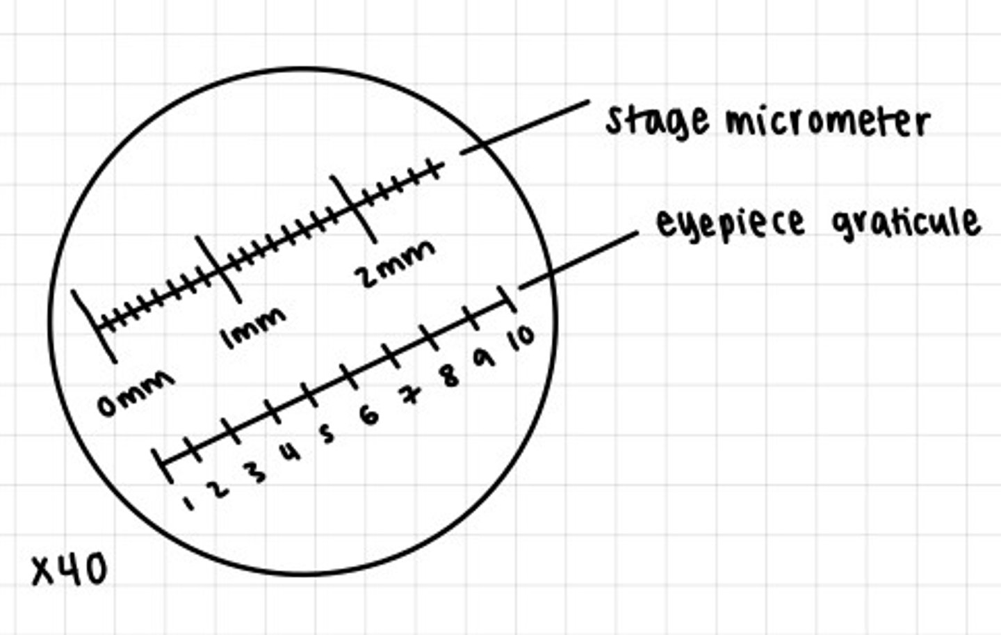
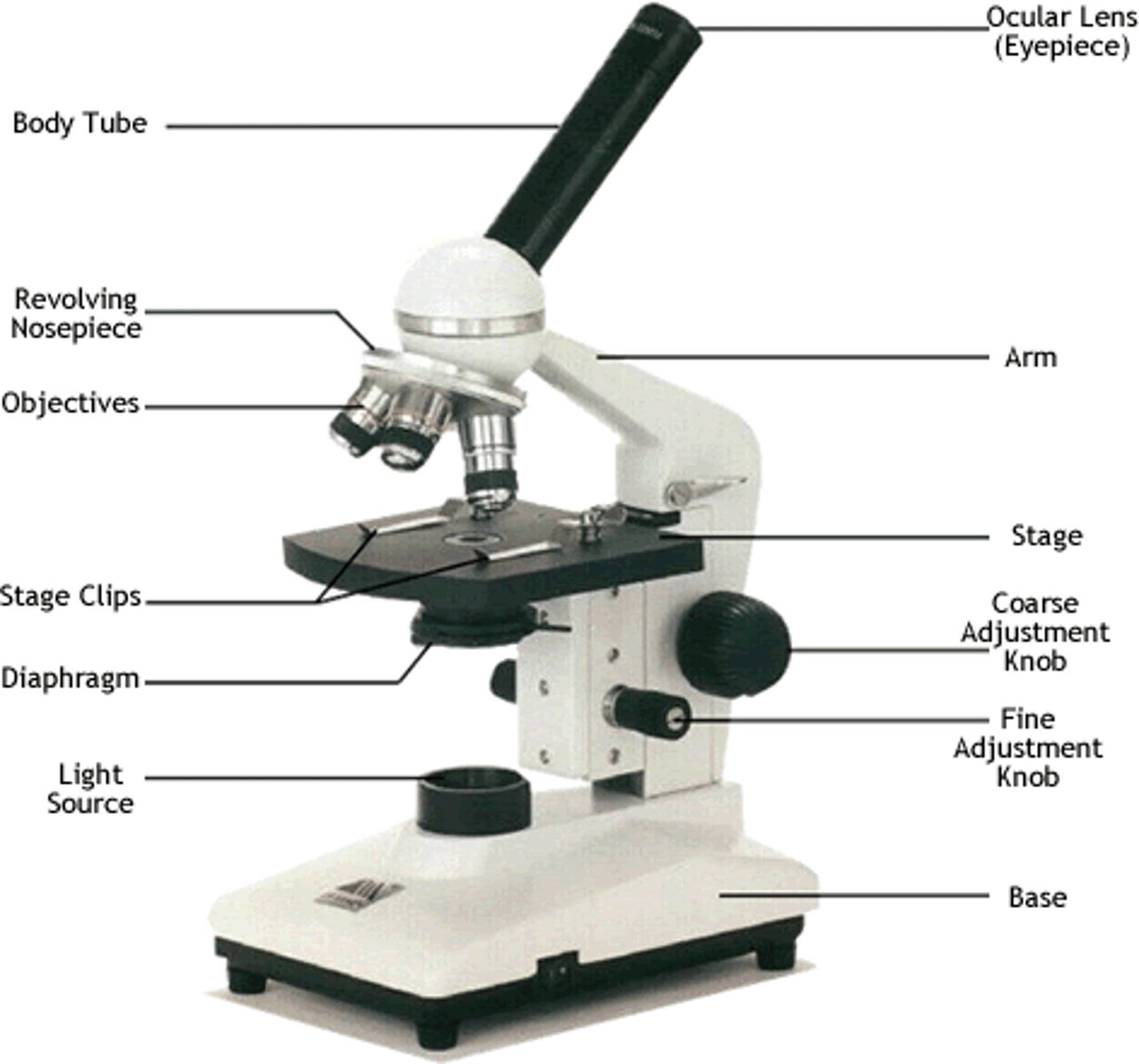
Diagram of microscope
Magnification
How many times bigger an object appears under a microscope compared to reality
Resolution
The distance between two objects which can be see distinctively in an image
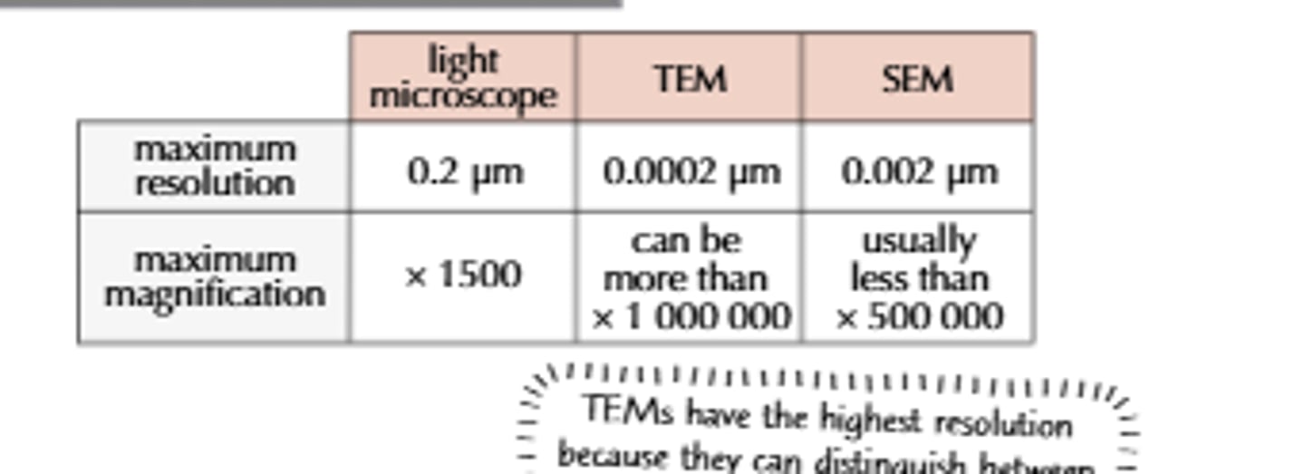
Why do electron microscopes have greater magnification and resolution
Electrons have much smaller wavelengths
How do light microscopes work?
light is focused through several layers of specimen, beam of light produces an image
Advantages of light microscopes
Inexpensive, easy to use, portable, observe dead and living specimens, natural colour can be observed, no vacuum needed, simple preparation by staining
Disadvantages of light microscopes
- resolution limited to 200 nm because it is impossible to resolve 2 objects less than half a wavelength's length of light away
- low magnification
- can only see 2D structures
How TEM microscopes work
- Beam of electrons are transmitted THROUGH the specimen with electromagnets
- denser parts show up as darker
Advantages of TEM microscopes
- higher magnification and resolution than SEM microscopes
- can see details of organelles
- can see differences in density
Disadvantages of TEM microscopes
- affected by magnetic fields
- black and white images only
- expertise required when operating and in preparation of specimen (complicated process)
- specimen can be distorted during preparation
- vacuum required
- expensive to buy and operate
- specimen must be dead
- 2D images only
How do SEM microscopes work
- electron beam transmitted ACROSS specimen by electromagnets
- electrons bounce of surface of specimen
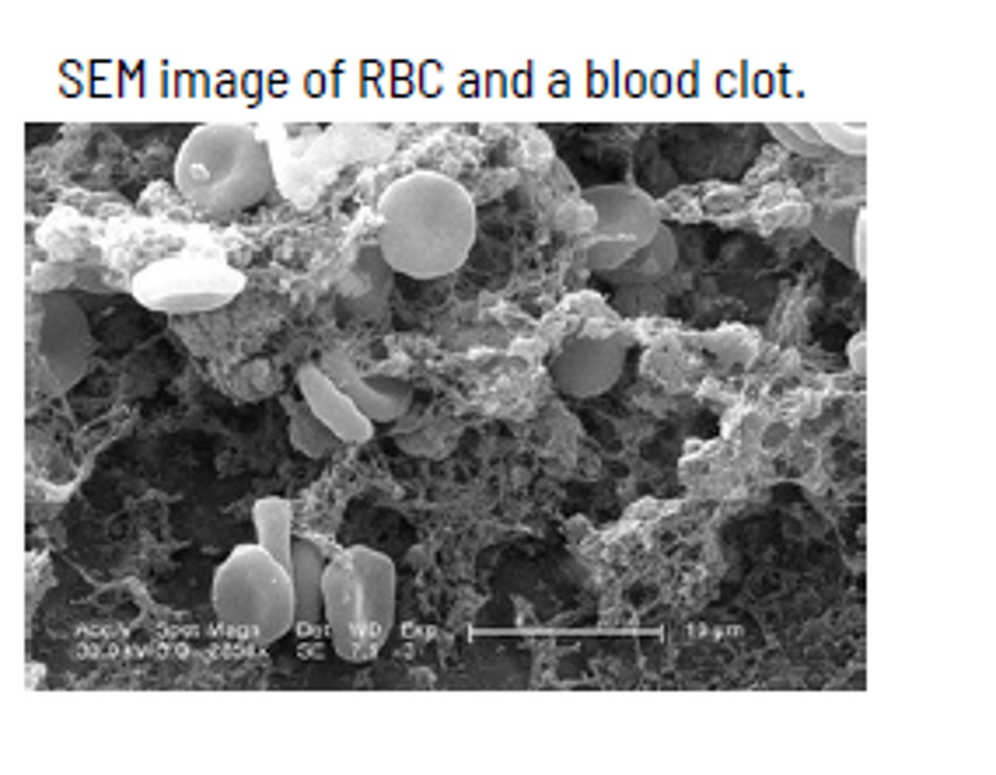
Advantages of SEM microscopes
- can see details of organelles
- 3D image produced
- higher magnification and resolution than light microscopes
Disadvantages of SEM microscopes
- lower magnification and resolution than TEM microscopes
- affected by magnetic fields
- black and white images only
- expertise required when operating and in preparation of specimen (complicated process)
- specimen can be distorted during preparation
- vacuum required
- expensive to buy and operate
- specimen must be dead
How do confocal microscopes work?
- cells are stained with fluroscent dye
- laser beam scans cells
- beam is reflected by the dye
- this occurs in multiple depths of cell tissue
Advantages of confocal microscopes
- 3D images produced
- 3D external structures can be seen
- high magnification and resolution
Disadvantages of confocal microscopes
- very slow, takes a long time
- specimen must be dead
- laser beam may cause photo damage to cells
Cytoskeleton
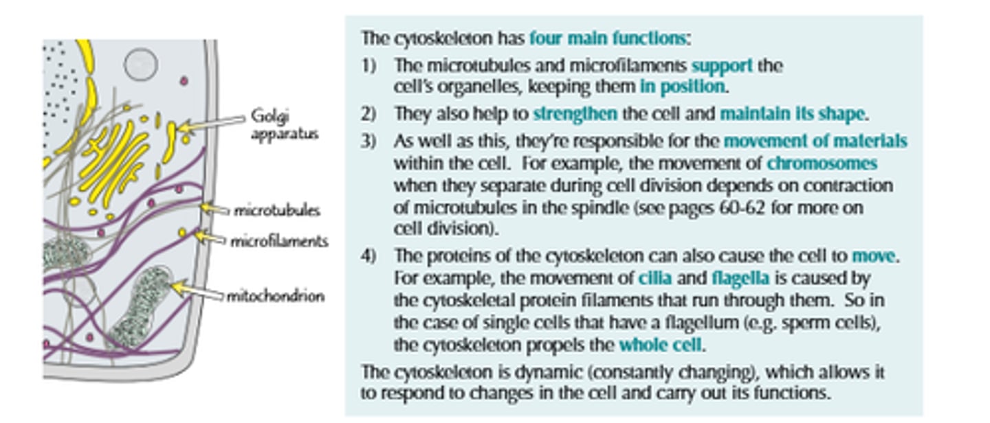
function of cytoskeleton
- changing cell shape
- moving materials
- moving the cell
- maintaining cell shape
- provides mechanical support
- enables cells to carry out essential functions like mitosis and meiosis
- connects cell surface membrane with every organelle
Microfilaments (in the cytoskeleton)
Contractile proteins formed from ACTIN
- 7mm in diameter
- important role in cell movement, cytoplasmic division during cytokinesis, phagocytosis, maintaining shape of cell, muscle contraction
Microtubules (in the cytoskeleton)
Tubular proteins (a type of globular protein) polymerise to form tubes from spindle fibres
- diameter 18-30cm
- forms scaffold to provide cell shape and structure
- acts as tracks for organelles and vesicles to move along
- forms spindle to move chromosomes in cell division
- make up cillia
Intermediate fibres (in the cytoskeleton)
Gives mechanical strength to cells and help maintain their integrity
- made up of a variety of proteins, 10mm in diameter
- are stable and do not grow or disassemble
- holds organelles in place
- provide tensile and mechanical strength to the cell
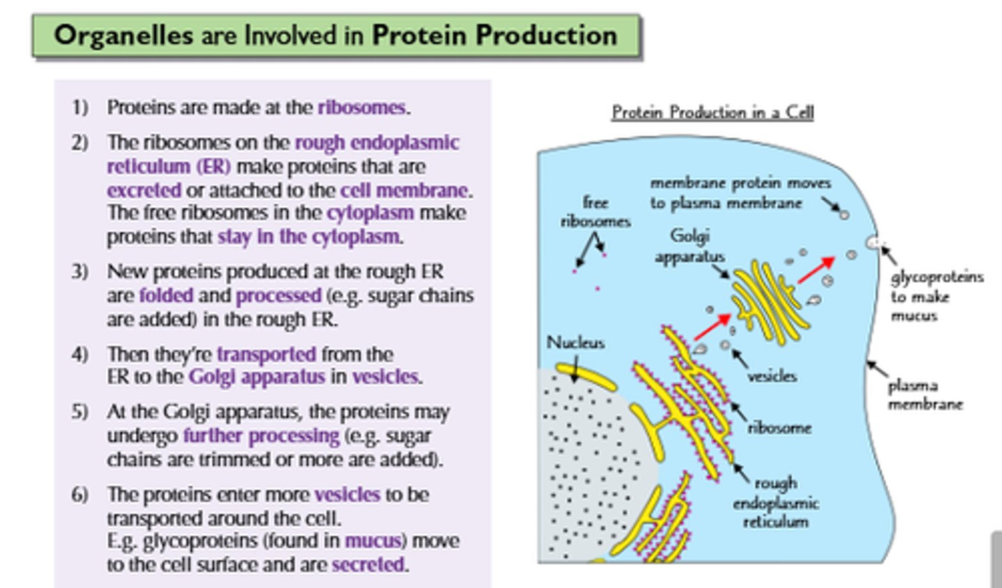
production and secretion of proteins
1. mRNA copy of the instructions for insulin made in the nucleus
2. mRNA leaves the nucleus through a nuclear pore
3. mRNA attatches to a ribosome and protein is assembled
4. insulin molecules are pinched off in vesicles and travel towards golgi apparatus
5. vesicle fuses with golgi apparatus
6. golgi apparatus processes and packages insulin molecules ready for release
7. packaged insulin molecules are 'pinched off' in vesicles from golgi apparatus and move towards the plasma membrane
8. vesicle fuses with plasma membrane
9. plasma membrane opens to release insulin molecules outside by exocytosis
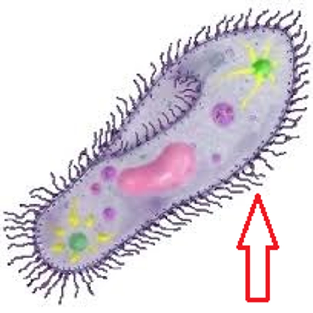
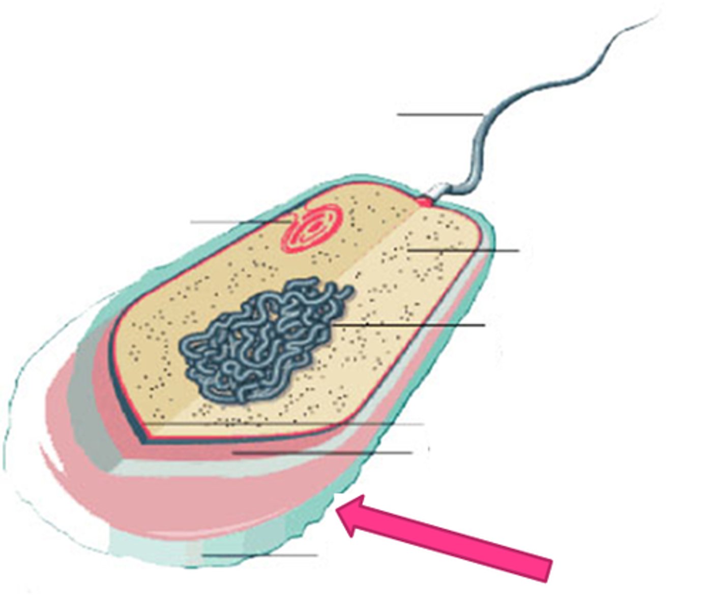
structures within a bacterial cell
flagella, pilli, nucleiod (single circular DNA), plasmids (small loops of DNA containing genes), ribosome, capsule (protects bacteria from drying out, cell wall, plasma membrane
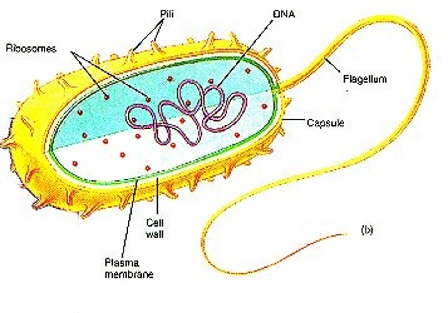
Prokaryote
no nucleus, no membrane bound organelles apart from ribosomes
- their ribosomes are structually smaller (70s)
- have single circular DNA instead that is free in the cytoplasm with no ribosomes
- has cell wall that contains murein (glycoprotein) and peptidoglycen
- 0.5-5 nanometre in diameter
- binary fission, no spindles involved
- unicellular organisms only
Eukaryote
A cell that contains a nucleus and membrane bound organelles.
- part of multicellular organism
- up to 40 nanometres in diameter
- has DNA associated with histones, formed into chromosomes
- mitosis and meiosis, spindle involved to seperate chromosomes
- 80s ribosomes
- cell wall made of chitin or cellulose
Plasmid
- short loop of DNA
- allow transfer of genes between different prokaryotic cells
- found in prokaryotic cells
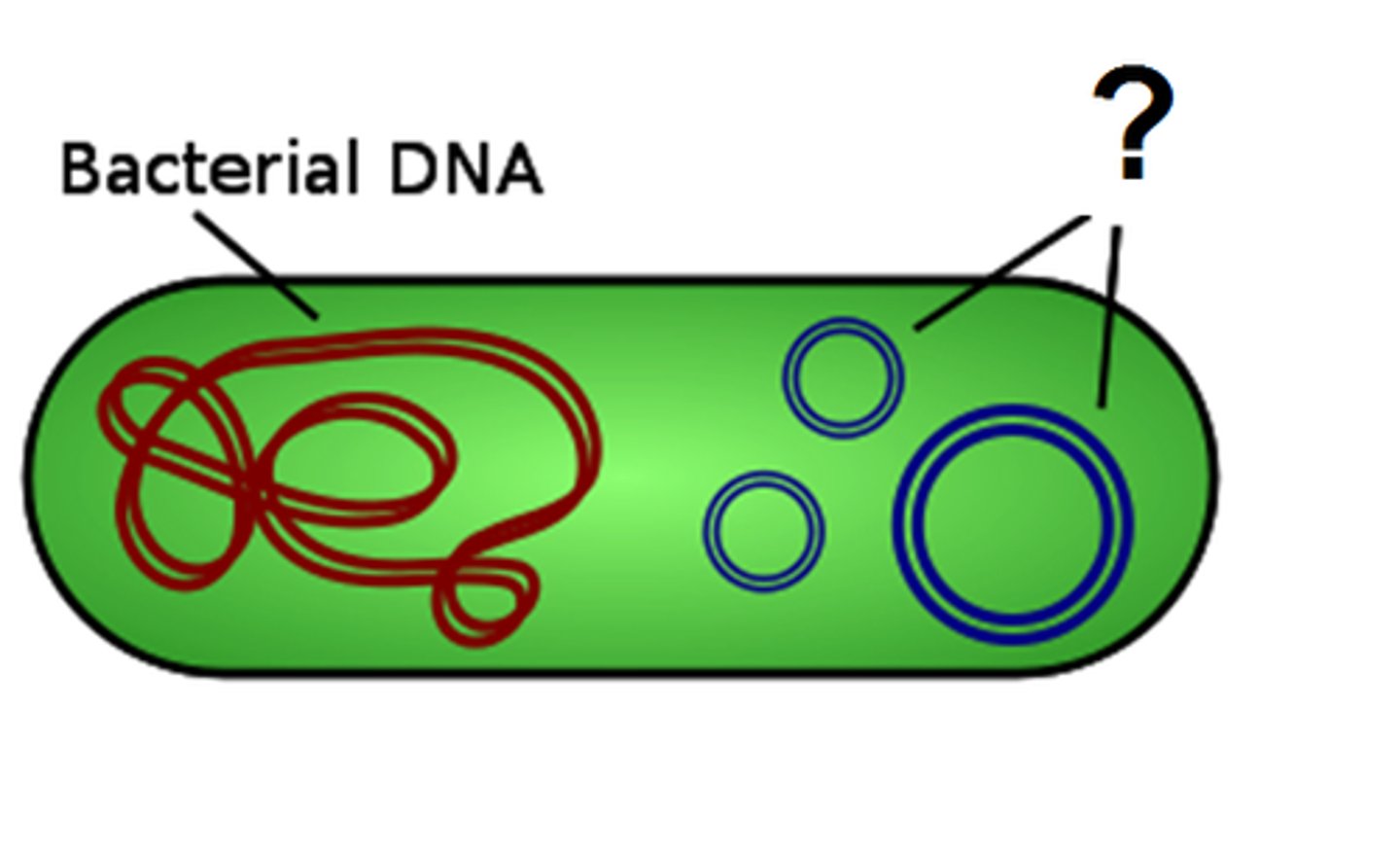
Capsule
- found in prokaryotic cells
- outer layer
- protects from attack and from drying out
compare the magnification and resolution of microscopes
What makes up an organism

Why would you need to use dry mount?
-solid/dry specimens e.g. hair
-avoid distortion
-easy preperation
rules when doing scientific drawings
Draw in pencil
Title the diagram to indicate what the specimen is
State the magnification that you are drawing it from
Annotate cell components, cells and sections of tissue
visible
Do not sketch. Only use solid lines that do not overlap
Do not colour in or shade
why do a wet mount?
-Viewing Living Specimens: Wet mounts are essential for observing living organisms such as protozoa, algae, and bacteria. The liquid medium keeps the organisms alive and active for observation
-Studying Cells and Tissues: Wet mounts are commonly used to study cells and tissues in their natural state, making it easier to observe cell morphology, arrangement, and interactions.
-Observing Natural Behavior: For living organisms, a wet mount allows for the observation of natural behaviors such as movement, feeding, and reproduction.
why do a squash mount?
-soften tissue
-better visablilty
-examining cell division
-better visability
why do a smear mount?
-consitency
-staining and contrast
-rapid prep
why do we stain?
improve contrast
improve visability
why do you need thin specimens?
so light can pass through
scale of units