Blastocyst GARDNER Grading
 Call Kai
 Learn
Learn Practice Test
 Spaced Repetition
 Match
Match Flashcards
 Knowt Play
1/37

Earn XP
Description and Tags
Monitoring Embryo Development (Lecture 3)
Name | Mastery | Learn | Test | Matching | Spaced | Call with Kai | Chat |
|---|
No analytics yet
Send a link to your students to track their progress
38 Terms
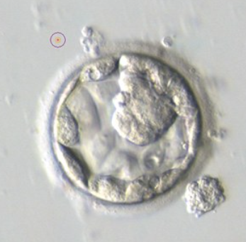
Grade 3:A:A
Blastocyst - Full Blastocoel with the cavity completely filling the embryo. Compact ICM
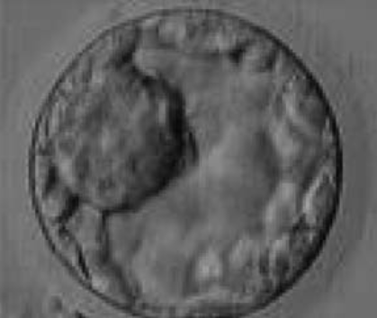
Grade 3:A:A
Blastocyst - Very large ICM at the 10’ position. The ICM is made up of many cells that are tightly compacted.

Grade 3:A:A
Blastocyst - With a dense ICM clearly visible at the 11’ position. The TE cells are variable in size but form a cohesive epithelium.
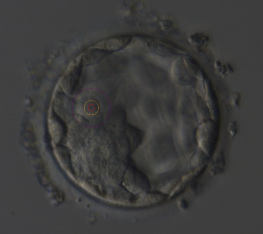
Grade 3:A:A
Blastocyst - With a dense ICM clearly visible at the base of the blastocyst
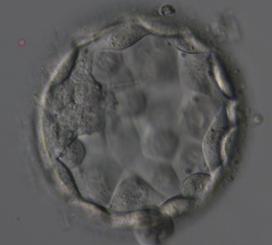
Grade 4:A:A
Expanded Blastocyst - Showing a large ICM made up of many cells that are tightly compacted.
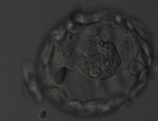
Grade 4:A:A
Expanded blastocyst - Large ICM. The blastocyst is now a greater volume than the original volume of the embryo and the ZP is thinned. There appears to be cytoplasmic strings extending from the ICM to the TE.
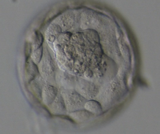
Grade 4:A:A
Expanded blastocyst - Showing a large ICM. The ICM is made up of many cells and is very compact. The blastocoel cavity is now larger than the original volume of the embryo and the ZP is very thin.
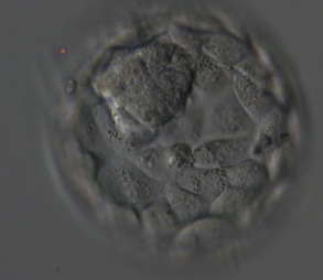
Grade 4:A:A
Expanded Blastocyst - Showing a large ICM at the top of the blastocyst in this view. There are very many TE cells forming a cohesive epithelium that lines the enlarged blastocoel cavity. The ZP is very thin.
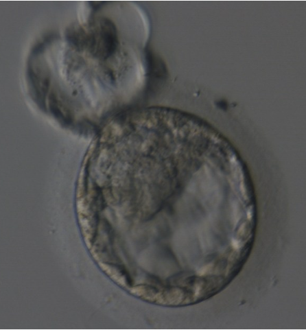
Grade 5:A:A
Hatching blastocyst - Showing a large, compact, crescent-shaped ICM retained within the ZP at the 12’ position. There are very many TE cells and almost 25% of the blastocyst has herniated out through a breach in the ZP at the 11’ positions in this view.

Grade 5:B:A
Hatching Blastocyst - Showing a small ICM being drawn out along with the herniating TE cells at the 1’ position. There are very many TE cells of similar size lining the blastocoel cavity and the ZP is thinned.

Grade 5:A:A
Hatching Blastocyst - Showing a large, compact ICM at the 12’. There are many TE cells of equivalent size lining the blastocoel cavity and several TE cells are herniating though a breach in the thinned ZP.
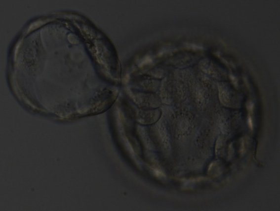
Grade 5:A:A
Hatching blastocyst - Approximately 25% of the blastocyst has herniated from a breach in the thinned ZP at the 10-11’ positions.

Grade 4:A:A
Expanded Blastocyst
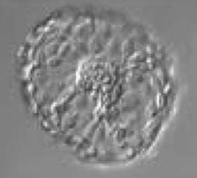
Grade 6:A:A
Hatched Blastocyst - That is now completely free of the ZP. The ICM positioned centrally at the base of the blastocyst appears to be connected or anchored to the TE by several bridges. There are very many TE cells of similar size forming a cohesive epithelium.

Grade 6:A:A
Hatched blastocyst - That is now only just free of the ZP. The ICM is large and compact at the base of the blasto. The TE is similarly made up of many cells forming a cohesive epithelium. There is some cellular debris discarded in the ZP

Grade 6A:A
Hatched Blastocyst - That is now completely free of the ZP. Some Cellular debris remains behind in the empty ZP. There are many TE cells making up a cohesive epithelium. Can clearly see the increase in diameter of the blastocyst from the diameter of the original embryo which was accommodated within the ZP.
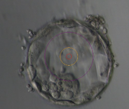
Grade 3:B:A
Blastocyst - Showing a compact ICM at the 6’ position. The ICM is small relative to the diameter of the blastocyst and probably made up of a few cells
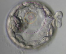
Grade 5:B:A
Early Hatching Blastocyst - Showing a compact ICM, small and flattened and made up of few cells relative to the size of the blastocyst.

Grade 3:B:C
Blastocyst - with sparse TE that does not form a cohesive epithelium.
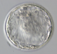
Grade 4:C:C
Expanded Blastocyst - With no clearly identifiable ICM. TE is made up of a few, sparse cells that do not form a cohesive epitelium
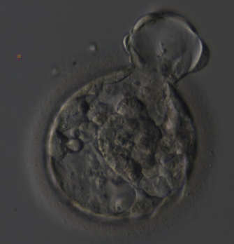
Grade 5:A:B
Hatching Blastocyst - The TE cells vary in size with some cells quite large forming a loosely cohesive epithelium. The ICM can be seen at the 3’.
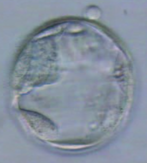
Grade 4:A:C
Expanded Blastocyst - With a very sparse TE that does not form a cohesive epithelium. The ICM is visible at the 10’ position.
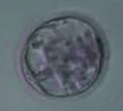
Grade 3:C:C
Blastocyst - with spare TE that does not form a cohesive epithelium. The ICM is not clearly identifiable.
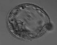
Grade 4:C:C
Expanded Blastocyst - With sparse TE that does not form a cohesive epithelium. The ICM is hardly distinguishable despite the expansion of the blastocoel cavity.
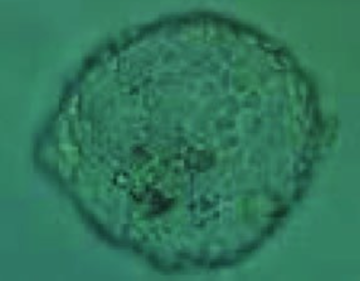
Grade 6:A:A
Hatched Blastocyst - Showing many cells in the TE making a cohesive epithelium. Many of the TE cells contain dark granules. The ICM is not clearly seen in this view and there are several degenerative foci (dark cells) associated with the ICM and the polar TE.

Page 43

p. 44
p. 45
p.46
p.47
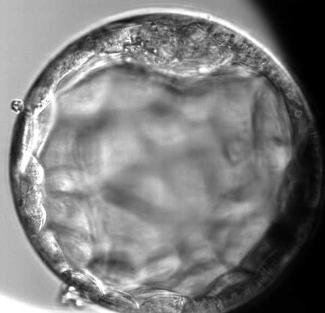
Grade 4:B:A
p.48
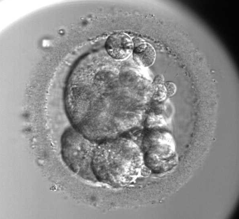
Activated Oocyte
p.49
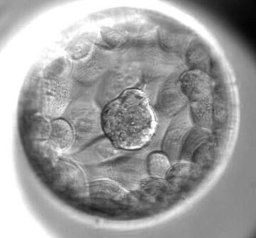
Grade 4:B:A

p.50
Grade 4:B:B
Small TE volume, not enough cells
p.51
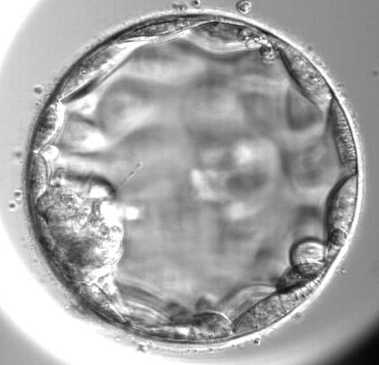
Grade 4:A:C
Big TE cells but very low in number, especially given the size of the blastocyst.
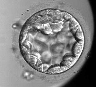
p.52
Grade 3:A:A
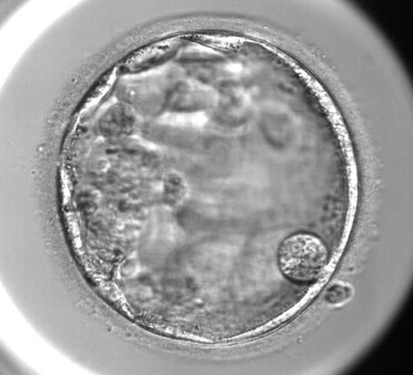
p.53
Grade 4:C:C
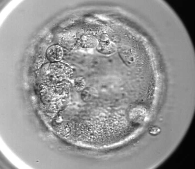
p.54
Grade 4:C:C
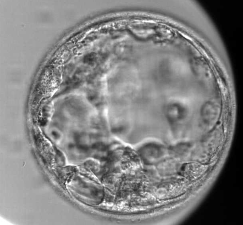
p.55
Grade 4:C:B