Exam 1 Objectives
 Call Kai
 Learn
Learn Practice Test
 Spaced Repetition
 Match
Match Flashcards
 Knowt Play
1/159

There's no tags or description
Looks like no tags are added yet.
Name | Mastery | Learn | Test | Matching | Spaced | Call with Kai | Chat |
|---|
No analytics yet
Send a link to your students to track their progress
160 Terms
Anatomy
study of strucutre
Physiology
study of function
Levels of organization in sequence
atoms, molecules, cells, tissues, organs, organ system, organism
Atoms
smallest units of matter
Molecules
two or more atoms combined
Cells
smallest living structures
Tissues
groups of similar cells with common function
Organs
two or more tissues working together to perform specific, complex functions
Organ system
related organs that work together for a common function
Organism
a single living organism
Anatomical position
stadning upright
feet parallel and on the floor
head level and looking foward
arms at sides
palms facing foward
thumb pointing away from body
Anatomical left & right
always refer to the patient's or subject's own left and right sides, never the observer's perspective
Supine
lying on back with face upwards
Prone
laying on stomach with face downwards
- Apply anatomical position to specimens, models, figures, and images in different orientations
Describe anatomical position, use it as a standard frame of reference, determine anatomical right and left, and distinguish supine from prone.
- Standard anatomical position
- Anatomical right vs. left
- Supine vs. prone
- Apply anatomical position to specimens, models, figures, and images in different orientations
Sagittal plane
divides body into unequal left and right
Midsagittal plane (median)
divides body into equal left and right
Parasagittal plane
a sagittal plane parallel to midline but shifted to one side
Coronal plane (frontal)
divides body into anterior and posterior parts
Transverse plane (cross-sectional, horizontal)
divides body into superior and inferior parts
Recognize that a structure can look different depending on how it is sectioned.
Identify and describe the major anatomical planes and use sectional orientation to interpret anatomical images.
- Sagittal
- Midsagittal
- Parasagittal
- Frontal/coronal
- Transverse
Recognize that a structure can look different depending on how it is sectioned.
Anterior
ventral; front
Posterior
dorsal; back
Superior
toward head
Inferior
toward feet
Cranial
toward head
Caudal
toward tail
Medial
toward midline
Lateral
away from midline
Proximal
close to trunk
Distal
far from trunk
Superficial
external
Deep
internal
Apply the terms to anatomical relationships, not just definitions
Correctly use directional terms to describe relationships between structures.
- Anterior / posterior
- Superior / inferior
- Cranial / caudal
- Medial / lateral
- Proximal / distal
- Superficial / deep
Apply the terms to anatomical relationships, not just definitions
Posterior aspect
consists of cranial cavity and vertebral canal; contains the brain
Cranial cavity
formed by skull bones
Vertebral canal
formed by vertebral column
Ventral canal
Contains two smaller cavities separated by diaphragm; consists of thoracic, abdominopelvic, abdominal, pelvic cavities; contains spinal cord
Thoracic cavity
superior to diaphragm
Abdominopelvic cavity
inferior to diaphragm; contains two cavities divided by pelvic brim of hip bones
Abdominal cavity
superior to pelvic brim
Pelvic cavity
inferior to pelvic brim
Upper limb
entire upper extremity from shoulder to hand
Arm
brachial
Forearm
antebrachial
Hand
manus
Thigh
femoral
Leg
crural
Armpit
axillary
Chest
pectoral
Cheek
buccal
Chin
mental
Wrist
carpal
Posterior of knee
popliteal
Calf
sural
Foot
Pes / Pedal

Toe
digital
The intermost digit of a forelimb
pollex
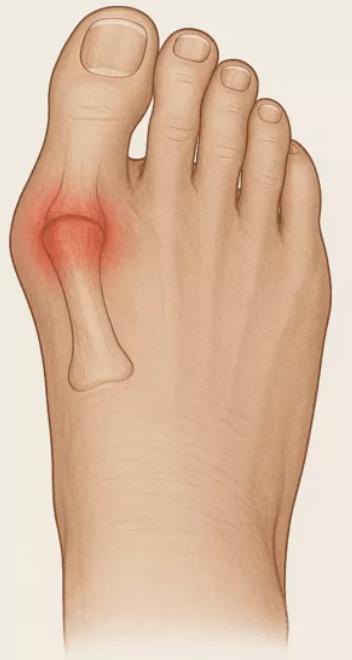
The innermost digit on a foot
hallux
Parietal serosa
lines internal surface of the body wall
Visceral serosa
covers external surface of organs (viscera)
Serous cavitites
Between layers; reduces friction between moving organs
Serous fluid
fluid between layers; reduces friction between moving organs
Organs
collection of different tissues that work together to perform a specific function in a living organism
Pleura
serous membranes around lungs; Body wall → parietal serosa → serous cavity/fluid → visceral serosa → organ
Pericardium
serous membranes around heart; Body wall → parietal serosa → serous cavity/fluid → visceral serosa → organ
Peritoneum
serous membranes around many abdominal organs; Body wall → parietal serosa → serous cavity/fluid → visceral serosa → organ

What is J?
Right Upper Quadrant
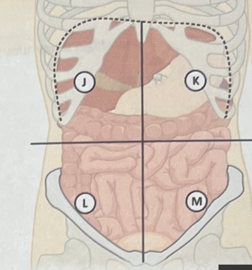
What is K?
Left Upper Quadrant

What is L?
Right Lower Quadrant
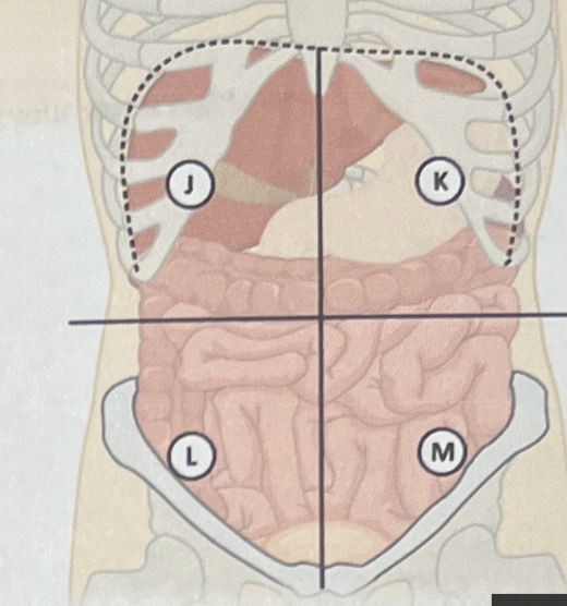
What is M?
Left Lower Quadrant

What is A?
Right Hypochondriac Region
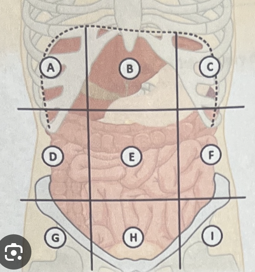
What is B?
Epigastric Region
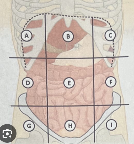
What is C?
Left Hypochondriac Region

What is D?
Right Lumbar Region

What is E?
Umbilical Region

What is F?
Left Lumbar Region
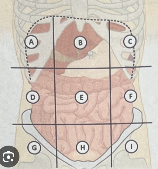
What is G?
Right Iliac Region
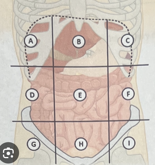
What is H?
Hypogastric Region

What is I?
Left Iliac Region
Anatomical Left
Your right
Anatomical Right
Your left
Tissue
a group of similar cells and extracellular products that perform a common function
Histology
he study of tissues
Epithelial Tissue
covers, lines, secretes

Connective Tissue
supports and binds
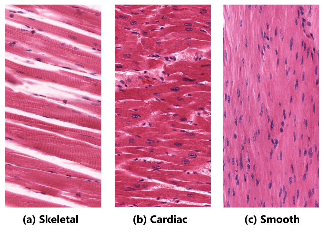
Muscle Tissue
contracts

Nervous Tissue
communicates
Cellularity
composed almost entirely of cells bound by intercellular junctions; little extracellular matrix between cells
Polarity
epithelia have an apical surface, lateral surface, and a basal surface
Apical surface
exposed and may have modifications (for example, microvilli)
Basal surface
attached to connective tissue underneath it
Basement membrane
a structure produced by both epithelial and neighboring connective tissues
Avascularity
Epithelial tissues lack blood vessels; receive nutrients by diffusion from underlying tissues
Regenerative capacity
Because exposed apical surface is frequently damaged, epithelial cells are quickly
replaced
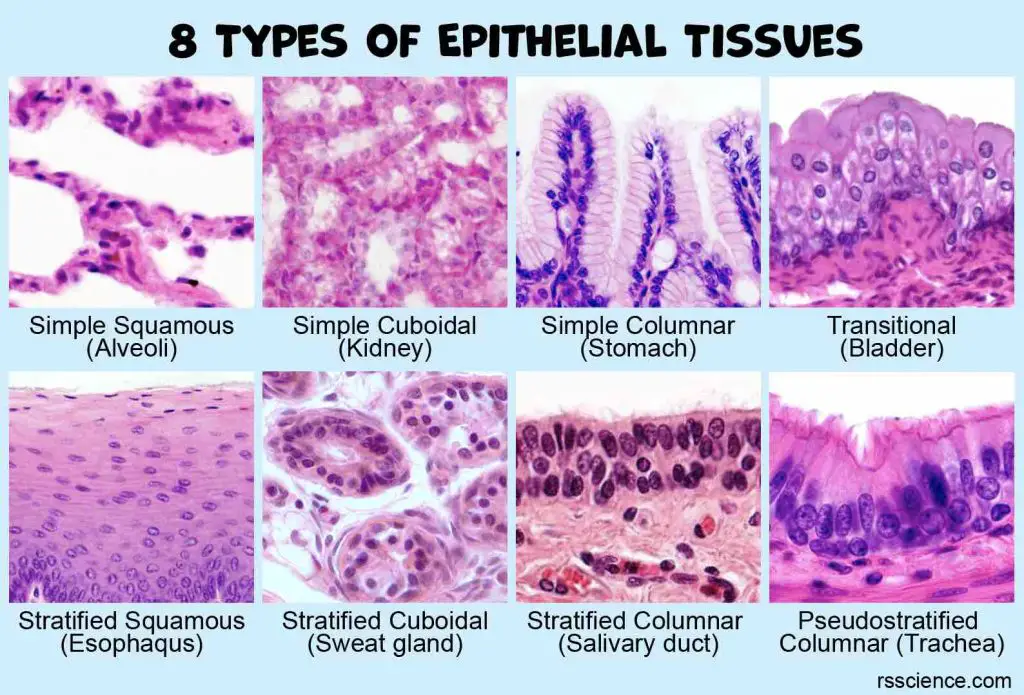
Classify epithelia by number of layers and cell shape and relate structure to function and location.
- Simple vs. stratified
- Squamous, cuboidal, columnar
- Simple squamous
- Simple cuboidal
- Simple columnar
- Pseudostratified columnar
- Stratified squamous
- Transitional
For stratified epithelia, classification is based on the apical cells.
Structure → Function → Representative Location
Endocrine
do not have ducts; secrete hormones (chemical messengers) directly into interstitial fluid or bloodstream