Dermatology Block 1, Week 1
 Call Kai
 Learn
Learn Practice Test
 Spaced Repetition
 Match
Match Flashcards
 Knowt Play
1/321

There's no tags or description
Looks like no tags are added yet.
Name | Mastery | Learn | Test | Matching | Spaced | Call with Kai | Chat |
|---|
No analytics yet
Send a link to your students to track their progress
322 Terms
A ______ is a circumscribed change of skin color without elevation and is up to 1cm in diameter
macule
A _______ is a large macule (greater than 1 cm diameter)
patch
A ______ is a raised, solid lesion that is LESS than 1 cm in diameter
papule
A ________ is a large papule that is greater than or equal to 1 cm in diameter
plaque
A ______ is a solid, palpable lesion originating in the dermis or subdermis that may or may not be elevated above the level of the surrounding skin and is up to 1 cm in diameter
nodule
A _______ is a large nodule that is greater than or equal to 1cm in diameter
tumor
A ______ is a circumscribed, elevated lesion containing fluid, less than 1 cm in diameter
vesicle
A _______ is a large vesicle that is greater than or equal to 1cm in diameter
bulla
A _______ is a circumscribed lesion containing pus ("pus-filled")
pustule
A _______ is a non-blanching, violaceous discoloration of the skin and it can be palpable or non-palpable
purpura
_________ refers to a purpura that is less than 1 cm in diameter
petechiae
_______ refers to a purpura that is greater or equal to 1 cm in diameter
ecchymosis
_____ refers to an evanescent, edematous (swollen), erythematous papule or plaque that is usually pruritic ("Hive")
wheal
_________ refers to small superficial blood vessels, dilated capillaries, blanchable
telangiectasia
An _______ refers to a superficial denudation of the skin, mainly the epidermis (loss of the epidermis)
erosion
An _____ refers to a denudation of the skin involving the dermis
ulcer
A ______ is an excess accumulation of "dead skin"
scale
A _________ is a linear crack in the epidermis
fissure
_______ can range from pink to deep red hue in the skin due to vasodilation and it indicates vascular inflammation or dilation
erythema
_____ refers to depression of the skin due to thinning of the epidermis, dermis or subcutaneous fat
atrophy
______ are permanent fibrotic changes that occur on the skin following damage to the dermis
scars
What are the functions of the skin?
-barrier
-immunologic
-temperature regulation
-protection from radiation
-nerve sensation
-injury repair
-appearance/quality of life
The _________ is the topmost layer of skin and consists of primarily _________
epidermis, keratinocytes
The ______ lies below the epidermis and consists primarily of fibroblasts, collagen and elastic fibers
dermis
What are the layers of the epidermis from inner to outer?
stratum basale,
stratum spinosum,
stratum granulosum,
stratum corenum
What are the key functions of the stratum basal epidermal layer?
-cell division
-source of epidermal stem cells
-keratinocytes start in this layer and move upwards
What are the key functions of the stratum spinosum epidermal layer?
has "spiny" appearance due to desmosomal junctions holding keratinocytes together
What are the key functions of the stratum granulosum epidermal layer?
lipids in this layer form a water barrier that keeps water in the skin
What are the key functions of the stratum corneum epidermal layer?
provides barrier against trauma and infection
Bullous Pamphigoid
Clinical Presentation:
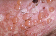
tense bullae on an erythematous base on the skin

Bullous Pamphigoid
Risk Factors:
Age (typically affects elderyly)
Bullous Pamphigoid
Cause:
Loss of Adhesion; autoantibodies form to antigens beneath the basal layer of the epidermis so epidermis forms roof of blister and dermis forms base of the blister
IgG autoantibodies bind to antigens that comprise the hemidesmosome adhesion complex in the basement membrane of the skin
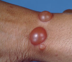
Bullous Pamphigoid is an __________ blistering disease
autoimmune
Pemphigus Vulgaris
Clinical Presentation:
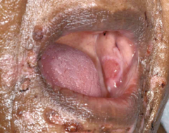
erosions or flaccid bullae
Pemphigus Vulgaris
Risk Factors:
young to middle aged adults
Pemphigus Vulgaris
Cause:
Loss of adhesion: autoantibodies form to desmosomes that attach keritonocytes to one another.
What causes Psoriasis?
over-proliferation of keratinocytes causing plaques

What are the four main types of cells that make up the epidermis?
kertainocytes
melanocytes
langerhans cells
merkel cells
________ are the main structural cell of the epidermis. They are held together by adhesion proteins called desmosomes.
Keratinocytes
_______ are pigment-producing cells that transfer a pigment called melanin to the keratinocytes in the basal cell layer
Melanocytes
__________ are dendritic cells found in the lower to mid-epidermis
Langerhans
_______ form a complex with afferent sensory nerve endings associated with touch, and are found more densely in touch-sensitive area of the skin
Merkel cells
What are the structural components of the dermis?
collagen type I (strength)
elastic fibers (elasticity)
extrafibrillar maxtrix
What are the cells of the dermis?
fibroblasts
macrophages
mast cells
Ehlers-Danlos Syndrome
Clinical Presentation:
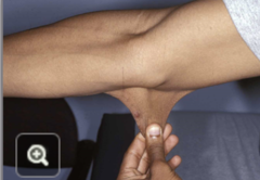
hyper-extensible joints, skin or vascular aneurysms
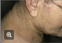
Solar Elastosis
Clinical Presentation:
degradation of collagen and elastic fibers
Solar Elastosis
Cause:
chronic UV exposure
________ is a disorder of keratin retention in the infundibulum of the hair follicle
keratosis pilaris

What are the types of skin cancers?
1) Basal Cell Carcinoma
2) Squamous Cell Carcinoma
3) Malignant Melanoma
What is the most common skin cancer?
Basal Cell Carcinoma
Basal Cell Carcinoma
______ (Most/Least) common skin cancer and rarely ________
Arises from the _____ layer of the epidermis
Basal Cell Carcinoma
Most common skin cancer and rarely metastasizes
Arises from the basal layer of the epidermis
Basal Cell Carcinoma
Cause:
UV radiation causing DNA damage
Basal Cell Carcinoma
Treatment Options:
Surgical:
Non-surgical:
Surgical:
Mohs Surgery: done mainly on facial lesions or cosmetically sensitive areas and on recurrent BCC; allows for maximal conservation of tissue
Cryosurgery
Curette and desiccation (scraping and burning affected area)
Excision with standard 3-4 mm margins
Non-surgical:
topical Imiquimod cream
topical 5-fluoruracil cream
Describe the clinical representation of Nodular Basal Cell Carcinoma
pearly,
white,
translucent papule or plaque
central ulceration
rolled borders
haphazardly organized telangiectasias (dilated capillaries)

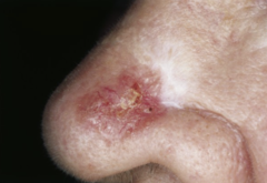
Describe the clinical representation of Superficial Basal Cell Carcinoma:
appears as a well-circumscribed, erythematous macule/patch or slightly raised, flat papule/plaque, mainly consists of a thinly-rolled border and a scant scale and haphazard telangiectasis

Describe the clinical representation of Morpheaform or Sclerosing Basal Cell Carcinoma
white, waxy, scar-like papule or plaque with indistinct borders, usually appears on the face
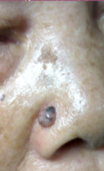
Describe the clinical representation of Pigmented Basal Cell Carcinoma
variable amounts of melanin within the lesion, globules of pigment
Basosquamous carcinoma is a mixed ________ cell and _______ cell carcinoma. These have an infiltrative growth pattern and can be more aggressive in their behavior
basal
squamous
When should you refer a patient that has Basal Cell Carcinoma to a dermatologist?
suspicion of BCC on the face or a cosmetically sensitive area
biopsy proven BCC
recurrent BCC, metastatic BCC
Basosquamous carcinoma
No or little experience in treating BCC
What causes actinic keratosis?
sun exposure
What can actinic keratosis develop into?
squamous cell carcinoma
Actinic Keratosis
Cell of Origin:
keratinocytes; typically premalignant SCC
Actinic Keratosis
Cause:
sun exposure
Actinic Keratosis
Clinical Presentation:
Clinical Presentation:
hyperkeratotic (thick, rough skin),
feels like "sandpaper,"
skin-colored to tan/brown colored,
irregularly shaped and ill-defined papules and plaques

Actinic Keratosis
Treatment Options:
Treatment Options:
cryotherapy, topical medications (5-FU, Imiquimod, Diclofenac, Tiranibulin), ED&C, Photodynamic therapy (PDT), chemical peels, laser surgery
Actinic Keratosis
Risk Factors:
fair skin (Fitzpatrick Type I-III), elderly, history of prolonged sun exposure, immunocompromised
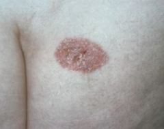
Squamous Cell Carcinoma
Clinical Presentation:
Clinical Presentation:
-erythematous, scaly, indurated papules and plaques, +/- ulceration, +/- pain, tenderness

Squamous Cell Carcinoma
Cause:
Cause:
-sun exposure
-mutations in keratinocyte DNA, usually caused by UVB
-Tobacco use
-Subtypes of HPV

Squamous Cell Carcinoma
Risk Factors:
Risk Factors:
-Age/Sex (common in elderly males)
-History of SCC or other skin cancers (BCC, MM)
-fair skin, blue eyes, blonde/red hair (FP I-III)
-h/o of smoking
-previous trauma to the area
-inherited syndroms (ex. albinism)
-exposures: ionizing radiation
Squamous Cell Carcinoma
Workup for suspected SCC:
Workup for suspected SCC:
-shave, punch or excisional biopsy (depending on site of lesion)
-CT/MRI for advanced stages of SCC
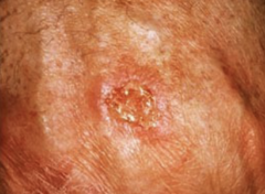
Squamous Cell Carcinoma
Treatment:
Treatment:
1) ED&C; low-risk, in-situ SCC of torso
2) Mohs surgery; for lesions of head/neck & for invasive SCC
3) Standard Excision; for low risk and primary SCC lesions in non-cosmetically sensitive areas
4) Radiation Therapy: used for patients who are poor surgical candidates
5) Chemotherapy/Immunotherapy; used for high risk /advanced cases of SCC
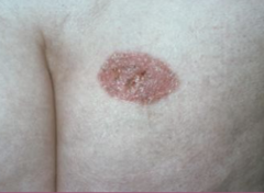
Can squamous cell carcinoma metastasize?
yes
When should you refer out a patient with SCC?
-biopsy proven SCC
-invasive SCC
-SCC of face/neck or cosmetically sensitive areas (refer for Mohs surgery)
-patients with h/o of skin cancer
-patients with history of excessive sun exposure and concomitant actinic damage
________ nevi consist of nests of nevus cells at the dermal-epidermal junction
Junctional
_______ nevi consist of nest of nevus cells within the dermis
Dermal
_______ nevi consists of nests of nevus cells both at the dermal-epidermal junction and within the dermis
Compound
A _________ nevus is a nevus with an appearance different from common nevi (atypical pigmentation, shape, behavior. This is also known as atypical nevus or nevus with architectural disorder and possibly with cytologic atypia
dysplastic
Dysplastic nevi are ________ (benign/malignant) lesions
benign (but they can indicate a greater risk for developing melanoma)
____________ is the predominant group of nevi in a person with multiple nevi
signature nevus
A ________ nevus has homogenous brown pigmentation
solid brown
A _______ nevus is present in fair-skinned individuals (Fitzpatrick I-II)
solid pink
A _______ nevus has a central area of dark pigmentation surrounded by rings of lighter and darker pigmentation
cockade
A ______ nevus is tan or pink in the central area with a surrounding ring of pigmentation
eclipse
A ______ nevus has a central raised papule with surrounding pigmentation. The central area may be lighter or darker than its surrounds.
fried egg
_____________ are hypopigmented areas surrounding hair follicles of a nevus
perifollicular
The "____________" refers to the one nevus among many that appears different from the others
ugly duckling
____________ __________ refers to a neoplasm of melanocytes (or of cells that have developed melanocytes)
malignant melanoma
Is malignant melanoma an aggressive form of cancer?
Yes, it can metastasize via direct extension, the lymphatic system or hematogenously
Describe the morphology of malignant melanoma:
irregularly shaped/pigmented macule, nodule, papule or plaque
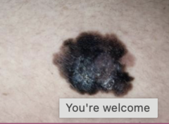
Superficial Spreading Melanoma is the ________ (most/least) common form of melanoma and the lesion is confined to the _________
most
epidermis
Describe the morphology of Superficial Spreading Melanoma:
growth is primarily horizontal (radial)

In ___________ melanoma, growth is vertical and the lesion has started to grow downward, penetrating the basement membrane (vertical growth phase)
nodular
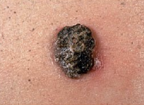
_______________ melanoma refers to a "changing freckle," in situ lesion. Growth is primarily horizontal and not vertical
Lentigo Maligna
__________ melanoma occurs on the acral surfaces (palms of hands, soles of feet, nail beds)
Acral Lentiginous
What is the most common type of melanoma in darker skin individuals?
Acral Lentiginous Melanoma
__________ melanoma is melanoma that does not produce pigment
Amelanotic
What mnemonic is commonly used clinically to identify melanoma and what does each letter represent?
ABCDEs
Asymmetry: lesion is asymmetric
Border: irregularly shaped or ill-defined borders
Color: variations of color within the lesion
Diameter: greater than 6mm
Evolution: lesion is changing in relation to any of the previous characteristics
*Note that not all lesions with these characteristics are melanoma nor will a melanoma exhibit all of these characteristics
_________ refers to lesion thickness of melanoma
Breslow Depth
_________ refers to involvement of skin compartment, I-V
Clark's level
The thicker the malignant melanoma, the ________ (worse/better) prognosis it is
worse