Dental Sciences & Prev Dentistry- CH 12- COMPLETE
1/55
Earn XP
Description and Tags
Name | Mastery | Learn | Test | Matching | Spaced | Call with Kai |
|---|
No study sessions yet.
56 Terms
are all anterior teeth succedaneous?
Yes
All anterior teeth have?
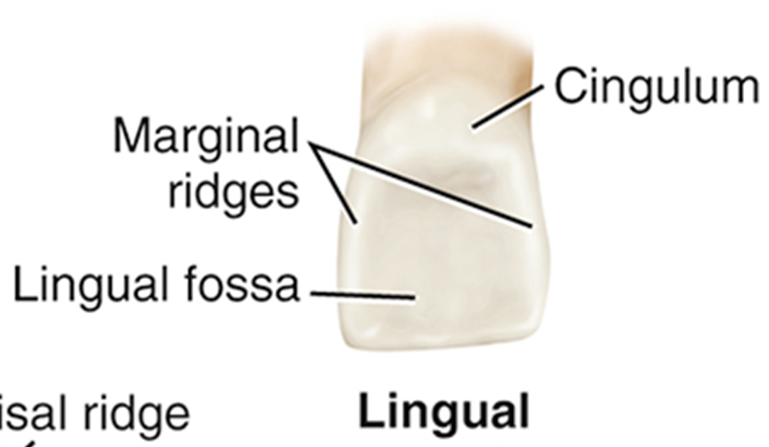
cingulum, a rounded, raised area on the cervical third of the lingual surface

The lingual surface on anterior teeth has?
rounded, raised borders on the mesial and distal surfaces called marginal ridges
Some anterior teeth have a?
fossa, which is a wide, shallow depression on the lingual surfaces

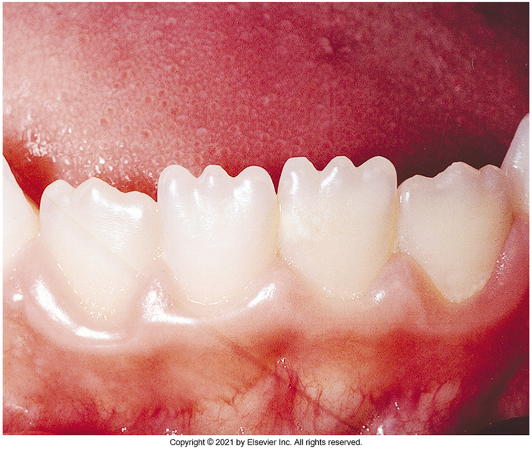
When newly erupted permanent incisors, the central and lateral incisors have
three mamelons, or rounded enamel extensions on the incisal ridge. These ridges will disappear over time after wear and occlusion.
The incisal edge of a permanent incisor is also known as
The incisal surface or incisal plane
The incisal edge of a permanent maxillary incisors have a?
lingual slant. The incisal edge of the mandibular incisors have a labial slant.
The incisal edge of the mandibular and maxillary incisors are parallel to each other and work together to create a cutting action.
The incisal edge of the mandibular and maxillary incisors are parallel to each other and work together to create a cutting action.
The incisal edge of the mandibular and maxillary permanent incisors are?
parallel to each other and work together to create a cutting action
Mesiodistally means?
The width of the tooth
Why is tooth morphology important to learn
It helps in understanding tooth function, placement, and the relationship between different types of teeth, which is essential for effective diagnosis and treatment in dentistry.
Also to:
•Mounting dental radiographs
•Assisting in charting a mouth with missing teeth and teeth that have “drifted”
•Selecting temporary crowns or orthodontic bands from a box with a variety of shapes
•Forming matrix bands before application
•Fabricating temporary crowns and bridges
How many teeth are in the anterior permanent dentition?
6 in the top maxillary jaw, 6 in the lower mandibular teeth, for a total of 12 teeth.
How many incisors does a permanent dentition have?
2 at the top, 2 at the bottom, for a total of 4 incisors.
How many incisors does a permanent dentition have?
4 at the top, 4 at the bottom. 2 central incisors (top and bottom), 2 lateral incisors (top and bottom), for a total of 8 incisors.
Characteristics of Permanent Anterior Teeth
•All anterior teeth have a cingulum, a rounded, raised area on the cervical third of the lingual surface
•The cingulum corresponds to the lingual developmental lobe
•The lingual surface on anterior teeth has rounded, raised borders on the mesial and distal surfaces called marginal ridges
•Some anterior teeth have a fossa, which is a wide, shallow depression on the lingual surfaces

Are the mandibular incisors are smaller than the maxillary incisors.
Yes
Maxillary central incisors (#8, #9) are?
•Maxillary central incisors (#8 and #9) are larger in all dimensions, especially mesiodistally (width), than a permanent mandibular central incisor
•Labial surfaces are more rounded from the incisal aspect, with the tooth tapering toward the lingual
•Root is short compared with the roots of other permanent maxillary teeth
Maxillary Lateral Incisors
•The maxillary lateral incisors (#7 and #10) are smaller than the central incisors in all dimensions except root length
•They usually erupt after the maxillary central incisors
•The crown of a maxillary lateral incisor has a single root that is relatively smooth and straight but may curve slightly distally
•Recognizing this feature is helpful in the mounting of radiographs
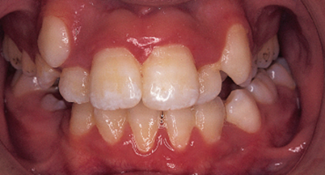
What is diastema?
Diastema is a gap or space between two teeth, commonly seen between the maxillary incisors. This spacing can occur naturally or may result from various factors including dental alignment issues.
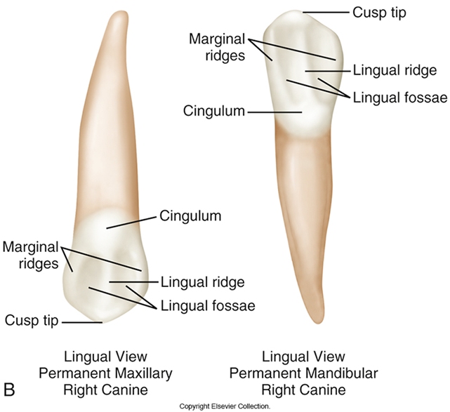
Permanent Canines
•The permanent canines are the longest teeth in the dentition. It has a particularly long, thick root.
•This large root is externally manifested by the bony vertical ridge called the canine eminence. This helps form facial contours.
•Patients commonly call the canines their “eye teeth” or "cuspids"
•The shape of the crown promotes cleanliness
Maxillary canines
•The maxillary canines (#6 and #11) usually erupt after the mandibular canines, after the maxillary incisors, and possibly after the maxillary premolars
•The cusp tip is sharper on a maxillary canine
•The mesial cusp slope is usually shorter than the distal cusp slope in both the maxillary and the mandibular canines when they first erupt
•The length of these cusp slopes, and the cusp tip can change with attrition
Mandibular canines
•The mandibular canines (#22 and #27) usually erupt before the maxillary canines and after most of the incisors have erupted
•Although the entire tooth is usually as long, a mandibular canine is narrower labiolingually and mesiodistally than a maxillary canine
•The lingual surface of the crown of the mandibular canines is smoother than that of the maxillary canines and has a less developed cingulum and two marginal ridges
Teeth in the permanent posterior dentition
Top (maxillary): 6 total molars, 4 total premolars. 3 molars in each quadrant, 2 premolars in each quadrant
Bottom (mandibular): 6 total molars, 4 total premolars. 3 molars in each quadrant, 2 premolars in each quadrant.
Total (Both mandibular and maxillary jaws): 12 molars, 8 premolars, resulting in 20 total teeth in the permanent posterior dentition.
Clinical Considerations with Canines
•The maxillary canines may erupt labially or lingually in relation to the surrounding teeth
•The maxillary canines may also fail to erupt fully and may remain impacted
•This occurs because the permanent maxillary canines erupt after the maxillary incisors and possibly after the premolars and their arch spaces have closed
Permanent premolars
Premolars are efficient at grinding food
•he premolars are succedaneous, and replace the primary molars
The crowns are shorter than crowns of anterior teeth
There are two types of premolars: First, Second
Maxillary First premolars
•A maxillary first premolar (#5 and #12) is larger than a maxillary second premolar
•Each maxillary first premolar has two cusps (buccal and lingual) and two roots (facial and lingual)
•Some first premolars have roots that are joined, or fused
•The roots are shorter in length and resemble the roots of the molars
•Both maxillary premolars erupt earlier than the mandibular premolars
•The buccal cusp is long and sharp to assist the canine with tearing.
•The central groove extends between the mesial and distal groves.
Maxillary Second Premolars
•Each maxillary second premolar (#4 and #13) has two cusps (buccal and lingual) and one root
•The mesiobuccal cusp slope is shorter than the distobuccal cusp slope on the second premolar
•The second premolar is wider buccolingually than mesiodistally
Differences Between Second and First Maxillary Premolars
•The cusps are closer in length on the second premolar
•The lingual cusp is slightly shorter, but not as short as the cusp on the maxillary first premolar
•The cusps of the secondary premolar are not as sharp as those of the maxillary first premolar
Mandibular First Premolars
•Each mandibular first premolar (#21 and #28) has a long and well-formed buccal cusp and a small, nonfunctioning lingual cusp
•The lingual cusp may be no larger than the cingulum on some maxillary canines
•The mandibular first premolars are smaller and shorter than the mandibular second premolars
•Has an equal buccolingual and mesiodistal widths when viewed from the occlusal area, making the outline almost round
•The crowns incline lingually, bringing the cusps into proper occlusion with the teeth on the opposing arch
Mandibular Second Premolars
•The permanent mandibular second premolars (#20 and #29) erupt distal to the mandibular first premolars
•They are the succedaneous replacements for the primary mandibular second molars
•There are two forms of the mandibular second premolar
•Three-cusp type, or tricuspidate form (more common). Has one large buccal cusp and 2 small lingual cusps
•Two-cusp type, or bicuspidate form
Permanent Molars
•The molar crowns have four or five short, blunt cusps, and each molar has two or three roots that help support the larger crown
•The permanent molars are the largest teeth in the dentition
•There are three types of molars: First, second, and third
•The first and second molars are also called the 6-year and 12-year molars because of the approximate ages at which they erupt
Maxillary Molars
•Usually the first permanent teeth to erupt into the maxillary arch
•Each maxillary molar usually has four major cusps, with two on the buccal portion of the occlusal table and two on the lingual
•Each maxillary molar has three well-separated and well-developed roots
•A tooth with three roots is said to be trifurcated, which means “divided into thirds”
•Maxillary molars are wider buccolingually than mesiodistally
Maxillary First Molars
•The maxillary first molars (#3 and #14) are the first permanent teeth to erupt into the maxillary arch
•They erupt distal to the primary maxillary second molars and are therefore nonsuccedaneous (do not replace the primary teeth)
•The maxillary first molar is the largest tooth in the maxillary arch and also has the largest crown in the permanent dentition
•This molar is composed of five developmental lobes, two buccal and three lingual
•The fifth cusp is called the cusp of Carabelli
Maxillary Second Molar
•The crown of the maxillary second molar is somewhat shorter than that of the first molar, and it usually has four cusps
•No fifth cusp is present
•There are three roots
•The roots of the secondary molars are smaller than those of the first molars
•The lingual root is still the largest and longest
Maxillary third molars
•The maxillary third molars (#1 and #16) differ considerably in size and contour
•Maxillary third molars are more likely than other teeth in the arch to be out of position.
•The crown of the maxillary third molar is smaller and the roots are usually shorter
•The roots of the maxillary third molar tend to fuse, and the result is a single tapered root
•People sometimes refer to the maxillary third molars as the “wisdom” teeth because they erupt last
Mandibular molars
•The mandibular molars erupt 6 months to 1 year before the corresponding permanent maxillary molars
•The crowns of the mandibular molars have four or five major cusps, with two lingual cusps always of about the same width
•All mandibular molars are wider mesiodistally than buccolingually, similar to anterior teeth
•Each mandibular molar has two well-developed roots, one mesial and one distal
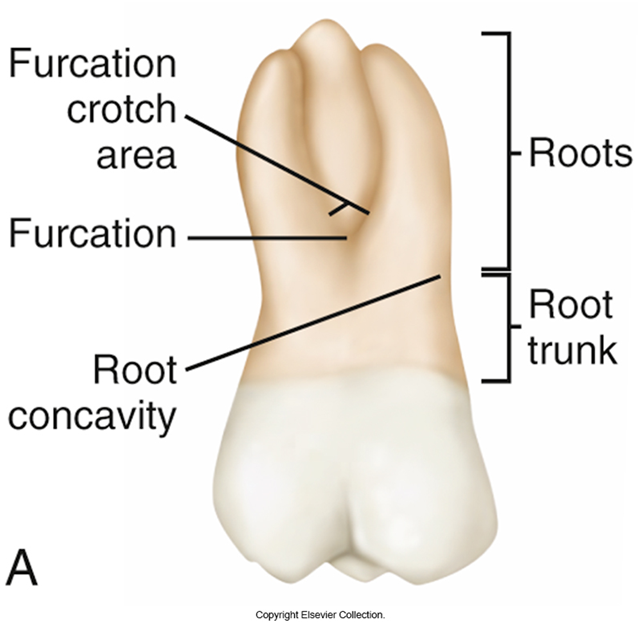
•A tooth with two roots is referred to as bifurcated, which means “divided into two”
•A bifurcation is the area at which the two roots divide
Mandibular First Molars
•The permanent mandibular first molars (#19 and #30) erupt between 6 and 7 years of age
•These teeth are commonly the first permanent teeth to erupt in the oral cavity
•They erupt distally to the primary mandibular 2nd molar and are nonsuccedaneous.
•The two roots, mesial and distal, of a mandibular first molar are larger and more divergent than those of a second molar
•The mesial root may have two root canals
Mandibular Second Molars
•The mandibular second molars (#18 and #31) erupt between 11 and 12 years of age
•These teeth erupt distal to the permanent first molars and therefore are nonsuccedaneous
•The crown of the mandibular second molar is slightly smaller than that of the first molar in all directions
•The crown has four well-developed cusps
Mandibular Third Molars
•The mandibular third molars (#17 and #32) are similar to the maxillary third molars in that they vary greatly in shape
•There is no typical mandibular third molar
•This molar is usually smaller in all dimensions than the second molar
•The third molar consists of four developmental lobes
•A mandibular third molar has two roots that are fused, irregularly curved, and shorter than those of a mandibular second molar
Primary Dentition
•There are 20 primary teeth: 10 in the maxillary arch and 10 in the mandibular arch
•Includes incisors, canines, and molars
•Numbered in the Universal Tooth Numbering System with the capital letters A through T
•Smaller overall and have whiter enamel than the permanent teeth do
•The crown of any primary tooth is short in relation to its total length
•The crowns are narrower at the cementoenamel junction (CEJ)
•The roots of primary teeth are narrower and longer than the crown
•The pulp chambers and pulp horns in primary teeth are relatively large compared with those of the permanent teeth
•There is a thick layer of dentin between the pulp chambers and the enamel, especially in the primary mandibular second molar
•The enamel layer is relatively thin
Primary Incisors
•The crowns and roots of deciduous incisors are smaller than those of their permanent successors
•The roots are twice as long as the crowns and taper toward the apex
Clinical Considerations with Primary Teeth
•Often parents do not understand the importance of the primary teeth
•Primary teeth hold the eruption space for the permanent teeth
•Because the enamel and dentin are thinner in primary teeth, decay can travel quickly through the enamel to the pulp, possibly causing loss of the tooth
•Early dental health education and dental care are essential in keeping the primary dentition
Primary Maxillary Central Incisors
•The crown of the primary maxillary central incisor (E and F) is wider mesiodistally than incisocervically
•It is the only tooth of either dentition with this crown dimension
•The primary maxillary incisors have no mamelons
•The cingulum and marginal ridges are more prominent than they are on the permanent successor, and the lingual fossa is deeper
Primary Maxillary Lateral Incisors
•The crown of the primary maxillary lateral incisor (D and G) is similar to that of the central incisor but much smaller in all dimensions
•The incisal angles on the lateral incisor are also more rounded than on the central incisor
•The lateral root is longer in proportion to its crown, and its apex is sharper
Primary Mandibular Central Incisors
•The crown of the primary mandibular incisor (O and P) resembles the primary mandibular lateral incisor more than it does its permanent central successor
•The mandibular central incisor is extremely symmetric
•It is also not as constricted at the CEJ as is the primary maxillary incisor
•The lingual surface of the mandibular central incisors appears smooth and tapers toward the prominent cingulum
•The marginal ridges are less pronounced than those of the primary maxillary incisors
Primary Mandibular Lateral Incisors
•The crown of the primary lateral incisor (Q and N) is similar in form to that of the central incisor in the same arch but is wider and longer
•The incisal edge of the mandibular lateral incisor slopes distally, and the distoincisal angle is more rounded
Primary Canines
•There are four primary canine teeth, two in each dental arch
•Differ from the outline of their permanent successors in the following ways:
•Maxillary canines
•The crown of the primary maxillary canine (C and H) has a relatively longer and sharper cusp than that of its permanent successor on eruption
•The mesial and distal outlines of the primary maxillary canine are rounder
Primary Maxillary Canines
•The crown of the primary maxillary canine (C and H) has a relatively longer and sharper cusp than that of its permanent successor on eruption
•The mesial and distal outlines of the primary maxillary canine are rounder
•The cingulum is well developed as are the lingual ridge and marginal ridge.
•The root is inclined distally and is twice as long as the crown
Primary Mandibular Canines
•The primary mandibular canine (M and R) resembles the primary maxillary canine, but this tooth is much smaller labiolingually
•The distal cusp slope is much longer than the mesial cusp slope
•The lingual surface of the primary mandibular canine is marked by a shallow lingual fossa
•The primary mandibular canine (M and R) resembles the primary maxillary canine, although some dimensions are different
•This tooth is much smaller labiolingually
Primary Molars
•The primary dentition consists of a total of eight primary molars
•Each quadrant includes a first primary molar and a second primary molar
•Each molar crown is wider than it is tall
•The permanent premolars replace the primary molars when they are exfoliated
Primary Maxillary First Molars
•The crown of the primary maxillary first molar (B and I) does not resemble any other crown of either dentition
•The height of contour on the buccal surface is at the cervical third of the tooth; on the lingual side, it is at the middle third
•The primary maxillary molars have three roots, which are thinner and have greater flare than do those of the permanent maxillary first molar
•The lingual root is the longest and most divergent
Primary Maxillary Second Molars
•The primary maxillary second molar (A and J) is larger than the primary maxillary first molar
•Closely resembles the permanent maxillary first molar but is smaller in all dimensions
•The second molar usually has a cusp of Carabelli, the minor fifth cusp
Primary Mandibular First Molars
•The crown of the primary mandibular first molar (L and S) is unlike any other tooth of either dentition
•The height of contour on the buccal surface is at the cervical third of the tooth; on the lingual side, it is at the middle third
•Has four cusps; the mesial cusps are larger
•Has two roots, which are positioned similarly to those of other primary and permanent mandibular molars
Mandibular Second Molars
•The primary mandibular second molar (K and T) is larger than the primary mandibular first molar
•Most closely resembles in form the permanent mandibular first molar in that it has 5 cusps
Which teeth are succedanous?
Central incisors, lateral incisors, canines, first premolar, second premolar
(the molars in the primary dentition are replaced with permanent 1,2 premolar)

which teeth are nonsuccedanous?
Molars in permanent dentition