carotid case studies
1/4
There's no tags or description
Looks like no tags are added yet.
Name | Mastery | Learn | Test | Matching | Spaced |
|---|
No study sessions yet.
5 Terms
During a routine cardiac work-up, a 65-year-old man with a his tory of smoking and hypertension admits he has experienced some intermittent loss of the use of his left arm. The cardiologist orders a carotid sonogram. Sonographic findings include decreased color flow in the right ICA with a high-pitched Doppler signal measuring a PSV of 600 cm/s and EDV of 140 cm/s. What do these findings suggest? What would be the next step in treatment?
70-99% stenosis; endarterectomy or carotid angioplasty with stent placement
A noncompliant 58-year-old woman with depression and hyper tension presents to the emergency department with aphasia, left hemiplegia, and possible left hemianopia. The patient communi cates that her symptoms started early in the morning. While wait ing for further testing that day, the neurologic deficits resolve. What is the most likely diagnosis?
TIA
A man stumbles into the emergency department showing signs of dysarthria and problems with gait and stance. The front desk personnel assume the man is severely intoxicated and do not assist him right away. Why is this possibly a detrimental mistake?
The man was having stroke-like symptoms; acting fast is crucial because the longer you wait, the more damage is done
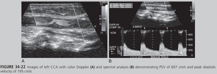
A 67-year-old man developed acute blindness in the left eye while at work. When he arrived at the hospital, he claimed to have got ten lost in the elevator. Staff noticed he appeared uncoordinated and confused and had trouble expressing himself. A carotid sono gram demonstrated the finding in Figure 34-22 (see Color Plate 53) in the left CCA. The left vertebral artery was found to be occluded. After 3 days, the patient’s symptoms completely resolved. What do these findings suggest?
the man was experiencing a reversible ischemic neurologic deficit
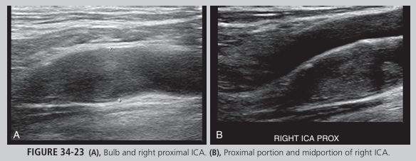
A 67-year-old man is taken to the hospital via ambulance. His symptoms include confusion, loss of memory, aphasia, and left sided hemiparesis. His history includes hypertension, hypercholes terolemia, and smoking. A carotid sonogram revealed echogenic material within the right internal carotid lumen. Images are pro vided in Figure 34-23. No flow was seen with spectral Doppler. What do these findings indicate?
right ICA occlusion