PNB 2264 Unit 2 Tissues
 Call Kai
 Learn
Learn Practice Test
 Spaced Repetition
 Match
Match Flashcards
 Knowt Play
1/59

There's no tags or description
Looks like no tags are added yet.
Name | Mastery | Learn | Test | Matching | Spaced | Call with Kai | Chat |
|---|
No analytics yet
Send a link to your students to track their progress
60 Terms
what is a tissue
collection of cells that are serving a common function inside the body
give rise to organs
Tissues are defined by differences in:
cellular composition, extracellular makeup (or matrix) and function
extracellular matrix
contains proteins, salts, water and other macromolecules
four primary tissue types
epithelial, connective, nervous and muscle
functions of epithelia
epithelial tissue
physical protection: water, chemicals, physical injury
selective permeability
secretion and absorption
sensation
characteristics of epithelia
cellularity, polarity, attachment, avascularity, regenerative capacity
epithelia classification
based on function or structure:
exchange, transporting, ciliated, protective or secretory
exchange epithelia
permit the rapid exchange of materials between the two compartments

transporting epithelia
prevent free movement between adjacent cells
substances must instead pass through the epithelial cell, crossing the membrane as they do so

ciliated epithelia
promote movement of extracellular particles
ex. line the airways
pathology: Kartagener’s syndrome, a primary ciliary dyskinesia

protective epithelia

secretory epithelia
are usually organized into glands
endocrine glands: release their secretions into blood or interstitial fluid
exocrine glands: release their secretions into ducts

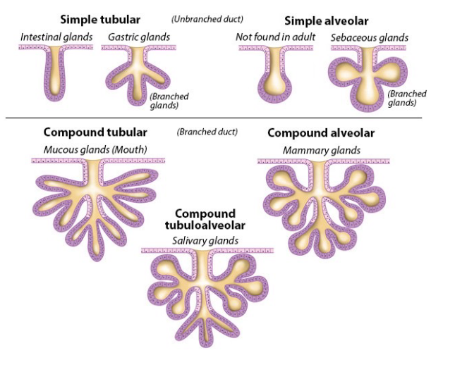
classification of glands: structure
based on number of ducts and architecture
unbranched duct: simple tubular, simple alveolar
branched duct: compound tubular, compound alveolar, compound tubuloalveolar
classification of glands: function
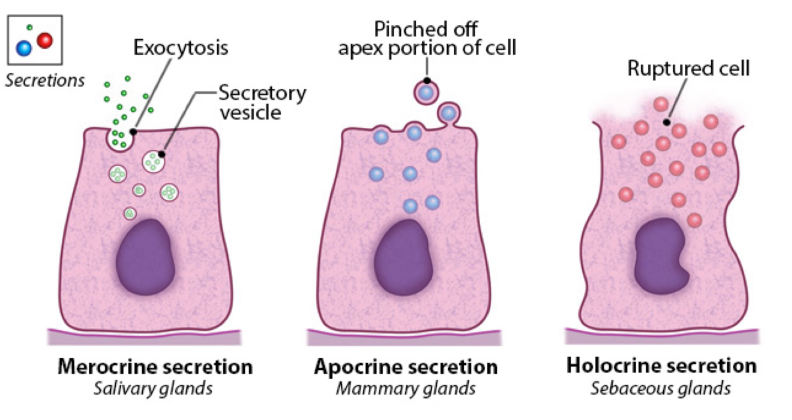
based on the mechanism of secretion
merocrine secretion (salivary glands): exocytosis
apocrine secretion (mammary glands): pinched off apex portion of the cell
holocrine secretion (sebaceous glands): ruptured cell
Intercellular junctions:
epithelial cells are strongly bound to each other on their lateral surface by membrane specialization called ___
types of intercellular junctions
tight junctions, adhering junctions, desmosomes, gap junctions
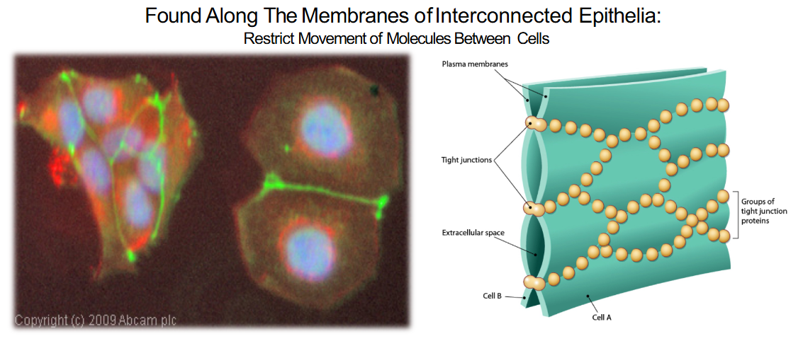
tight junctions
found along the membrane of interconnected epithelia
restrict movement of molecules between cells
ex. food poisoning
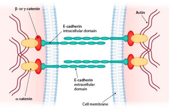
adhering junctions
found along the membrane of interconnected epithelia
attach to the cytoskeleton
often found on the basal membrane
desmosomes
found along the membranes of interconnected epithelia
physical connection between adjacent cells
flexibility and support
pathology: pemphigus vulgaris, an autoimmune disorder characterized by separation of epidermal layers and blistering
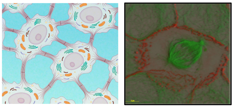
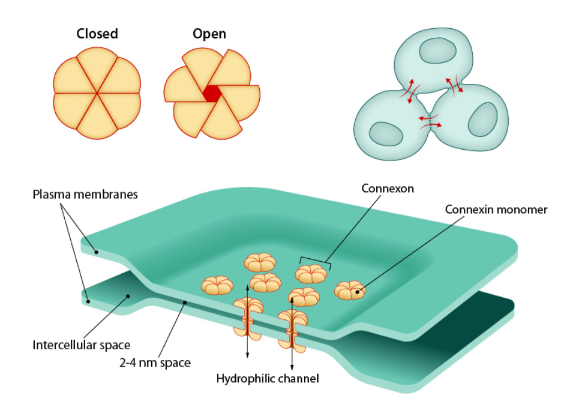
gap junction
found along the membrane of interconnected epithelia
electrochemical connection between adjacent cells
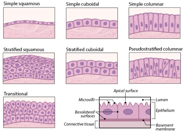
classification of epithelia: anatomical
based upon the shape of the cell and number of layers
simple squamous, simple cuboidal, simple columnar, stratified squamous, stratified cuboidal, pseudostratified columnar, transitional
pathology: cystic fibrosis
caused by a mutation in CFTR (cytic fibrosis transmembrane receptor)
connective tissue
most diverse and abundant tissue type
connects other tissues together
cells, protein fibers (collagen, elastin, etc), ground substance (water and ions)
these are often referred to collectively as the ECM (extracellular matrix)
connective tissue classification
connective tissue proper: loose ( 1. areolar, 2. adipose, 3. reticular) and dense (1.regular, 2.irregular, 3. elastic)
supporting connective tissue: cartilage (1. hyaline, 2. fibrocartilage, 3. elastic) and bone (1. compact, 2. spongy)
fluid connective tissue: blood and lymph
general function of connective tissue
physical protection, support and structure, storage, binding of structures, transport, immune protection
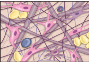
areolar tissue
loosely packed assembly of all fiber types, fibroblasts and immune cells; primary function is to cushion organs; contains key mediators of immunity to minimize invasion by infectious pathogens
location: supporting under epithelial layers and surrounding organs
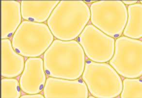
adipose tissue
matrix is crowded by adipocytes which are tightly packed; serve to insulate and protect organs
location: around major organs and within the subcutaneous layer of the skin (under dermis)
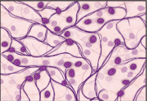
reticular tissue
meshwork of reticular fibers loosely organized to form a flexible meshwork to support tissue and immune cells of few organs
location: lymph nodes, bone marrow and splenic pulp
regular tissue
fibroblasts embedded within a regularly ordered assembly of collagen fibers; functions to resist stress of being pulled in a given direction
location: attach muscle to bone (tendon) or bone to bone (ligament)

irregular tissue
irregularly arranged collagen fibers with fibroblasts embedded within; irregular organization provides tissue the ability to resist forces in many directions

blood contains:
platelets, erythrocytes (red blood cells), lymphocytes (white blood cells) and neutrophils (white blood cells)
bone
heterogenous structure
two kinds: compact/ cortical bone and trabecular/ cancellous/ spongy/ diploe bone
compact/ cortical bone
absorbs a great deal of mechanical energy before fracture
organized in an osteon haversion system: bones is a system of tubes within tubes — key vocab: periosteum, lamellae, osteon, osteocyte, lacunae, central canal, canaliculi
trabecular/ cancellous/ spongy/ diploe bone
can be deformed without fracture
bone matrix is a ___ material.
at any one time, most of the calcium in the body exists as ____
bones are a ___ and ____ reservoir
composite
hydroxyapatite (Ca10(PO4)6(OH)2)
calcium; phosphate
bone cell types:
stem cells (osteoprogenitors), osteoblasts, osteocytes, and osteoclasts
osteoprogenitor cells develop into osteoblasts; some osteoblasts (form the bone matrix) differentiate into osteocytes (maintains bone matrix)
resorption
breakdown of bone
Cartilage
many similarities with bone
found throughout the entire body
semi-rigid connective tissue, not as strong as bone but more flexible
chondroblasts
begin to produce extracellular matrix
chondrocytes
cells that continue to produce matrix and become surrounded by the matrixh
chondroclasts
breakdown cartilage
hyaline
flexible, firm gel matrix secreted by chondroblasts which become embedded in lacunae (chondrocytes); cushions, supports and reinforces other tissue and organs
location: nose, trachea, ribs, ends of long bones, and embryonic skeleton

fibrocartilage
firm hyaline matrix but with more organized collagen fibers; capable of resisting compressive force
location: components of joints with limited flexibility such as intervertebral discs, menisci and pubic symphysis

elastic cartilage
organization is same as hyaline cartilage enriched with elastin fibers to promote greater degree of flexibility and stretch and allowing tissue to return to original shape
location: outer ear and epiglottis

Models of cartilage growth
interstitial and appositional growth
interstitial growth
chondrocytes housed in lacunae undergo mitotic division, 2 chondrocytes occupy one lacuna
the cells begin to synthesize and secrete their own extracellular matrix and are now separated from each other by a matrix border
the result is two chondrocytes, two lacunae and more extracellular matrix secretion
appositional growth
stem cells along the internal edge of the perichondrium begin to divide into chondroblasts
chondroblasts begin to produce extracellular matrix and differentiate into chondrocytes and from new lacunae and add to the already existing matrix
cartilage is ____
avascular
this gives it only limited repair capacity
Four tissue types:
epithelial, connective, muscle, and nerve
epithelial tissue characteristics
minimal matrix, basal lamina matrix, no direct blood supply, microvilli and cilia
location: covers body surface; lines cavities and hollow organs and tubes; secretory glands
cell arrangements and shapes: variable number of layers from one to many; cells= flattened, cuboidal or columnar
connective tissue characteristics
extensive matrix, varied matrix (protein gibers in ground substance that ranges from liquid to gelatinous to firm to calcified), cartilage has no blood supply
location: supports skin and other organs; cartilage, bone and blood
cell shape and arrangement: cells not in layers; usually randomly scattered in matrix; cell shape irregular to round
muscle tissue characteristics
minimal matrix, external lamina matrix, able to generate electrical signals, force and movement
location: makes up skeletal muscles, hallow organs and tubes
cell arrangement and shape: cells linked in sheets or elongated bundles; cells shaped in elongated, this cylinders; heart muscle cells may be branched
nerve tissue characteristics
minimal matrix, external lamina matrix, able to generate electrical signals
location: throughout body; concentrated in brain and spinal cord
cell arrangement and shape: cells isolated or networked; cell appendages highly branched and / or elongated
Metaplasia
one tissue type is replaced by another
hypertrophy
cell growth in the absence of cell division
hyperplasia
tissue growth due to “normal” cell growth
neoplasia
tissue growth due to “abnormal” cell growth
apoptosis
programmed cell death
necrosis
uncoordinated cell division