Lecture 2: Equine Lameness Exam
 Call Kai
 Learn
Learn Practice Test
 Spaced Repetition
 Match
Match Flashcards
 Knowt Play
1/62

There's no tags or description
Looks like no tags are added yet.
Name | Mastery | Learn | Test | Matching | Spaced | Call with Kai | Chat |
|---|
No analytics yet
Send a link to your students to track their progress
63 Terms
what is the order of events for a lameness exam?
history
PE
motion exam
flexion tests
hoof testers
localize lameness
diagnostic imaging
history for lameness exam
signalment
use of the horse
duration of lameness
worsened or improved
history of trauma
management changes
medications administered
which leg owner thinks is affected
last farrier visit
PE for lameness exam
quick TPR
observation → conformation, swellings, how horse is standing, hoof care, muscle symmetry
limb palpation → joints, tendons/ligaments, digital pulses
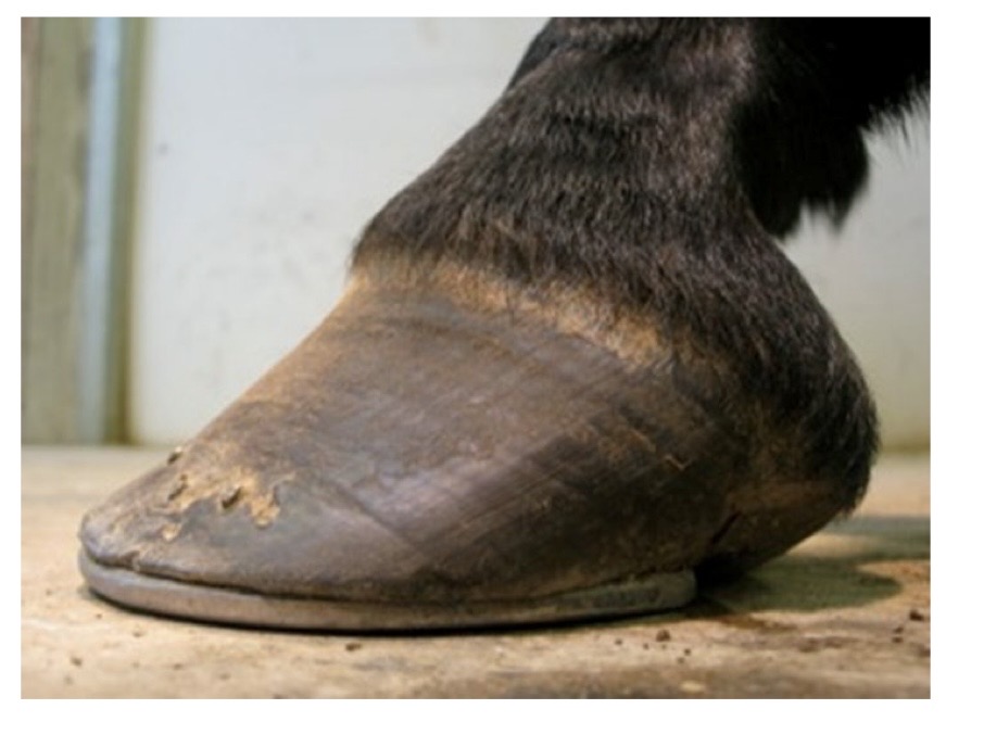
is this poor or good quality hoof trimming/shoeing?
poor
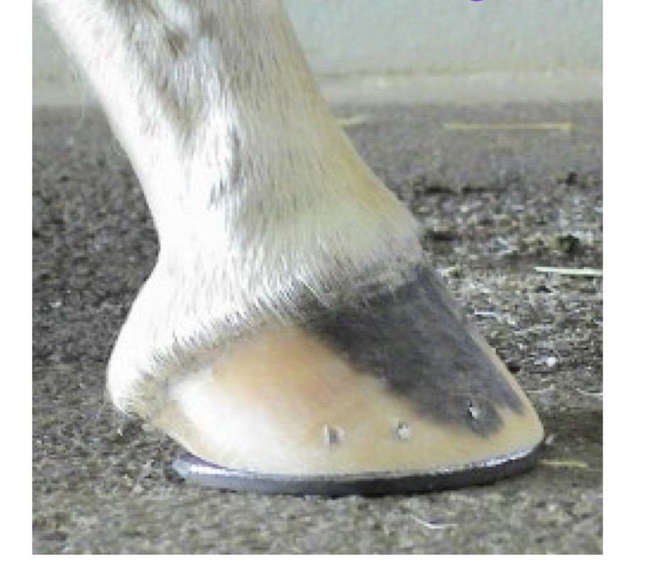
is this poor or good quality hoof trimming/shoeing?
good
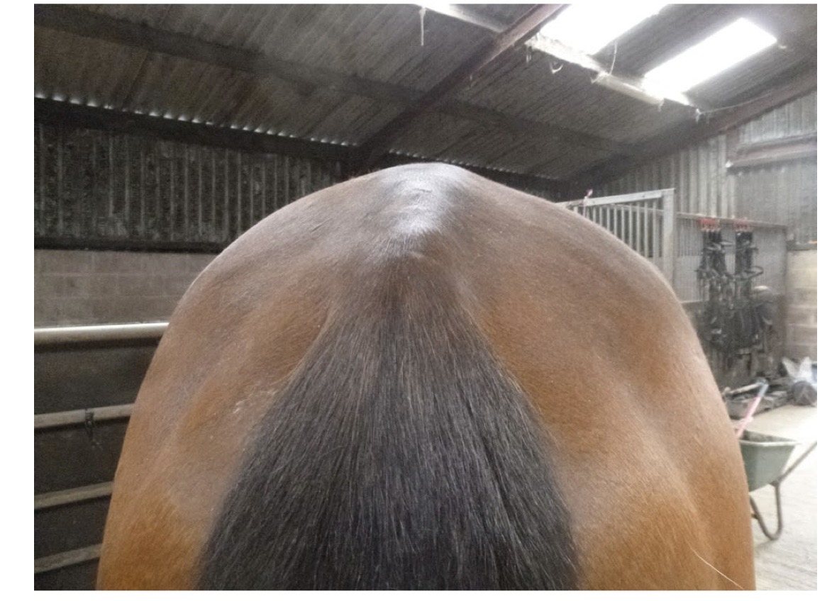
which side of this horse is hollow and abnormal?
the right
limb palpation is to check for
effusion, edema, bony proliferation
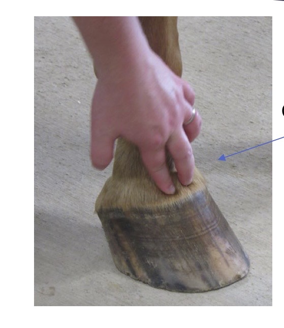
what joint is being palpated?
coffin joint
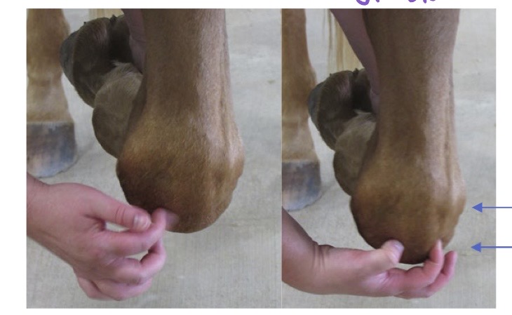
what joint is being palpated?
carpus
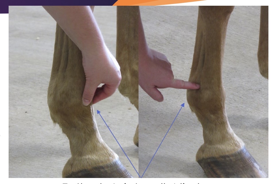
what joint is being palpated?
fetlock joint of all 4 limbs
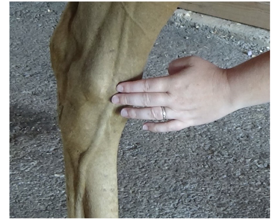
what joint is being palpated?
tibiotarsal joint
what joint is being palpated?
femoropatellar
what is the most common site of joint palpation in horses?
medial femorotibial joint
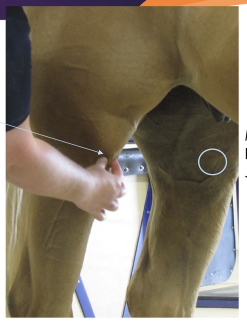
what is circled?
medial femorotibial joint
what is this?
pastern OA
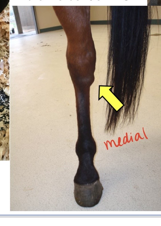
what is this?
bone spavin, distal tarsal joint OA
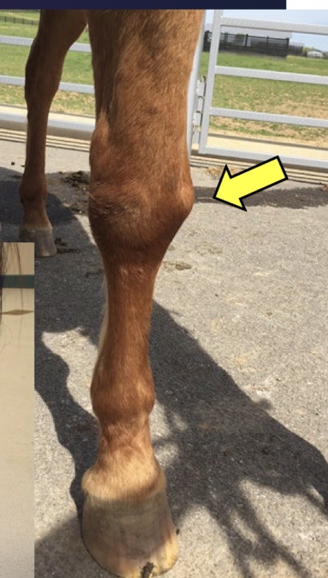
what is this?
carpal OA
what are you looking for during tendon and ligament palpation?
enlargement, thickening, pain
digital pulses
every horse should have them → faint in normal horse
increases in ability to palpate are significant
should not be hot
what is the most appropriate conclusion to make from palpating increased digital pulses?
the horse has inflammation in the foot
back pain is often secondary to
hock pain/hind limb lameness
hoof testers should be used before
blocking the horse
basic motion exam
walking in straight line
trotting in straight line
trotting in a circle → hard and soft surface
advanced motion exam
canter and under saddle
walking gait evaluation
4 beat gait
evaluating stride length
tracking up
trot gait evaluation
2 beat gait
limbs move in diagonal pairs
best gait for seeing lameness
what is the best gait for seeing lameness?
trot
AAEP lameness grade 0
lameness is not present under any circumstances
AAEP lameness grade 1
lameness is difficult to observe and is not consistently apparent
AAEP lameness grade 2
lameness is difficult to observe at a walk or when trotting in a straight line but consistently apparent under certain circumstances
AAEP lameness grade 3
lameness is consistently observable at a trot under all circumstances
AAEP lameness grade 4
lameness is obvious at a walk
AAEP lameness grade 5
lameness produces minimal weight bearing in motion and/or a rest
sound
the “not” lame leg/legs
head bob
refers to movement of the horse’s head and neck when lame in the front limb → down on sound
hip hike
lame limb has increased motion in the pelvis as the horse tries to bear less weight on the lame hind limb
fetlock drop
sound limb fetlock drops further with increased weight bearing to compensate for lame limb
tracking up
hind limb lands in or beyond front limb hoof print
stride length
comparable between sound and lame limbs
lameness detection
evaluating for asymmetry in the gait
sound limb lameness detection
drops fetlock
head nod → head moves down when horse bears more weight
lame limb lameness detection
head bob → head moves up when lame leg hits the ground
hip hike → hind limb lameness, increased pelvic movement
asymmetric movement → shorter stride
flexion tests
put stress on a joint to see if it changes the horse’s lameness
pretty subjective
learn to do these ergonomically
variation in medical records
forelimb flexion tests
distal limb → coffin, pastern, fetlock
carpus → will flex elbow some
upper limb → shoulder and elbow
coffin joint hyperextension → stress on navicular apparatus
hindlimb flexion tests
challenge of the reciprocal apparatus
distal limb → same as forelimb, will flex hock some
upper limb → hock, stifle, hip, will flex fetlock some
caudal extension → mostly stifle
distal limb flexion test
typically hold for 30s
carpus/tarsus flexion test
hold for 45 seconds
nerve blocks
local anesthetic injected SQ over nerves
numbs the area distal to the injection site
principles of nerve blocks
start distal and work proximal
typically skip the palmar digital in the hind limb
principles of joint blocks
local anesthetic within a joint to numb the joint
order of joints is not as important as nerves
local anesthetic migrates so
time → wait 5 mins
volume → least possible
location → accurate
mepivicaine
most commonly used
less tissue irritation
fast acting
duration is 2-3 hours
lidocaine
stings
fastest acting
duration is 1.5-3 hours
bupivicaine
intermediate onset of action
duration is 3-6 hours
palmar digital nerve block
called PD
insert needle over neurovascular bundle proximal to the collateral cartilages
1-2ml carbocaine per site
usually 25g needle
what structures are blocked with a PD nerve block?
caudal 1/3 of the foot, sole, navicular apparatus
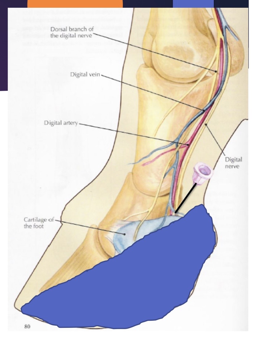
what nerve block is this?
PD
abaxial sesamoid block
commonly called abaxial
insert needle SQ over the neurovascular bundle at the abaxial surface of the sesamoid bones
1-3ml carbocaine at each site
what structures are blocked with an abaxial sesamoid block?
all structures below the fetlock, ± the fetlock itself
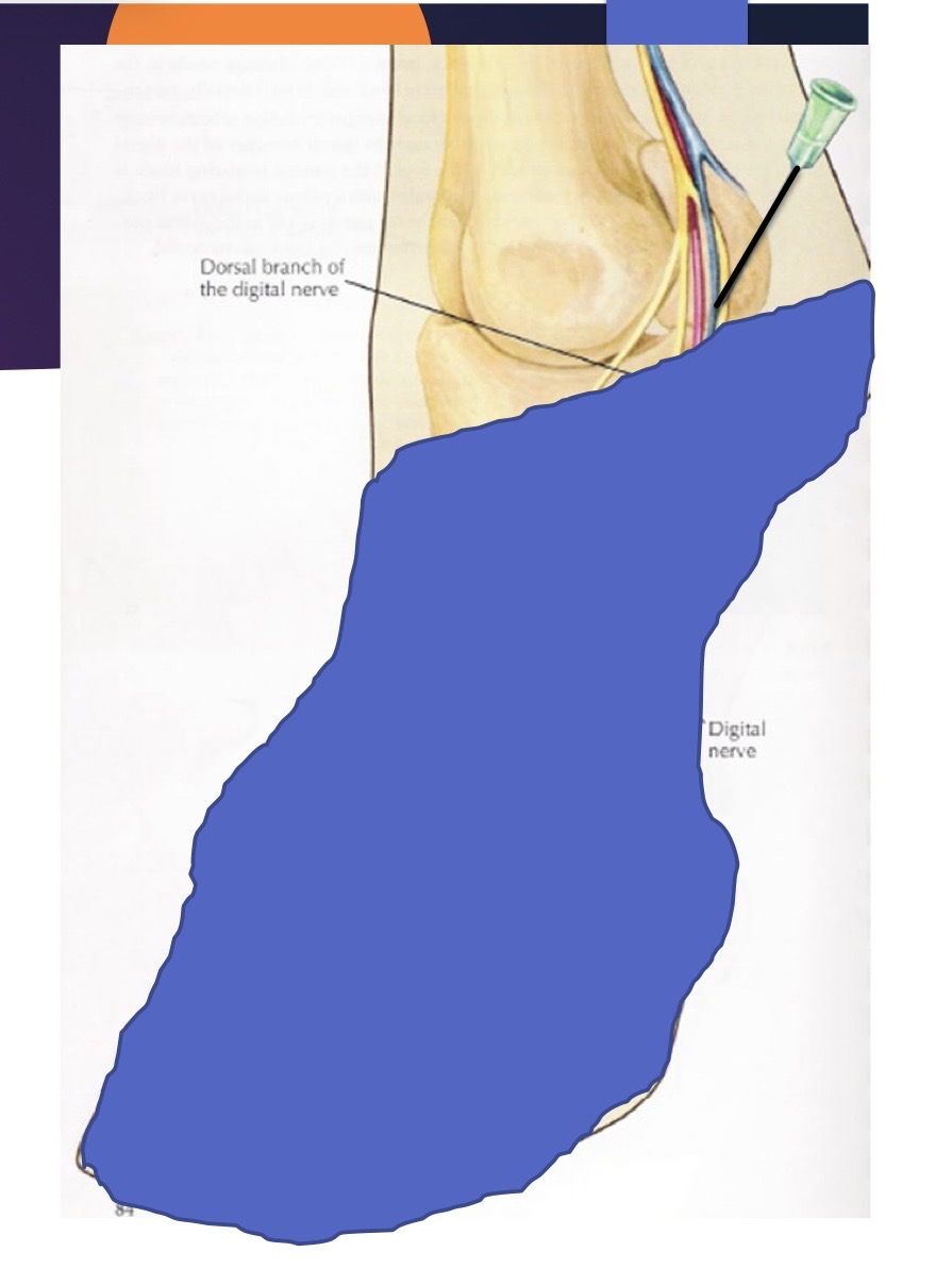
what block is this?
abaxial sesamoid
low 4 point block
inject under the button of the splint bone (medial and lateral)
inject over the neurovascular bundle between the DDF and the suspensory ligament
deposit 2-3ml per site
what structures are blocked with the low 4 point block?
fetlock joint and distal → inconsistent loss of skin sensation dorsally on the hind limb
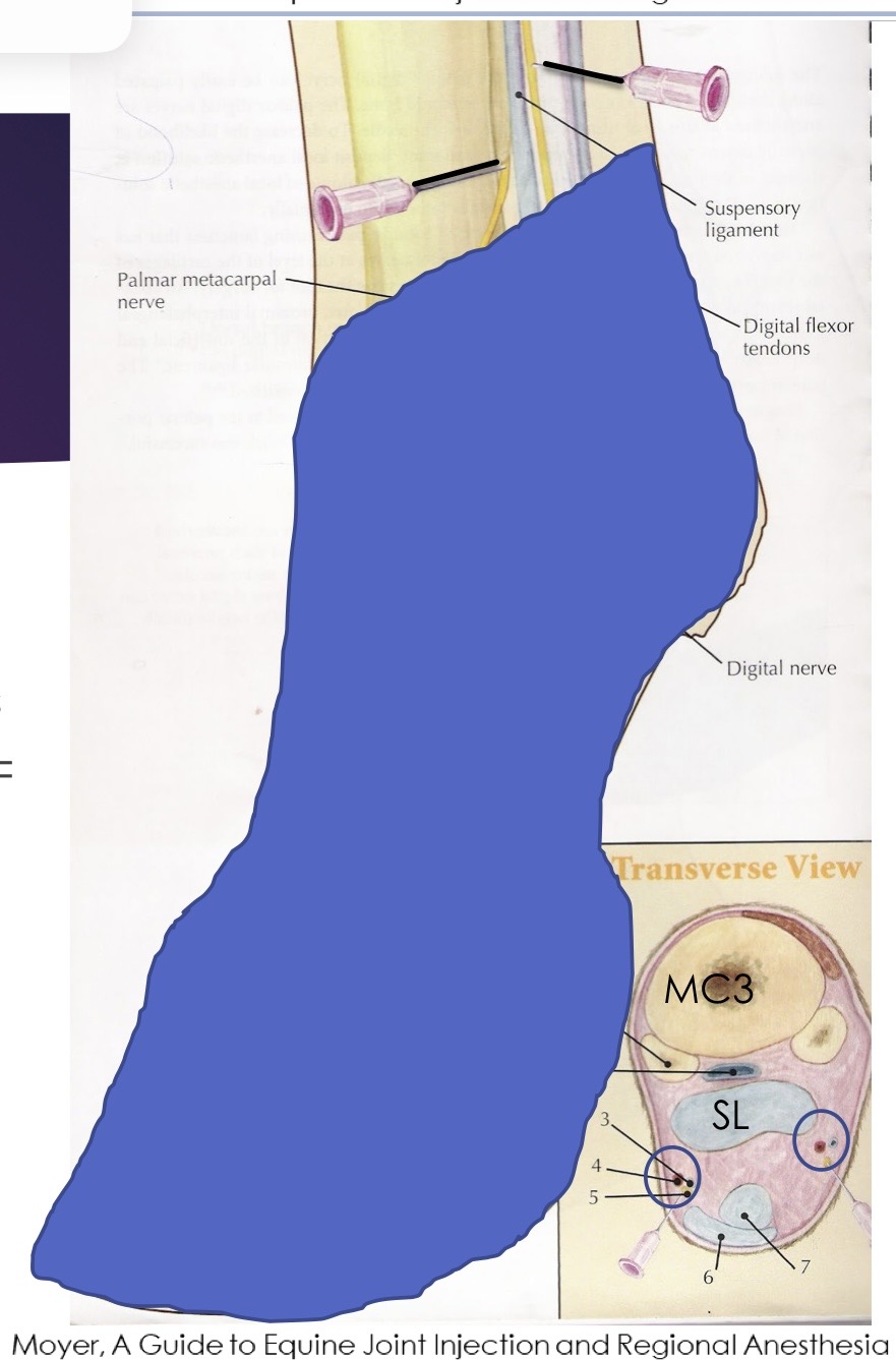
what block is this?
low 4 point block