Causes of Female Infertility
 Call Kai
 Learn
Learn Practice Test
 Spaced Repetition
 Match
Match Flashcards
 Knowt Play
1/40

There's no tags or description
Looks like no tags are added yet.
Name | Mastery | Learn | Test | Matching | Spaced | Call with Kai | Chat |
|---|
No analytics yet
Send a link to your students to track their progress
41 Terms
Infertility is defined as the inability to conceive a child after — of unprotected intercourse
1 year
— is a common cause of infertility
PID
—, and their impact on female infertility
Congenital uterine malformations (especially septate uterus), endometriosis, polycystic ovarian syndrome, tubal causes, Asherman syndrome, and uterine leiomyomas
Congenital uterine malformations, also referred to as —, have been linked with female infertility
Müllerian anomalies
uterine malformations often lead to repeated abortions as a result of —
structural abnormalities within the uterus
— is a common structural defect of the uterus that could lead to fertility troubles
Bicornuate uterus
— frequently requires surgical intervention to reduce the division of the uterine cavity.
a septate uterus
For patients with a septate uterus, sonography can also aid in the resection of the septum during a —
hysteroscopic uterine septoplasty
— is defined as functional, ectopic endometrial tissue located outside the uterus
Endometriosis
— may be the result of endometrial tissue being passed through the fallopian tubes during menstruation or may result from scarring from surgery, such as after a cesarean section
Implantation of ectopic endometrial tissue
Hemorrhage of this tissue often occurs, resulting in focal areas of bloody tumors known as —
endometriomas or chocolate cysts
. Endometriosis can be located anywhere throughout the pelvis, with the most common location being —
the ovaries
The age of the patients at the time of diagnosis for endometriosis is typically between 25 and 35 years, with the common clinical findings being —
pelvic pain, dyspareunia, and infertility

Various locations of endometriosis.
— are commonly cystic masses with lowlevel echoes that may or may not contain fluid–fluid levels
Endometriomas
one valid argument states that infertility results from the—, thus preventing ovum pickup or causing tubal obstruction
development of pelvic adhesions that may alter normal anatomy
Polycystic ovary syndrome (PCOS), also referred to as Stein–Leventhal syndrome, is an —
endocrinologic ovarian disorder linked with infertility.
PCOS has been cited as the most common cause of —, which is termed hyperandrogenism, and hirsutism in women
androgen excess
clinical findings of endometriosis
asymptomatic
pelvic pain
infertility
dysmenorrhea
menorragia
dyspareunia
painful bowel movements
sonographic findings of an endometriosis
Predominantly cystic mass with low-level internal echoes (may resemble a hemorrhagic cyst)
Anechoic or complex mostly cystic mass with posterior enhancement and may contain a fluid–fluid level
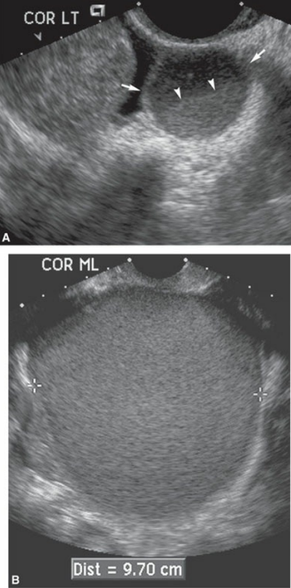
Endometrioma. A. Fluid–fluid level in an endometrioma. Coronal scan of the left ovary revealing a cystic adnexal mass (arrows) containing a fluid–fluid level (arrowheads). B. Transvaginal image of a homogeneous mass containing lowlevel echoes that was determined to be an endometrioma.
for the imaging diagnosis of PCOS—it has been suggested that one or both ovaries should contain— follicles that measure between 2 and 9 mm in diameter, and the ovarian volume should exceed 10 mL
12 or more
—, as seen in patient suffering from PCOS, has also been linked to the subsequent development of endometrial and breast cancer
high levels of unopposed estrogen stimulation on the endometrium

Polycystic ovary. Longitudinal sonogram demonstrating an enlarged round ovary (between arrows) with multiple small follicles less than or equal to 8 mm in diameter located around the periphery of the ovary (string of pearls sign). The stroma (S) of the ovary also appears to have increased echogenicity.
clinical findings of polycystic ovary syndrome
Stein–Leventhal syndrome (amenorrhea, hirsutism, and obesity)
infertility
oligomenorrhea
hyperandrogenism
sonographic findings of infertility
“String of pearls” sign or “necklace” sign describes the presence of many small cysts measuring along the periphery of the ovary
Many small cysts scattered throughout the ovary
Bilateral enlargement of the ovaries
Increased stroma and increased stromal echogenicity
One or both ovaries should contain 12 or more follicles that measure between 2 and 9 mm in diameter
Ovarian volume greater than 10 mL
Threshold of 25 small follicles can also be used
—is often the result of obstruction of the fimbriated end of the fallopian tube by adhesions
hydrosalphinx
The presence of adhesions within the tube can prevent the —
normal peristaltic motion that typically occurs.
— can occur as a result of long-standing PID, endometriosis, and tubal surgery
Adhesion development
— can be used to evaluate the patency of the fallopian tubes, as well as hysterosalpingosonography or hysterosalpingocontrast-sonography
Hysterosalpingography
The endometrium may not develop appropriately in the luteal phase of the endometrial cycle as a result of reduced progesterone production by the ovary; this is termed —
“luteal phase deficiency.”
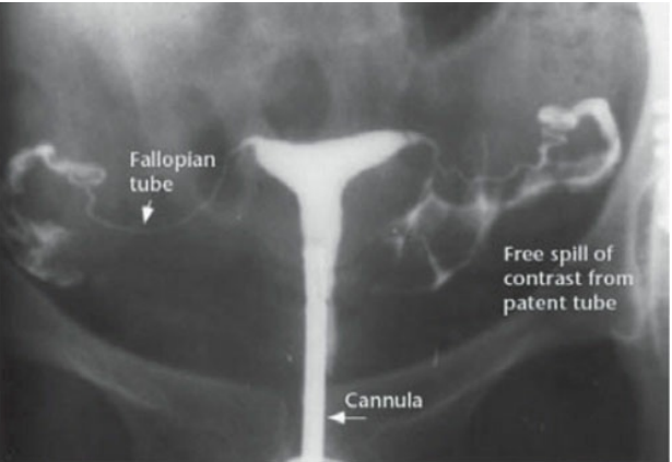
Hysterosalpingography. The hysterosalpingography procedure is a radiographic procedure used to evaluate the patency of the fallopian tubes with contrast.
— is the presence of intrauterine adhesions or synechiae within the uterine cavity that typically occur as a result of scar formation after uterine surgery, especially after a D&C.
Asherman syndrome
Adhesions within the — often prevent implantation or lead to recurrent early pregnancy loss
uterine cavity
clinical findings of asherman syndrome
History of D&C, trauma, and uterine surgery
Recurrent pregnancy loss
Amenorrhea or hypomenorrhea
sonographic findings of asherman syndrome
Bright areas within the endometrium
Sonohysterography findings include bright bands of tissue traversing the uterine cavity
fibroids that are — in location may distort the endometrium, thus preventing implantation of the early products of conception
intracavitary or submucosal
Fibroids may also impair — because of obstruction, and they may also enlarge during pregnancy.
tubal transport
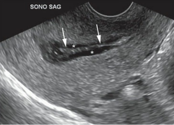
With saline infusion (*) during a saline infusion sonohysterogram, an endometrial adhesion is noted (arrows).
clinical findings of a uterine leiomyoma
pelvic pressure
menorrhagia
palpable abdominal mass
enlarged, bulky uterus
urinary frequency
dysuria
constipation
infertility
sonographic findings of a uterine leiomyoma
Hypoechoic mass within the uterus
Posterior shadowing from mass
Degenerating fibroids may have calcifications or cystic components
Multiple fibroids appear as an enlarged, irregular shaped, diffusely heterogeneous uterus