Advanced strength + conditioning (Exam 1)
 Call Kai
 Learn
Learn Practice Test
 Spaced Repetition
 Match
Match Flashcards
 Knowt Play
1/185

There's no tags or description
Looks like no tags are added yet.
Name | Mastery | Learn | Test | Matching | Spaced | Call with Kai | Chat |
|---|
No analytics yet
Send a link to your students to track their progress
186 Terms
what is strength?
the ability to produce force or torque agains an external stimulus.
shows up in our daily life
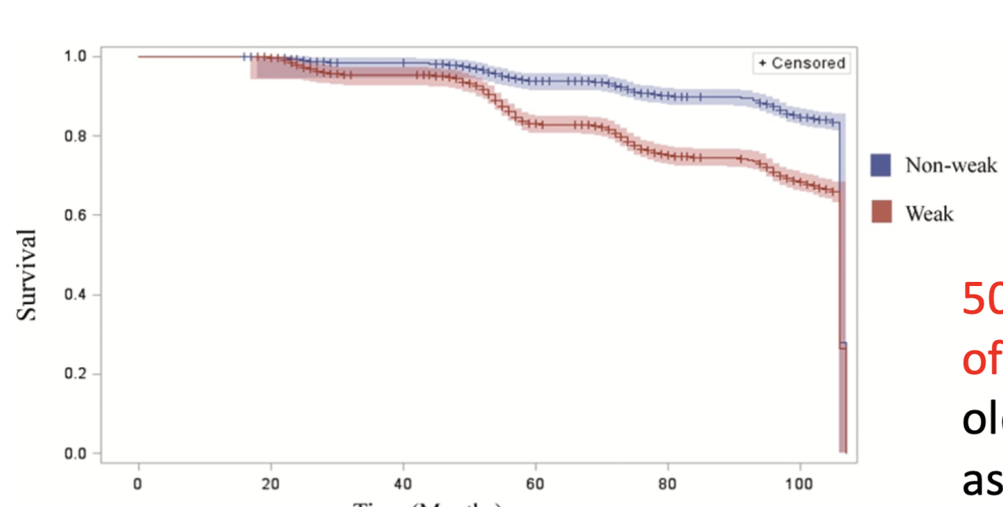
Strength = survival
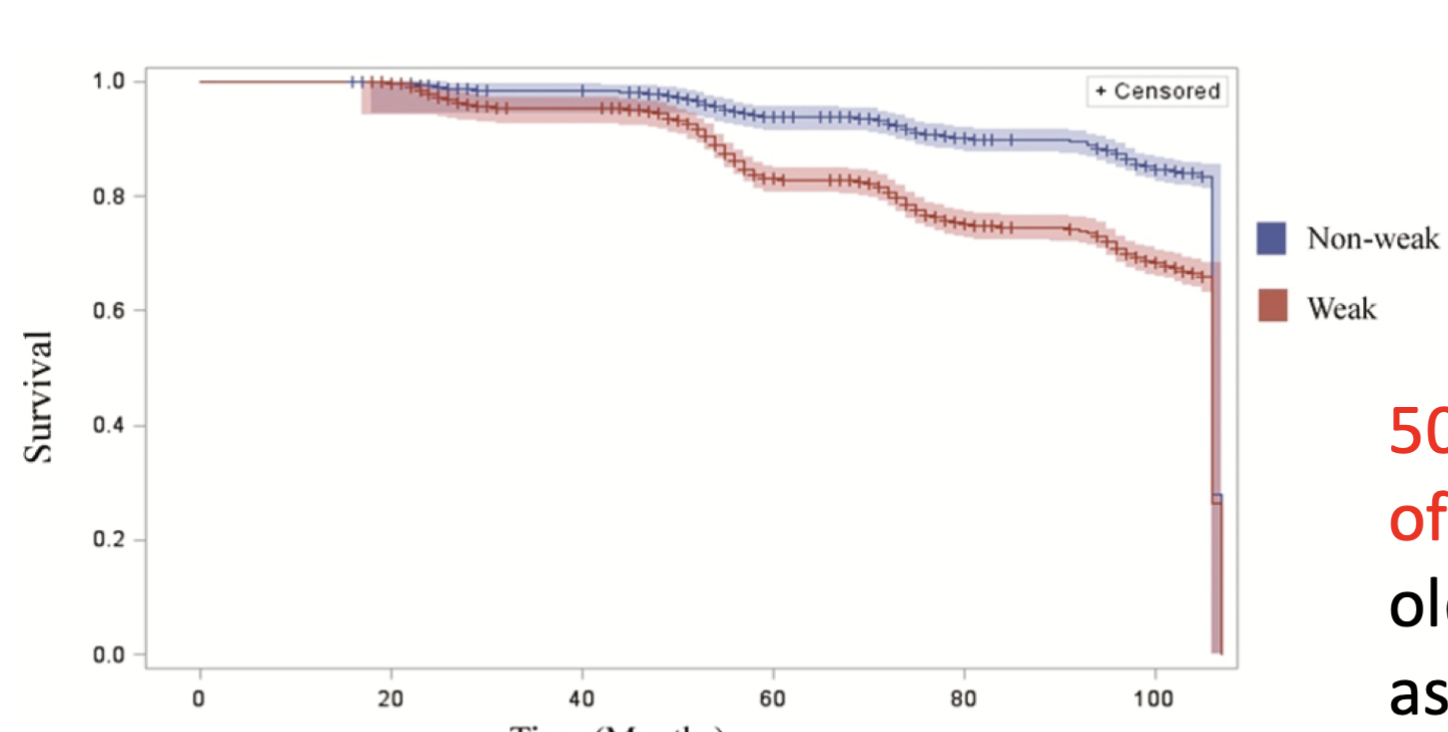
50% higher likelihood of early death among older adults classified as “weak”
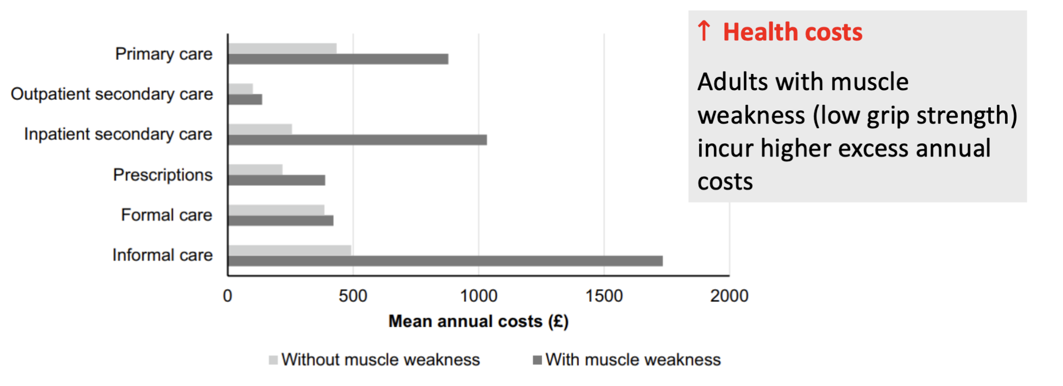
How does muscle weakness affect health-care costs?
Adults with muscle weakness (low grip strength) have higher annual health-care costs.
Muscle weakness is associated with increased costs for:
Primary care
Secondary/inpatient care
Prescriptions
Formal care
Informal care
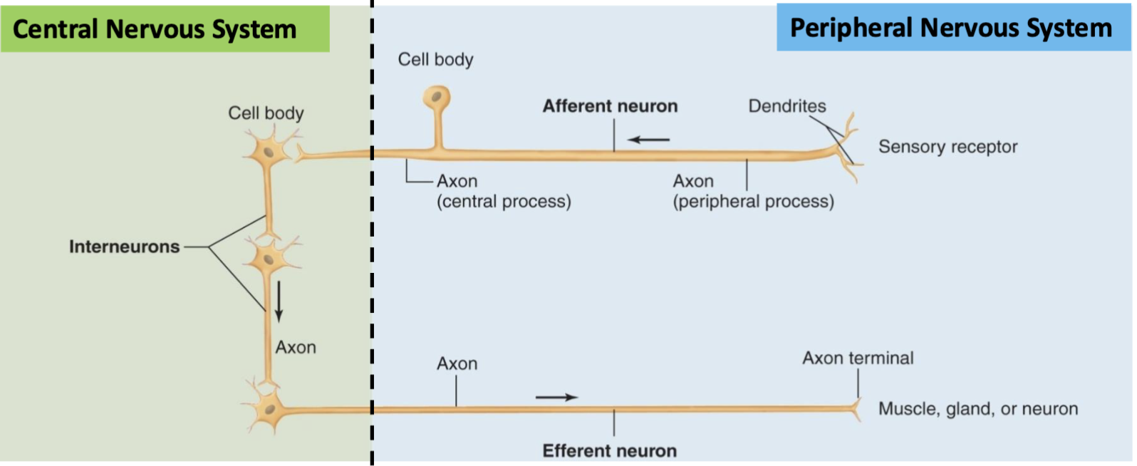
What are the 3 functional types of neurons?
Afferent (sensory) neurons
Receptors → CNS
Cell body located in PNS
Bring sensory information IN to CNS
Efferent (motor) neurons
CNS → muscles/glands/other neurons
Cell body located in CNS
Carry commands OUT from CNS
Interneurons
Located in CNS
99% of all neurons
Integrate/connect afferent and efferent neurons
Memory: Afferent = Arrives at CNS; Efferent = Exits CNS.
What are the basic components of a neuron and their functions?
Dendrites → receive information
Cell body (soma) → processes/integrates information; contains the nucleus → “command module”
Axon → sends information away from the cell body to other neurons or target cells (e.g., muscle fibers)
Memory: Dendrites IN → Soma PROCESSES → Axon OUT

How does the axon conduct an action potential?
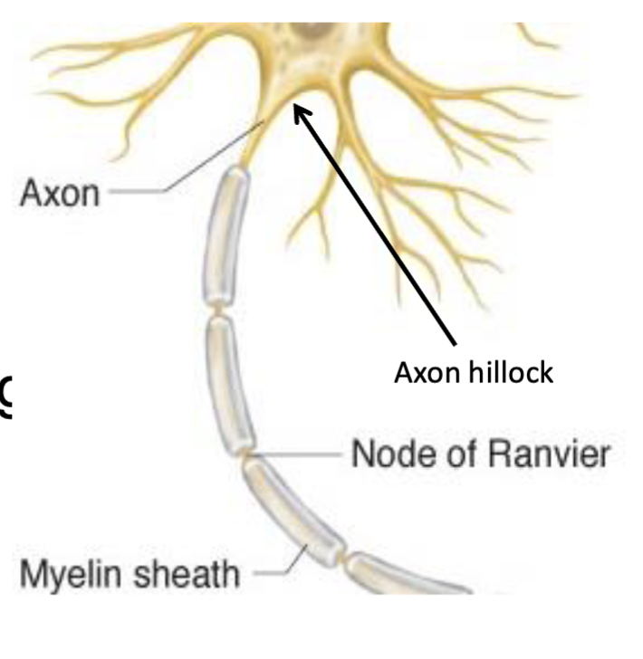
Axon → conducts the action potential (AP) away from the cell body, beginning at the axon hillock.
Myelin sheath → fatty insulating layer covering the axon that increases conduction speed.
Nodes of Ranvier → gaps between sections of myelin along the axon.
Saltatory conduction → AP “jumps” from one Node of Ranvier → the next.
Result → increases conduction velocity without increasing axon diameter.
Memory: Myelin = insulation → AP jumps node-to-node → faster conduction.
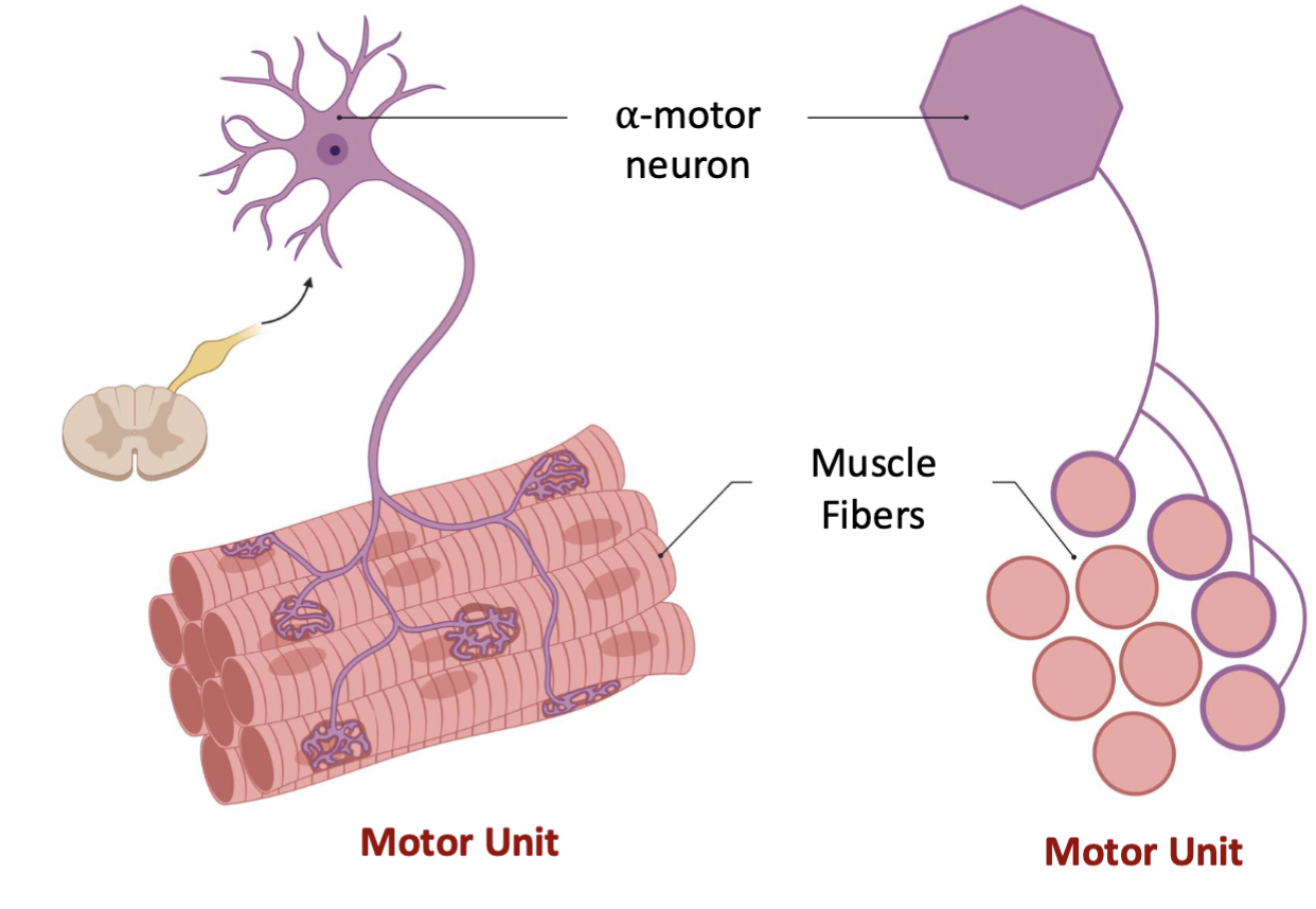
What is a motor unit?
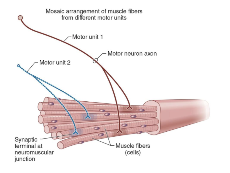
Motor unit = 1 alpha-motor neuron + ALL muscle fibers it innervates
One motor neuron can innervate 100s–1000s of muscle fibers
Each muscle fiber has only ONE neuromuscular junction (NMJ)
All muscle fibers within a motor unit contract together when stimulated
Memory: 1 motor neuron → many muscle fibers → all contract together
What is the innervation ratio, and how does it relate to movement precision?
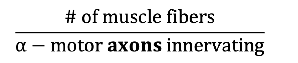
Innervation ratio = # of muscle fibers ÷ # of α-motor axons innervating them
It represents the average motor unit (MU) size.
Large innervation ratio → many fibers per motor unit → large/gross movements
Ex: Gastrocnemius ≈ 1,934 fibers/MU
Small innervation ratio → few fibers per motor unit → small/precise movements
Ex: Rectus lateralis (eye) ≈ 5 fibers/MU
Memory: More fibers = more force, less precision; fewer fibers = more precision.
What is an action potential (AP), and what are its main phases?
Action potential = a rapid, large change in membrane potential used for fast, long-distance communication.
Occurs in excitable cells → nerves, muscles, glands.
Resting membrane potential ≈ −70 mV
Depolarization → voltage-gated Na⁺ channels open → Na⁺ enters → membrane becomes more positive
Repolarization → voltage-gated K⁺ channels open → K⁺ exits → membrane becomes more negative again
May briefly hyperpolarize before returning to resting potential.
Memory: Na⁺ IN = Depolarize → K⁺ OUT = Repolarize

How does an action potential (AP) propagate through a neuron?
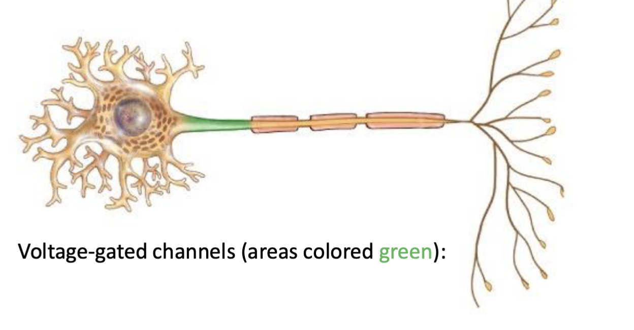
AP is initiated at the axon hillock (trigger zone).
AP propagates along the axon → axon terminals.
Propagation occurs through activation of voltage-gated ion channels along the axon.
Memory: Axon hillock → Axon → Axon terminals
What is the all-or-none principle for motor units?
Motor unit (MU) activation depends on reaching a voltage threshold.
Threshold reached → ALL muscle fibers in that motor unit contract
Threshold NOT reached → NONE of the muscle fibers contract
You cannot activate only some fibers within a single motor unit.
A “larger” action potential does NOT cause a stronger contraction; APs are all-or-none.
Memory: Hit threshold = ALL. Miss threshold = NONE.

How does the CNS increase muscle force?
Muscle force is primarily controlled by 2 mechanisms:
Motor unit recruitment → activate more motor units
Rate coding → increase the firing rate of active motor units
↑ Recruitment + ↑ firing rate → ↑ muscle force
Memory: More motor units + faster firing = more force.
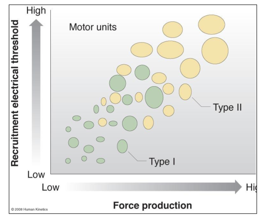
What is the motor unit size principle?
Motor units are recruited in an orderly fashion as force demand increases:
Low-threshold Type I → high-threshold Type II
Type I: recruited first → lower force demands
Type II: recruited later → higher force demands
At a given force, firing rate and recruitment threshold are inversely related → lower-threshold MUs tend to fire faster than higher-threshold MUs (“onion skin” phenomenon).
Memory: Small/slow first → large/fast last.
How does the size principle determine motor unit recruitment?
Motor units are recruited from low → high threshold as force demand increases:
Type I → Type IIa → Type IIx
Low-threshold MUs: recruited first, lower force production
High-threshold MUs: recruited later, greater force production
To recruit high-threshold MUs, the lower-threshold MUs must already be recruited.
↑ force demand → ↑ number/size of motor units recruited
Memory: You must recruit the small ones before the big ones.
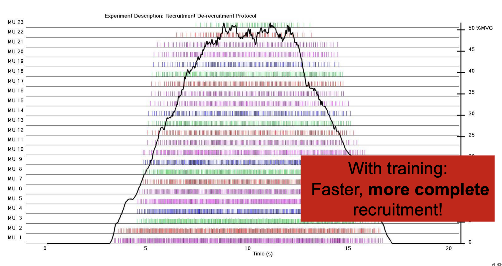
How does resistance training affect motor unit recruitment?
Resistance training causes a neural adaptation that allows:
Faster recruitment of motor units
Earlier recruitment of high-threshold MUs
More complete recruitment of available MUs
This helps explain why strength can increase before significant muscle hypertrophy occurs.
To recruit high-threshold, high-force fibers → load/effort must be high enough to reach their activation threshold.
Memory: Training → faster + more complete recruitment → early strength gains.
How does firing frequency affect motor unit force?
As firing frequency increases → force increases:
Single twitch → twitch summation → unfused tetanus → fused tetanus
Single twitch = one stimulus/contraction
Summation = twitches overlap → ↑ force
Unfused tetanus = repeated contractions with partial relaxation
Fused tetanus = sustained contraction with no relaxation → maximal force
Overall muscle force is increased by:
Recruiting more motor units
Increasing firing rate (rate coding)
Memory: ↑ frequency → ↑ summation → ↑ force → tetanus.
Nueromuscular physicology (Part 2) …
What is a motor unit?
A motor unit = 1 alpha-motor neuron + ALL the muscle fibers it innervates (ON QUIZ)
The α-motor neuron controls when its muscle fibers contract.
When the motor neuron fires → all fibers in that motor unit contract (all-or-none).
One motor neuron can innervate many muscle fibers.
Memory: 1 neuron → all of its fibers = 1 motor unit.
What is the neuromuscular junction (NMJ)?
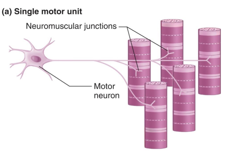
The NMJ = the synapse/interface between an α-motor neuron and a muscle fiber.
NMJ signaling is always excitatory → initiates muscle contraction, never inhibits.
Each muscle fiber has only ONE NMJ.
The NMJ allows the motor neuron's electrical signal to activate the muscle fiber → excitation-contraction coupling.
Think: Motor neuron → NMJ → muscle fiber → contraction.
What are the steps of excitation-contraction coupling at the NMJ?
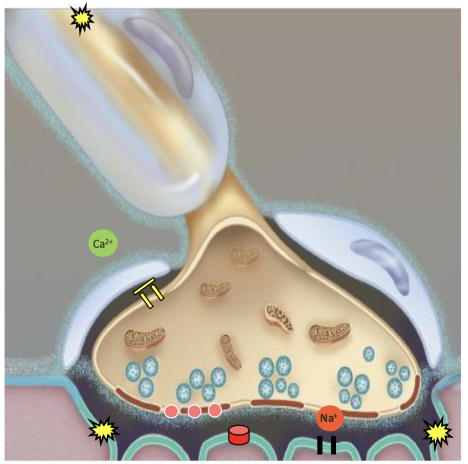
AP travels down the motor neuron → reaches axon terminal
Voltage-gated Ca²⁺ channels open → Ca²⁺ enters axon terminal
Ca²⁺ triggers synaptic vesicles → ACh released into synaptic cleft
ACh binds receptors on the motor end plate → Na⁺ channels open → Na⁺ enters
Creates a large end-plate potential (EPP) → depolarizes the sarcolemma
Sequence:
AP → Ca²⁺ IN → ACh release → Na⁺ IN → EPP → muscle depolarization
What happens after the muscle fiber is depolarized during excitation-contraction coupling?
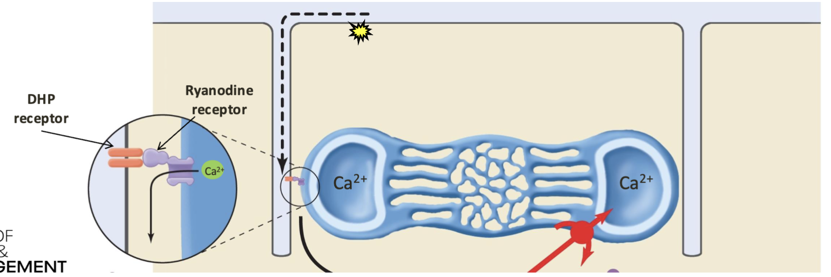
AP travels along the sarcolemma → T-tubules
T-tubule DHP receptor (voltage sensor) activates the ryanodine receptor (RyR) on the sarcoplasmic reticulum
RyR opens → Ca²⁺ released from SR → sarcoplasm
Ca²⁺ is now available to bind troponin → initiate contraction
Sequence:
AP → T-tubule → DHP → RyR → Ca²⁺ out of SR → troponin → contraction
What are the 3 connective tissue layers of skeletal muscle?
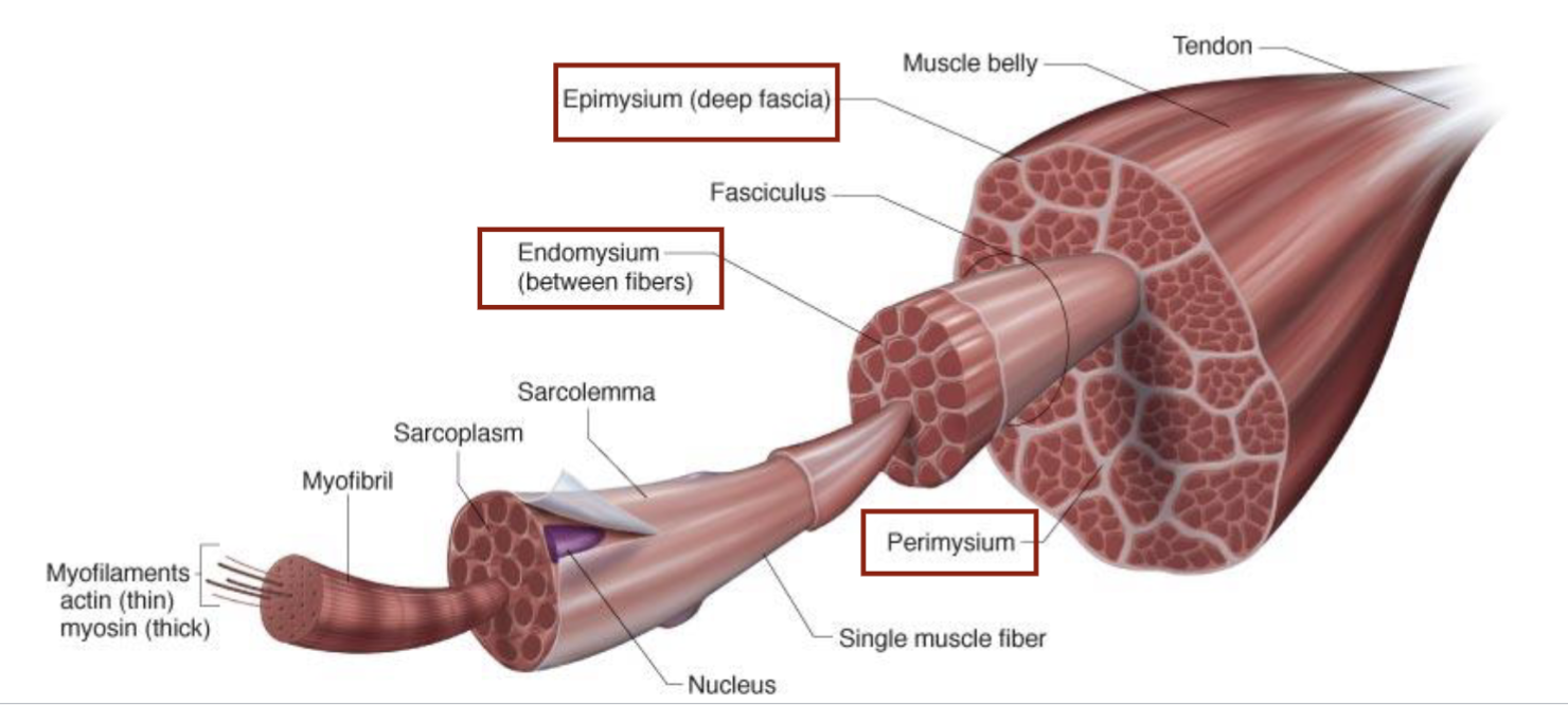
Epimysium → surrounds the entire muscle (outer layer)
Perimysium → surrounds each fascicle (bundle of muscle fibers)
Endomysium → surrounds each individual muscle fiber
Outside → inside:
Epimysium → Perimysium → Endomysium
What are the major structural components of a muscle fiber?
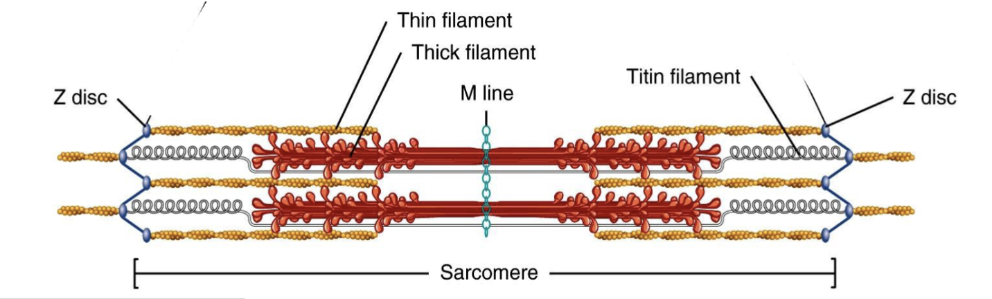
Myofibrils → cylindrical bundles of thick + thin filaments within muscle fibers
Sarcomere → functional/contractile unit of muscle; extends Z-line → Z-line
Z-line → defines sarcomere boundaries; anchors thin filaments
Titin → connects myosin (thick filament) → Z-line
Cross-bridges → globular myosin heads that project toward/bind thin filaments
Thick filament → myosin
2 heavy chains + 4 light chains
Thin filament → actin + tropomyosin + troponin
High-yield:
Thick = myosin
Thin = actin + tropomyosin + troponin
What is the A band of a sarcomere?
A band → spans the entire length of the thick (myosin) filaments
Includes areas where myosin overlaps with actin
Centered around the M line
Does NOT change length during contraction
High-yield: A band = All of the thick filament.

What is the I band of a sarcomere?
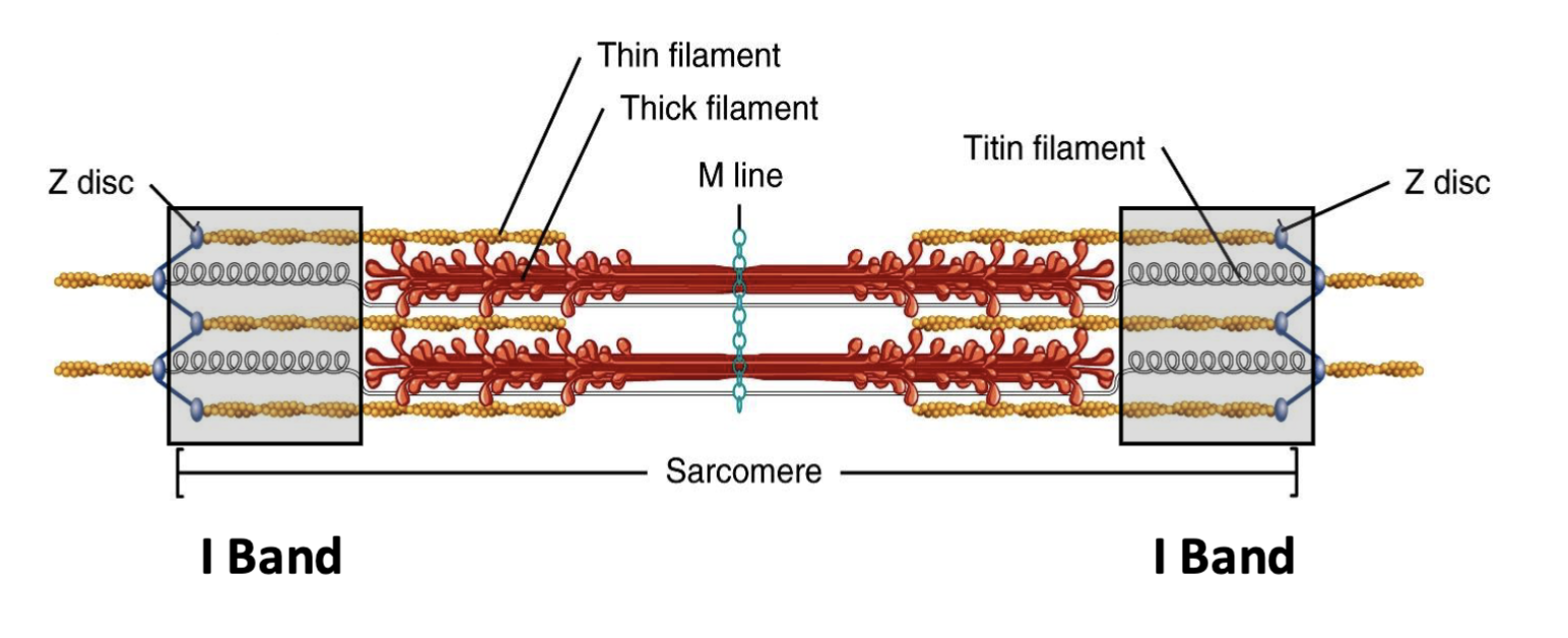
I band → lighter region containing only thin (actin) filaments
No thick/myosin filaments
Contains the Z-line
Shortens during muscle contraction
High-yield: I band = thIn filaments only.
hat is the H zone of a sarcomere?
H zone → contains only thick (myosin) filaments
No thin/actin filaments
Located in the center of the A band
Contains the M line
Shortens/decreases during contraction
High-yield: H zone = tHick only.
What is the M line of a sarcomere?
M line → anchors thick (myosin) filaments together
Located at the center of the sarcomere
Located in the center of the H zone
Helps keep thick filaments aligned
High-yield: M = Middle + Myosin.
What happens during the sliding filament theory of muscle contraction?
Myosin cross-bridges pull actin (thin filaments) toward the center/M-line → sarcomere shortens.
During contraction:
I band ↓
H zone ↓
A band stays SAME
Z-lines move closer together
Actin and myosin do NOT shorten → they slide past each other
High-yield: A = Always same; I + H shrink.

only H and I change sizes during contraction…
NO CHANGE in sarcomere during isometric
eccentric is Active
What is the role of ATP in cross-bridge cycling?
ATP has 2 key roles:
ATP binds myosin → myosin DETACHES from actin
ATP hydrolysis → energizes/cocks the myosin head for the next contraction
⚠ No ATP → myosin cannot detach from actin → rigor mortis.
Memory: ATP = Detach + Recharge.
What happens in Step 1 of cross-bridge cycling?
ATP → ADP + Pi via myosin ATPase
→ ADP + Pi remain bound to myosin
→ Energy from ATP hydrolysis cocks/energizes the myosin head
→ Myosin is ready to bind actin
Step 1 = ATP hydrolysis → cock the myosin head.
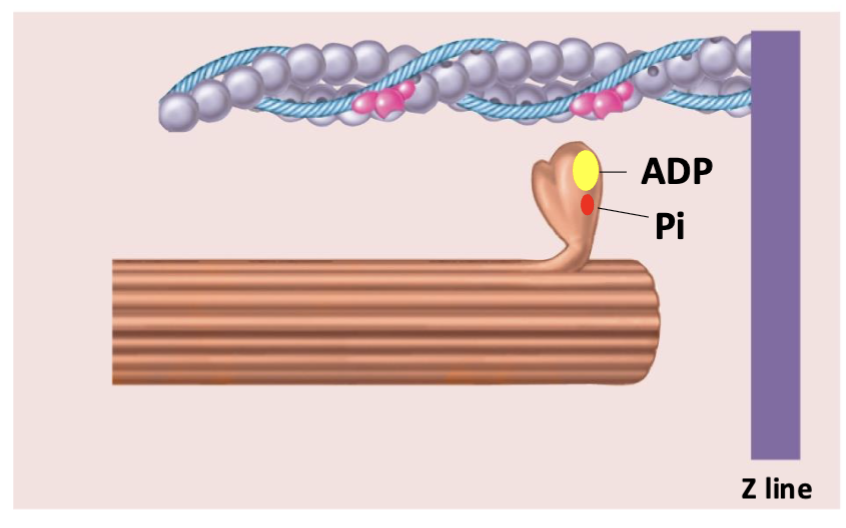
What happens in Step 2 of cross-bridge cycling?
↑ Ca²⁺ released from SR → Ca²⁺ binds troponin → tropomyosin moves → exposes myosin-binding sites on actin → energized myosin binds actin → cross-bridge forms
Memory: Ca²⁺ → Troponin → Tropomyosin moves → Actin exposed → Myosin binds.
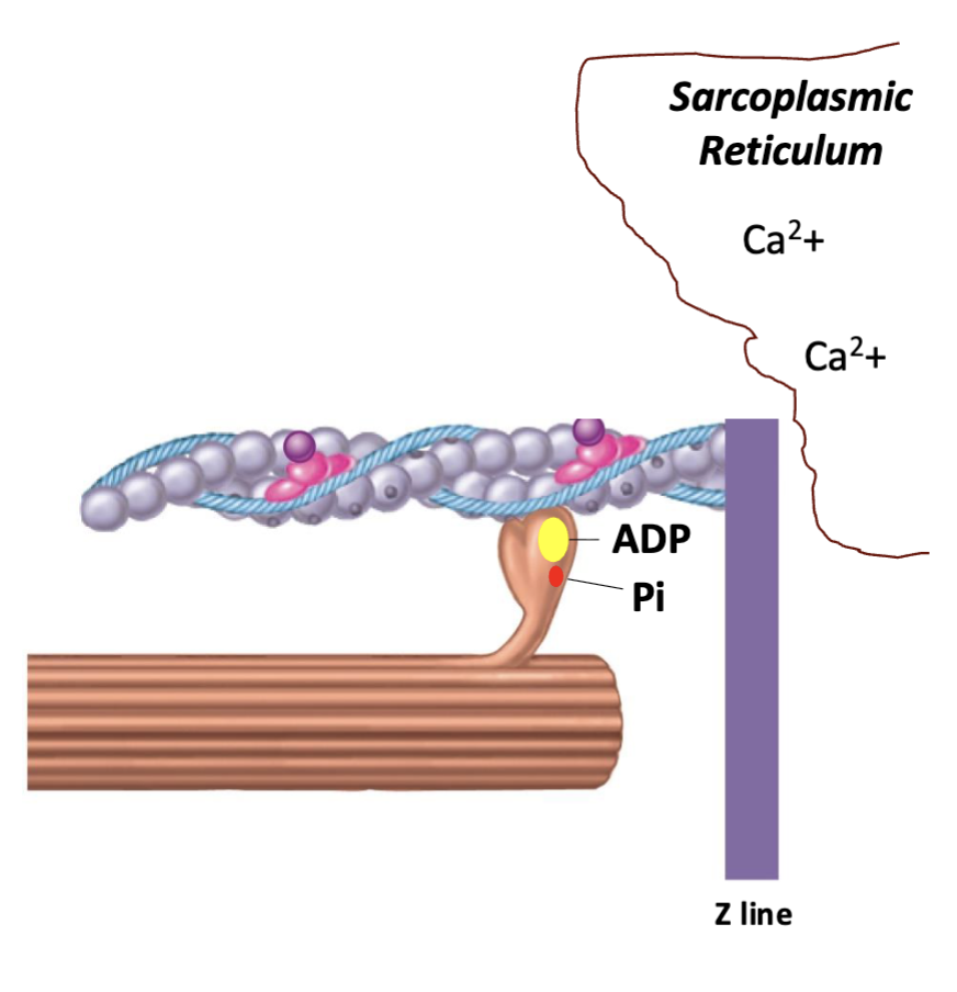
What happens in Step 3 of cross-bridge cycling (power stroke)?
Myosin head bends/pivots → pulls actin toward the center (M-line) of the sarcomere → ADP + Pi are released.
Step 3 = Power stroke → Pull actin → Release ADP + Pi.
⚠ Small correction to the slide: actin is pulled toward the M-line, not the Z-line.

What happens in Step 4 of cross-bridge cycling (detachment)?
New ATP binds myosin → myosin DETACHES from actin.
Then ATP is hydrolyzed → myosin head re-cocks/energizes → cycle repeats if Ca²⁺ remains present.
Memory: New ATP = LET GO of actin.

What happens during muscle relaxation?
Nerve stimulation stops → Ca²⁺ pumped back into SR → Ca²⁺ leaves troponin → tropomyosin re-covers actin binding sites → cross-bridge cycling stops → muscle relaxes.
Memory: ↓ Ca²⁺ → actin sites covered → no myosin binding → relaxation.
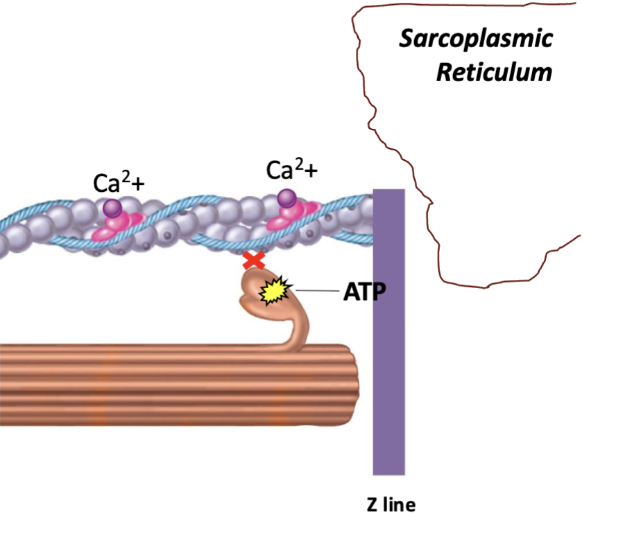
Why does rigor mortis occur?
After death → ATP runs out → myosin cannot detach from actin → actin-myosin cross-bridges remain locked → muscle becomes rigid.
Memory: No ATP = No detachment = Rigor mortis.

Bioenergetics…
Why is bioenergetics important for human movement?
Food (carbohydrates, fats, proteins) provides chemical energy
Chemical energy is used to produce ATP
ATP provides energy for cellular activity and mechanical work (movement)
ATP → ADP + Pi releases energy
Some energy is released as heat
Energy = capacity to do work
What is bioenergetics, and how does the first law of thermodynamics apply to it?
Bioenergetics = flow of energy in a living system; conversion of macronutrients into biologically usable energy
First law of thermodynamics = energy cannot be created or destroyed, only transformed
Chemical energy from food can be converted into:
Work
Heat
Stored energy molecules
What are catabolism and anabolism?
Catabolism = breakdown of larger molecules → smaller molecules + releases energy
Anabolism = synthesis of smaller molecules → larger molecules + requires energy
Key distinction:
Catabolism = breaks down, releases energy
Anabolism = builds up, requires energy
What are exergonic vs. endergonic reactions, and how do they relate to metabolism?
Exergonic = releases energy → generally catabolic
Example: glycolysis
Endergonic = requires energy → generally anabolic
Example: protein synthesis
Energy released by exergonic reactions is used to drive endergonic reactions.
Metabolism = total sum of all chemical reactions occurring in the body’s cells.
Why is bioenergetics important when designing training programs?
Identify the predominant energy system for the activity → design an effective, individualized training program.
Energy system determines:
Intensity
Duration
Work-to-rest ratio
Exercise mode
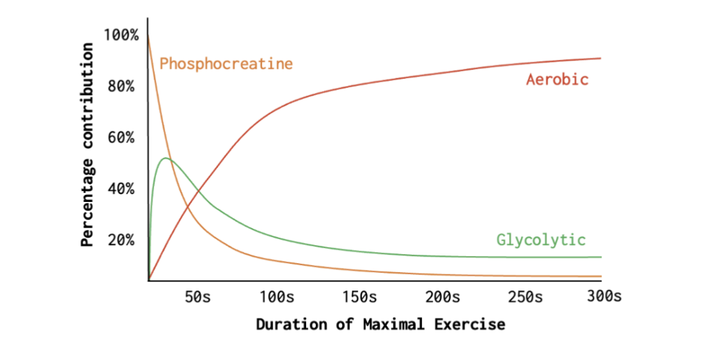
As maximal exercise duration increases:
Phosphocreatine → dominates first, rapidly decreases
Glycolytic → increases early, then decreases
Aerobic → gradually increases and becomes predominant
Key idea: Train the specific energy pathways required by the sport/activity.
What is the role of ATP in the body?
ATP (adenosine triphosphate) = universal energy currency of cells
Directly provides energy for cellular processes
All energy systems ultimately function to resynthesize ATP
ATP → ADP + Pi releases usable energy
ATP must continually be resynthesized because cellular activities constantly use it
Key idea: Food fuels cannot directly power cellular work → their energy is transferred to ATP → ATP powers cellular work.
How does ATP provide energy for muscle contraction?
ATP fuels the actomyosin cross-bridge cycle
Energy is associated with the bond involving the terminal (3rd) phosphate
ATPase breaks down ATP
ATP → ADP + Pi + energy
Released energy is used for muscle contraction.
What occurs during ATP hydrolysis, and why must ATP be continually resynthesized?
ATP hydrolysis = breakdown of ATP to release energy
ATP + H₂O → ADP + Pi + H⁺ + energy
Catalyzed by ATPase
Energy released fuels muscle contraction and other cellular processes
Body stores only ~80–100 g of ATP
Because ATP stores are limited but demand is constant → ATP must be continually resynthesized
How do the energy systems contribute during exercise, and what is the rate vs. capacity tradeoff?
All energy systems are always active → exercise demand determines which dominates.
Phosphocreatine (PCr) = fastest ATP production, lowest capacity → dominates first
Glycolytic = intermediate rate and capacity → contributes most during short-duration exercise
Aerobic/oxidative = slowest ATP production, highest capacity → dominates prolonged exercise
Key idea: Faster ATP production = lower capacity; greater capacity = slower ATP production.
How does the phosphagen (PCr) system rapidly resynthesize ATP?
Phosphagen system = fastest ATP-producing system
Creatine phosphate (CP/PCr) donates a phosphate to ADP → rapidly reforms ATP
ADP + PCr → ATP + creatine
Enzyme = creatine kinase (CK)
Very high rate of ATP production
Very low capacity because PCr stores are small
CK highest in Type IIx muscle fibers
Important for short, high-intensity strength/power activities
Key idea: PCr acts as an immediate energy reserve to rapidly replenish ATP.

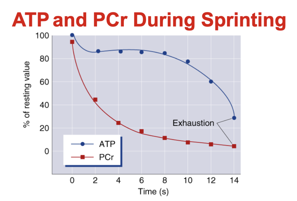
What happens to ATP and PCr during maximal sprinting?
ATP is continuously broken down but rapidly resynthesized → levels initially remain relatively stable
PCr rapidly donates phosphate to regenerate ATP → PCr levels fall quickly
PCr stores are limited → PCr depletion becomes a limiting factor during maximal sprinting
As PCr becomes depleted → ATP eventually declines → contributes to fatigue/exhaustion
Key idea: PCr drops much faster than ATP because PCr is being used to maintain ATP levels.
What is glycolysis?
Glycolysis = breakdown of carbohydrates to resynthesize ATP
Fuel sources:
Muscle glycogen
Blood glucose
Does NOT require O₂ → anaerobic
Occurs in the sarcoplasm
Key idea: Glycolysis breaks down glucose/glycogen to rapidly produce ATP without
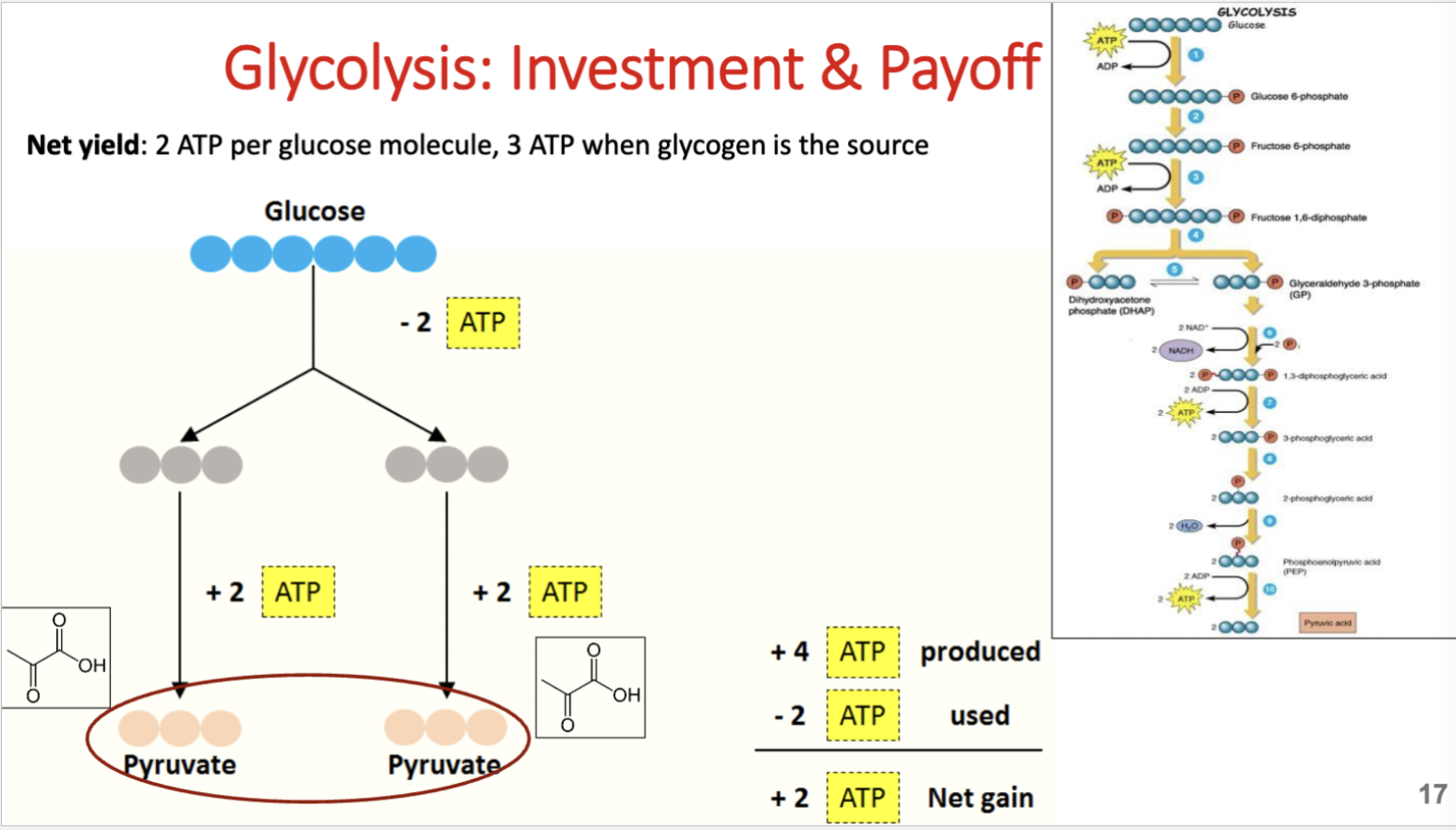
What is the ATP investment and payoff of glycolysis?
Glucose:
Investment = 2 ATP used
Payoff = 4 ATP produced
Net gain = 2 ATP
Produces 2 pyruvate
Muscle glycogen:
Net gain = 3 ATP per glycogen-derived glucose
Key idea: Glycolysis from glycogen yields 1 extra ATP because it bypasses the first ATP-requiring step.
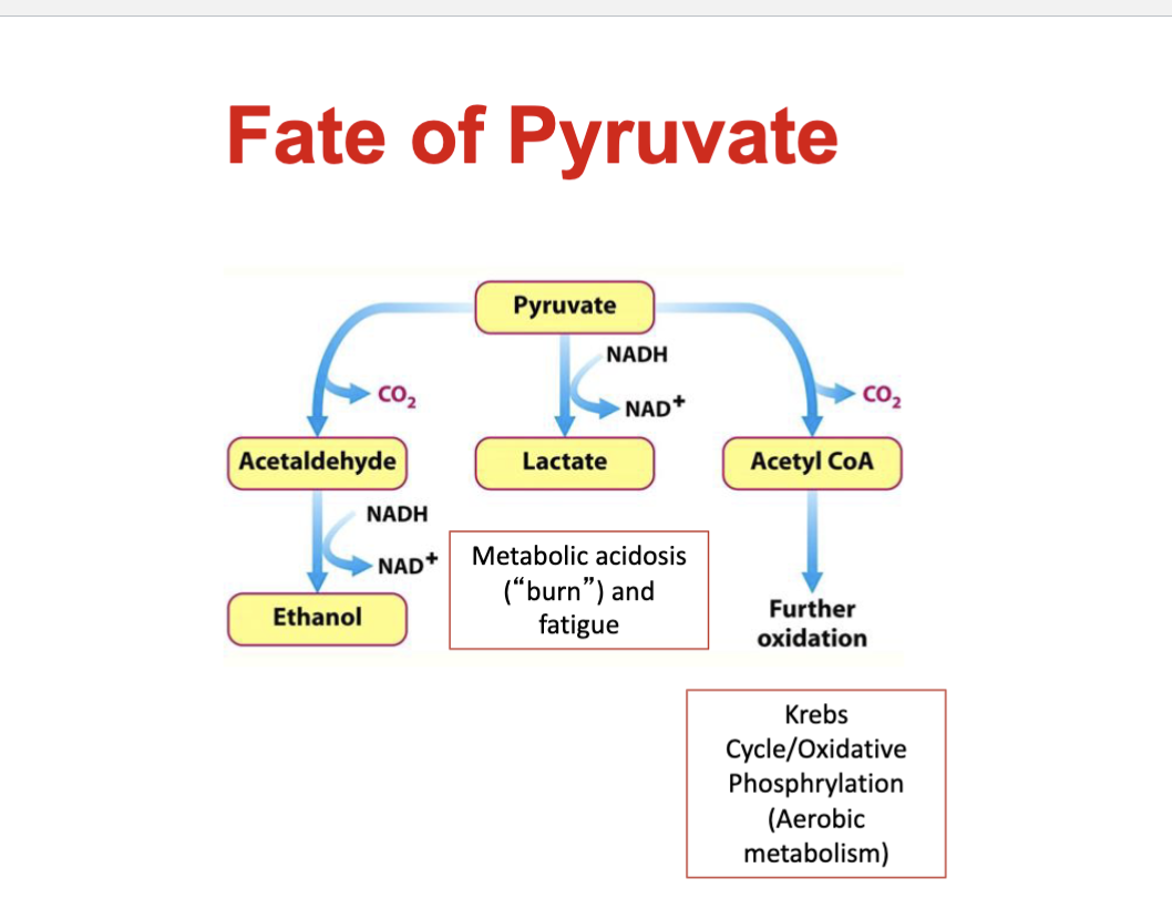
What determines the fate of pyruvate after glycolysis?
Depends on O₂ availability and ability of mitochondria to metabolize pyruvate.
Fast (anaerobic) glycolysis:
Glycolysis exceeds aerobic metabolism
Pyruvate → lactate
Associated H⁺ accumulation → ↓ pH → contributes to fatigue
Slow (aerobic) glycolysis:
Glycolysis matches aerobic metabolism
Pyruvate + NADH enter mitochondria
Pyruvate proceeds through Krebs/TCA cycle and ETC
Key idea: Fast glycolysis → lactate; slow glycolysis → mitochondrial oxidation.
What happens to lactate produced during glycolysis?
Pyruvate → lactate via lactate dehydrogenase (LDH)
Lactate is associated with fatigue but is not necessarily the cause
Highest lactate concentrations occur in Type II muscle fibers
Blood lactate = balance between lactate production and clearance
Greatest accumulation occurs with high-intensity, intermittent exercise
Cori cycle:
Lactate → bloodstream → liver → converted back to glucose via gluconeogenesis
Key idea: Lactate is not simply a waste product; it can be recycled into glucose.
How does H⁺ accumulation contribute to muscle fatigue?
↑ H⁺ from ATP hydrolysis → ↓ pH (acidosis)
Muscle pH:
Resting ≈ 7.1
Exercise can ↓ to 6.4–6.6
H⁺ contributes to fatigue by:
↓ enzyme activity
↓ actin-myosin binding affinity
↓ O₂-carrying capacity of hemoglobin
Key idea: H⁺ accumulation, rather than lactate itself, contributes to fatigue during high-intensity exercise.
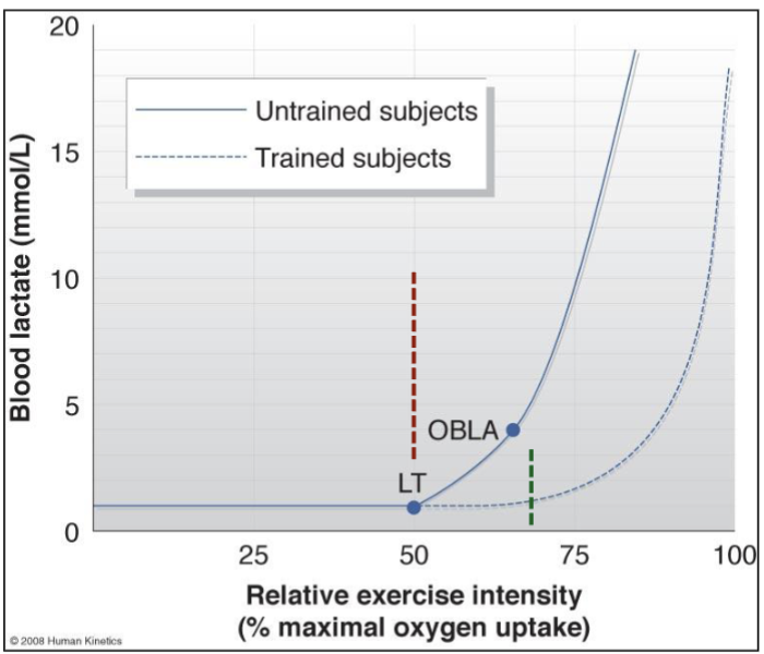
What are lactate threshold (LT) and onset of blood lactate accumulation (OBLA)?
Lactate Threshold (LT) = exercise intensity where blood lactate begins to abruptly rise above baseline
Indicates increased reliance on anaerobic metabolism
OBLA = point where blood lactate reaches 4 mmol/L
Lactate production > lactate clearance
Training effect:
Trained individuals reach LT at a higher exercise intensity
Higher LT = can sustain greater intensity before rapid lactate accumulation
Key distinction: LT = initial abrupt rise; OBLA = 4 mmol/L.
Does lactate cause DOMS, and how is lactate cleared after exercise?
Lactate does NOT cause DOMS.
Lactate fate:
~70% oxidized → used as fuel by heart/skeletal muscle
~20% → converted back to glucose
~10% → converted to amino acids
Lactate clearance:
Light exercise clears lactate faster than complete rest
Optimal active recovery ≈ 30–40% VO₂ max
Key idea: Lactate is rapidly cleared/reused after exercise and does not cause delayed-onset muscle soreness.
What is the oxidative energy system and how does it produce ATP?
Oxidative system = primary ATP source at rest and during low-intensity/prolonged activity
Fuel:
Carbohydrates + fats
Protein = minimal contribution
Fat pathway:
Triglycerides → free fatty acids → mitochondria → β-oxidation → acetyl-CoA
Then:
Acetyl-CoA → Krebs cycle → NADH/FADH₂ → ETC → ATP
Key idea: Oxidative system = slow ATP production but very high capacity; requires mitochondria and O₂.
What determines oxidative capacity?
3 main factors:
Oxidative enzyme levels → ↑ enzyme activity = faster Krebs cycle + ETC
Fiber-type composition → Type I fibers have high oxidative capacity
Oxygen availability → depends on cardiac output, capillary density, and hemoglobin
Key idea: Oxidative capacity depends on the muscle’s ability to use O₂ and the body’s ability to deliver O₂.
What is cardiorespiratory endurance and VO₂ max?
Cardiorespiratory endurance = ability of the whole body to sustain prolonged aerobic exercise
VO₂ max = highest rate of O₂ consumption during maximal exercise
Major measure of aerobic fitness
Important for exercise performance + cardiovascular health
Assessed using a graded exercise test (GXT)
Key idea: Higher VO₂ max = greater capacity to take in, transport, and use oxygen during maximal exercise.
What are the major requirements for endurance success?
High VO₂ max
High economy of effort
High lactate threshold
High percentage of slow-twitch (Type I) muscle fibers
Key idea: Endurance success requires high aerobic capacity + efficient movement + ability to sustain high intensity aerobically.
What is the relationship between the rate and capacity of ATP production?
Rate = how fast ATP can be produced
Capacity = total amount of ATP that can be produced
Inverse relationship: ↑ rate → ↓ capacity
Fastest rate → slowest:
Phosphagen > fast glycolysis > slow glycolysis > carbohydrate oxidation > fat/protein oxidation
Capacity is the opposite order.
Key idea: Phosphagen = fastest but lowest capacity; fat oxidation = slowest but highest capacity.
What is the relationship between the rate and capacity of ATP production?
Rate = how fast ATP can be produced
Capacity = total amount of ATP that can be produced
Inverse relationship: ↑ rate → ↓ capacity
Fastest rate → slowest:
Phosphagen > fast glycolysis > slow glycolysis > carbohydrate oxidation > fat/protein oxidation
Capacity is the opposite order.
Key idea: Phosphagen = fastest but lowest capacity; fat oxidation = slowest but highest capacity.

How do exercise duration and intensity determine the primary energy system used?
All energy systems are always active, but intensity + duration determine which predominates.
0–6 sec, extremely high intensity → Phosphagen
6–30 sec, very high → Phosphagen + fast glycolysis
30 sec–2 min, high → Fast glycolysis
2–3 min, moderate → Fast glycolysis + oxidative
3 min, lower intensity → Oxidative
Key idea: Shorter + higher intensity → anaerobic systems; longer + lower intensity → oxidative system.
How are phosphagen and glycogen stores depleted and replenished during exercise?
Phosphagens:
CP ↓ ~50–70% within first 5–30 sec of high-intensity exercise
Can be nearly depleted during maximal exercise
ATP fully resynthesized within ~3–5 min
CP fully resynthesized within ~8 min
Muscle/liver glycogen:
Glycogen depletion ↑ as exercise intensity ↑
Above ~60% VO₂ max → muscle glycogen becomes increasingly important
Glycogen stores can become depleted during prolonged exercise
Key idea: Phosphagens deplete rapidly but recover quickly; glycogen depletion depends largely on exercise intensity.
Where is glycogen stored in the body and approximately how much is stored?
Glycogen is stored as granules in the cytoplasm of muscle and liver cells.
Muscle glycogen ≈ 400 g
Liver glycogen ≈ 100 g
Key idea: Most of the body’s glycogen is stored in skeletal muscle.
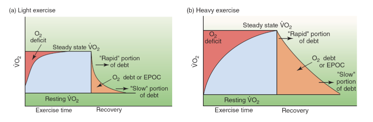
What is oxygen deficit during the beginning of exercise?
Oxygen deficit = initial period when aerobic ATP production cannot yet meet ATP demand.
At exercise onset:
O₂ uptake gradually increases
ATP demand initially met by anaerobic pathways → phosphagen + glycolysis
O₂ reaches steady state within ~1–4 min during light-moderate exercise
At steady state → ATP demand is primarily met aerobically
Key idea: Oxygen deficit = gap between ATP demand and aerobic ATP production before steady state is reached.

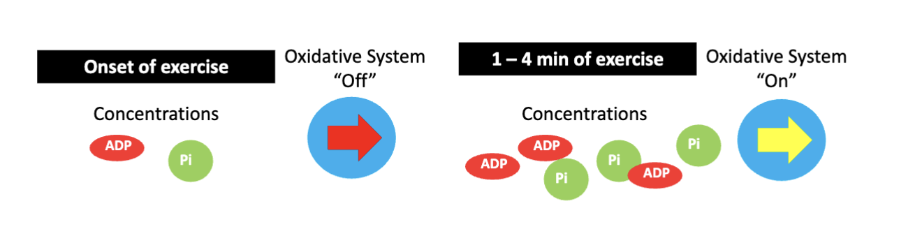
what causes the lag that produces an oxygen deficit at the start of exercise?
Oxidative phosphorylation does not immediately reach full activity.
ADP + Pi stimulate oxidative phosphorylation in the ETC
ADP and Pi take time to accumulate to sufficient levels
This delays full activation of aerobic ATP production
Key idea: The oxidative system needs time to “turn on,” so anaerobic systems initially help meet ATP demand.
How does aerobic training affect oxygen deficit?
Trained individuals:
Reach steady-state VO₂ faster
Have a smaller O₂ deficit
Activate aerobic ATP production earlier
Rely less on anaerobic pathways
Produce less lactate and H⁺ at exercise onset
Key idea: Training → faster aerobic response → smaller O₂ deficit + less anaerobic reliance.

What is EPOC during recovery from exercise?
EPOC = excess post-exercise oxygen consumption
After exercise stops:
VO₂ does NOT immediately return to resting levels
O₂ consumption remains elevated during recovery
Two components:
Rapid component = first ~2–3 min
Slow component = >3 min
Key idea: EPOC = elevated O₂ consumption after exercise as the body recovers toward resting conditions.

Why does oxygen consumption remain elevated during EPOC?
Rapid component (first 2–3 min):
Resynthesize ATP + creatine phosphate
Replenish muscle + blood O₂ stores
Slow component (>3 min):
HR + breathing remain elevated
Body temperature remains elevated
Gluconeogenesis → converts lactate back to glucose
Key idea: EPOC provides the extra O₂ needed to restore the body to its pre-exercise state.

How does exercise intensity affect EPOC?
Higher exercise intensity → greater and longer EPOC
Why?
Higher body temperature
Greater phosphocreatine (PCr) depletion
Greater blood lactate accumulation
Key idea: High-intensity exercise creates greater recovery demands → more post-exercise O₂ consumption.
What is metabolic specificity of training (SAID principle)?
Training should match the specific metabolic demands of the activity.
Exercise intensity + rest intervals determine which energy systems are emphasized
Training a specific energy system → specific adaptations
Sprinters → train sprinting/high-intensity energy systems
Distance runners → train prolonged aerobic energy systems
Key idea: The body adapts specifically to how it is trained.
What is interval training and what is its purpose?
Interval training = alternating predetermined periods of exercise and rest.
Promotes bioenergetic adaptations → more efficient energy transfer
Allows more training to be performed at higher intensities
Work-to-rest ratios can be manipulated to target specific energy systems
Key idea: Alternating work + recovery allows repeated bouts of higher-intensity exercise.
Adaptations to anaerobic training programs I…
What is anaerobic training, and which energy systems does it stress?
Anaerobic training = high-intensity exercise relying primarily on anaerobic energy systems, typically above the lactate threshold.
Requires ATP to be regenerated faster than oxidative metabolism can provide it
ATP-PCr system:
Activity <10 seconds
Requires near-complete recovery between sets: ~5–7 min
Examples: sprints, plyometrics
Glycolytic system:
Longer-duration, high-intensity interval training
Work intervals typically <1 minute
Key:
<10 sec → ATP-PCr
Longer high-intensity efforts (<1 min) → Glycolysis
How does resistance training improve muscular strength, and how does training status affect strength gains?
Average strength gains:
Untrained → ~40%
Moderately trained → ~20%
Trained → ~16%
Advanced → ~10%
Elite → ~2%
Key: The more trained you already are → the smaller the expected strength gains.
Adaptations that improve strength:
↑ Recruitment of higher-order motor units
Type IIx → Type IIa fiber shift → ↑ fatigue resistance at similar force output
↑ Muscle myofibrillar content → contributes to greater force production
Work = Force × Distance
Key: Strength gains occur through both neural and muscular adaptations.
What is power training?
Light-to-moderate loads moved at high velocities
↑ Force at high movement speeds
↑ Rate of force development (RFD)
Peak power:
Lower body: 30–60% 1RM
Upper body: 46–62% 1RM
Power = Work / Time
What are the local muscular endurance and body composition adaptations to strength/anaerobic training?
Local muscular endurance:
↑ Oxidative & buffering capacity
↑ Capillaries & mitochondria
↑ Metabolic enzyme activity
Body composition:
↑ Fat-free/lean mass
↓ Body fat
↑ Basal metabolic rate
↑ Energy expenditure during exercise
How does anaerobic/strength training affect flexibility and mobility?
Can ↑ flexibility, especially if baseline flexibility is poor
Strength training + stretching = most effective for improving flexibility while ↑ muscle mass
Training through full ROM → enhances/maintains mobility
flexibility = muscle stretching
mobility is the joint movment
Key: Full ROM strength training + stretching = best for flexibility & mobility
What major neuromuscular adaptations occur with resistance training?
Resistance training causes adaptations in:
Higher brain centers → motor cortex
Descending corticospinal tract
Muscle fibers
Key: Resistance training causes adaptations from the brain → spinal pathways → muscle.
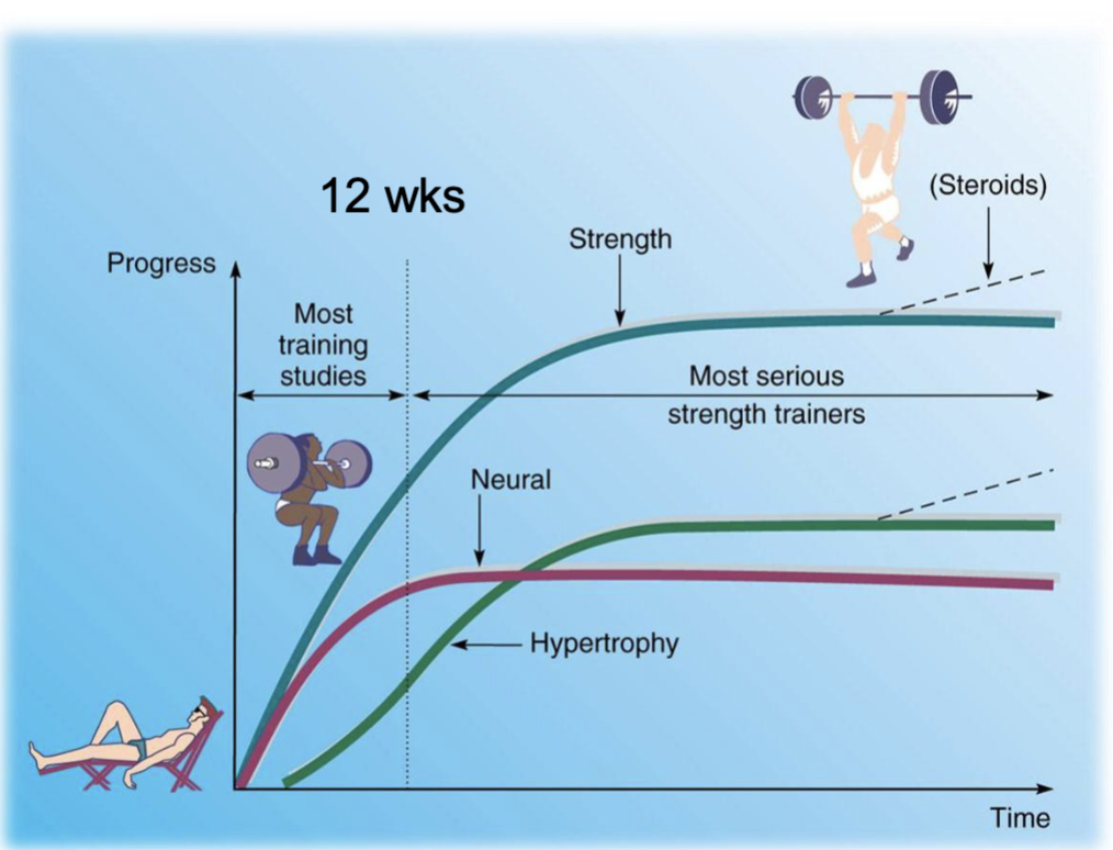
Why does strength increase rapidly when beginning resistance training?
early strength gains = Neural adaptations
Occur BEFORE increases in muscle size
↑ Descending neural drive
↑ Neuromuscular junction area
↑ Total neuromuscular activation
After ~3–4 weeks = Morphological adaptations
Muscle size begins to increase
Hypertrophy = muscle enlargement
MOST CHANGE at 10 weeks
Key: Early strength gains = neural → Later gains = hypertrophy
What central neural adaptations occur when force development increases or new movements are learned?
↑ Motor cortex activity
↑ Corticospinal excitability
↓ Corticospinal inhibition
Key: Resistance training → brain sends stronger, less inhibited signals to the muscles.
What motor unit adaptations increase strength and power with resistance training?
↑ Motor unit recruitment → lower recruitment threshold
↑ Firing rate (rate coding) → motor units fire faster
↑ Synchronization → motor units fire together
Key: Strength & power improve through better motor unit recruitment, firing rate, and synchronization.
How does heavy resistance training affect muscle fibers?
All muscle fiber types can become larger (hypertrophy)
Fibers are recruited in order from smaller → larger motor units
Heavy loads recruit more/all fiber types to produce high force
What changes occur at the neuromuscular junction (NMJ) with resistance training?
↑ Total NMJ area
↑ Nerve terminal branching
↑ End-plate perimeter and area
ACh receptors become more dispersed
Synapses become more dispersed and irregularly shaped
How does training affect neuromuscular reflexes?
Training enhances the reflex response → ↑ reflex magnitude and ↑ rate of force development (RFD).

What are cross-education and the bilateral deficit?
Cross-education:
Training one side → ↑ strength on the opposite (contralateral) side due to neural adaptations.
Bilateral deficit:
Untrained individuals produce less force with both limbs together than expected from each limb individually → reduced neural drive.
Training → ↑ agonist activation during bilateral movements = bilateral facilitation.
How does strength training affect antagonist muscle activation?
Strength training → ↓ antagonist coactivation → ↑ agonist force production.
Untrained individuals = higher antagonist coactivation.
Example: ↓ hamstring activation → ↑ leg extensor force, even without hypertrophy.
what the last slide means -
It means that when you first start strength training, your body learns to coordinate your muscles better.
For example, during knee extension:
Quadriceps = agonist → contracts to straighten the knee
Hamstrings = antagonist → oppose knee extension
In an untrained person, the hamstrings may contract more than necessary while the quads are working. This fights against the quads and reduces the force produced.
After training:
↓ Hamstring activation → less opposition → ↑ quadriceps force
So you can become stronger even before your muscles get bigger, simply because your nervous system gets better at activating the right muscles and relaxing the opposing muscles.
What is EMG and what does it tell us about muscle activation?
EMG (electromyography) measures neural drive to skeletal muscle.
↑ EMG signal = ↑ muscle activation due to:
↑ Number of active motor units
↑ Motor unit firing rate
Training → ↑ neural activation, especially early in beginners.
What is the time course of strength adaptations to resistance training?
Early strength gains → primarily neural adaptations/learning
After ~3–5 weeks → muscle hypertrophy becomes more evident
Key idea: Beginners can gain strength quickly before significant muscle growth occurs.
What happens to the window of adaptation as training progresses?
As training status ↑ → window of adaptation ↓
Beginners → large gains from the same stimulus
Advanced → smaller gains from the same stimulus
Performance approaches a genetic ceiling
Training must evolve/progress to continue adaptations.

Adaptations to Anaerobic Training Part 2…
What is muscle hypertrophy and how does it occur?
Increase in the cross-sectional area (CSA) of existing muscle fibers due to:
Increased actin and myosin protein synthesis
Increased number of myofibrils
Addition of new myofilaments → increased myofibril diameter
Occurs only when protein synthesis > protein degradation
What is muscle hyperplasia?
Increase in the number of muscle fibers via longitudinal fiber splitting. Scientific evidence suggests it may not occur in humans.
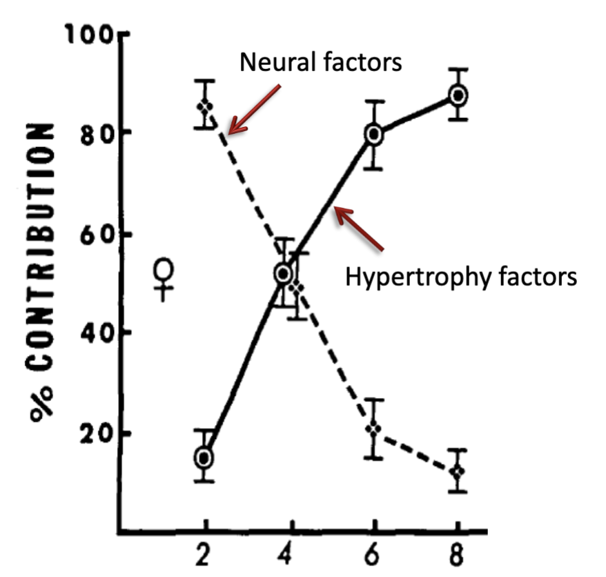
How do neural and hypertrophic factors contribute to strength gains over time?
Early training → neural factors dominate strength gains.
Later training → neural contribution decreases while muscle hypertrophy contributes increasingly.
How do Type I and Type II muscle fibers respond to heavy resistance training?
Both Type I and Type II fibers increase in size, but Type II fibers have greater increases in size than Type I fibers.
Why do individuals with more Type II muscle fibers have a greater potential for hypertrophy?
Type II fibers have a greater capacity to increase in size with resistance training.
What muscle fiber type transition occurs with resistance training?
Type IIx fibers shift toward Type IIa fibers: IIx → IIax → IIa. This involves changes in myosin ATPase and myosin heavy chains.

Can training cause conversion between Type I and Type II muscle fibers?
No. Type I ↔ Type II conversion does not occur. High-volume training may reduce truly fast-twitch Type IIx fibers, which can be undesirable for power athletes.
What structural adaptations occur in skeletal muscle with training?
Increased myofibrillar volume, cytoplasmic density, sarcoplasmic reticulum, T-tubule density, and Na⁺/K⁺ ATPase activity. Sprint training can also enhance Ca²⁺ release.
