[ODOD1] Introduction & Ocular Adnexa
 Call Kai
 Learn
Learn Practice Test
 Spaced Repetition
 Match
Match Flashcards
 Knowt Play
 Podcast
1/67

There's no tags or description
Looks like no tags are added yet.
Name | Mastery | Learn | Test | Matching | Spaced | Call with Kai | Chat |
|---|
No analytics yet
Send a link to your students to track their progress
68 Terms
Pathology
medical science that studies the causes, mechanisms, development, and effects of diseases. It acts as a bridge between basic science and clinical medicine.
analyze cells, tissues, organs, and bodily fluids to diagnose illnesses
Disease
an abnormal condition that negatively affects the structure or function of all or part of a living organism, and is not caused by an immediate external injury.
Sickness
a person’s subjective experience of being unwel, representing how they feel and function in daily life.
while "disease" is the objective medical condition, (BLANK) is the personal and social experience of that condition.
Ocular disease
refers broadly to any medical condition or disorder that affects the health, structure, or function of the eye and its surrounding visual system.
Ocular Adnexa
In eye care, the (BLANK) refers to the accessory structures that protect, hydrate, and move the eye.
Eyelids
Specialized skin and sweat/sebaceous glands that shield the globe
Lacrimal System
The tear glands, tear ducts, and drainage systems that keep the eye lubricated
Orbit
The bony eye socket that houses the eyebal
Normal Ocular Adnexa
means that all of the supporting structures surrounding your eyeball are healthy, properly positioned, and functioning correctly.
In a clinical charting note, an eye care professional wil typicaly document this finding as "(BLANK): Normal," "(BLANK): Clear," or "WNL" (Within Normal Limits)
eyelashes
specialized, highly sensitive hairs that grow in multiple rows along the upper and lower margins of the eyelids.
their primary biological function is to protect the eye by deflecting airborne debris, sweat, and dust, and triggering a reflexive blink when touched.
Upper Eyelid
Approximately 90 to 160 lashes
organized in 5 to 6 uneven rows.
They curve upward and reach lengths between 8 and 12 mm
Lower Eyelid
Approximately 75 to 80 lashes
arranged in 3 to 4 rows.
They curve downward and are shorter, measuring 6 to 8 mm
Texture & Shape (Eyelashes)
exhibit a smooth texture, tapered tips, and normal curvature.
Natural density and curl direction vary based on genetics and ethnic background.
Glands of Zeis
Glands of Moll
The base of each eyelash follicle is supported by (2)
Glands of Zeis
sebaceous glands producing lubricating sebum
Glands of Moll
modified sweat glands
Eyebrows
symmetrical patches of hair situated on the bony ridge above the eye sockets (the supraorbital ridge).
They serve a vital biological purpose by channeling sweat, rain, and debris away from the eyes toward the side of the face.
They also play a crucial role in human communication, non-verbal expression, and facial recognition.
250 to 600
average person has roughly (BLANK) hairs per eyebrow.
1,100
Brows that have never been plucked or altered can feature up to (BLANK) total hairs.
adult men
In (BLANK), brows are generally lower, thicker, and flatter.
adult women
In (BLANK), they sit higher above the eye ridge and display a more defined arch.
0.14 mm - 0.15 mm
Eyebrow hairs grow at a slow rate of about (BLANK) per day.
normal lacrimal puncta
small, pale openings located at the inner corners of the upper and lower eyelids that serve as the entry points for the eye's tear drainage system.
one upper and lower
Each eye normally features two puncta (BLANK), which actively pump and siphon tears away from the ocular surface into the nose every time you blink.
0.2 mm and 0.5 mm
A normal punctal opening typicaly measures between (BLANK) in diameter.
lacrimal papillae
Lacrimal puncta sit on top of slightly elevated, tiny mounds of tissue called (BLANK)
Because this specific tissue has fewer blood vessels compared to the surrounding eyelid, the normal punctum naturally looks pale or flesh-colored
Lacrimal Puncta
The physical orifice varies from round, oval, linear, to a "fish-mouth" shape. This structure naturaly shifts in geometry as a person ages.
medial canthus / inner canthus
the anatomical junction where the upper and lower eyelids meet on the side closest to the nose.
It serves as a vital structural anchor for the eyelids and houses the primary mechanical components of the ocular tear drainage system
Lacus Lacrimalis (Tear Lake)
The small, triangular reservoir where tears collect before draining.
Lacrimal Caruncle
A small, pinkish, globular nodule of modified skin sitting inside the tear lake. It contains sebaceous and sweat glands and may sprout fine hairs.
Plica Semilunaris
A crescent-shaped fold of conjunctiva lateral to the caruncle. It is a vestigial remnant of the nictitating membrane (third eyelid).
Lacrimal Puncta
Tiny, oval openings on a slight elevation (papilla) near the medial margin of both the upper and lower eyelids. These serve as the entrance points for tear drainage.
lacrimal sac
dilated, upper reservoir of the nasolacrimal drainage system that collects tears from the surface of the eye before pumping them into the nasal cavity.
in its normal, healthy state, it sits hidden behind the skin and musculature of the inner corner of the eye, remaining completely flat, unpainful, and un palpable
Lacrimal Sac
Length: Approximately 12 to 15 mm tall.
Width: Roughly 4 to 8 mm wide when moderately distended.
Normal Volume: Holds roughly 20 microliters (μL) of fluid at rest, though it is highly distensible.
nasolacrimal duct (or tear duct)
is the terminal, conduit portion of the lacrimal apparatus that drains tears away from the lacrimal sac and into the nasal cavity.
in its normal, healthy state, it provides an unobstructed, one-way passage for fluid, preventing tears from overflowing onto the cheeks.
NasoLacrimal Sac
Length: Typically measures 12 to 18 mm long total.
Diameter: The internal lumen spans roughly 2 to 4 mm wide under normal conditions.
Direction: It travels from the eye socket downward, slightly backward, and outward (inferiorly, posteriorly, and laterally) along the side wall of the nasal cavity.
eyelid
serves as a vital barrier that protects the eye from debris, regulates light entry, and maintains the tear film.
Eyelid
Positioning: In a primary forward gaze, the upper (BLANK) typically covers about 1 to 2 milimeters of the top of the cornea. The lower (BLANK) rests precisely at or just above the lower border of the cornea (the inferior limbus).
Eyelid skin
Skin Thickness: (BLANK) is highly elastic and stands as the thinnest skin on the entire human body, measuring less than 1 milimeter in thickness. It contains no subcutaneous fat.
Eyelid
Natural Creases: Depending on genetic background, a normal (BLANK) may feature a distinct supratarsal crease dividing it into two sections (double eyelid), a smooth transition without a crease (monolid), or a partial fold of skin (hooded eyelid). All of these are completely normal variations.
Eyelid
Lash Alignment: Eyelashes point outward and away from the eyebal, working to sweep away dust and debris. The upper (BLANK) generally contains 100 to 150 lashes, while the lower lid contains 50 to 75.
lacrimal gland
an almond-sized exocrine gland responsible for producing the aqueous (watery) portion of the tear film, which keeps the eye moist, clean, and nourished.
Each person has two (BLANK), with one positioned behind the upper outer corner of each eye socket.
Lacrimal Gland
The gland is roughly 2 cm long and is bisected by the tendon of the levator palpebrae superioris muscle.
Orbital Lobe
Palpebral Lobe
2 continuous segments (Lacrimal Gland)
Orbital Lobe
The larger portion that sits deeper within the bony orbit. (lacrimal gland)
Palpebral Lobe
The smaller, flatter portion located closer to the inner surface of the upper eyelid. (lacrimal gland)
normal lateral canthus
sharp, V-shaped outer corner of the eye where the upper and lower eyelids meet toward the temple.
In a healthy, structurally sound eye, it holds a precise position and angle that preserves eye function and creates a natural, almond-shaped appearance.
Lateral Canthus
Sharp Angle: The upper and lower lids meet cleanly to form a crisp, triangular intersection.
Positive Tilt: A straight, horizontal line drawn from the inner corner of the eye will naturally sit slightly below the outer corner.
Globe Apposition: The corner rests snugly against the surface of the eyeball without gaps.
Lid Position: The lower eyelid rests right at or slightly above the lower edge of the iris (the inferior limbus). No white sclera is visible beneath the iris.
Structural Support: It is anchored firmly to the bone (Whitnall's tubercle) by the lateral canthal tendon, keeping the lower eyelid taut and preventing drooping.
meibomian glands
specialized oil glands located vertically within the tarsal plates of the upper and lower eyelids.
Their primary function is to secrete meibum, a clear, fluid lipid layer that coats the ocular surface.
This crucial oil layer stabilizes the tear film, prevents watery tears from evaporating too quickly, and ensures the eyes remain lubricated and comfortable
Meibomian Glands
Quantity: There are approximately 25 to 40 distinct glands in the upper eyelid and 20 to 30 glands in the lower eyelid. The upper lid glands are naturally longer and thinner than those in the lower lid.
Structure: When viewed via specialized infrared imaging (meibography), normal glands appear as long, straight, parallel columns neatly arranged in a row. They exhibit zero "dropout" (missing sections) or significant shortening.
Secretions: Healthy meibum has a clear, thin, fluid consistency similar to olive oil or baby oil. It flows easily from the gland orifices with normal blinking.
Orifice Appearance: The tiny openings of the glands sit evenly along the waterline of the lid margin, just behind the eyelashes. In a normal eye, these openings are completely clear, open, and free of capping or crusting
Outer Lipid Layer
Made by the meibomian glands in the eyelids.
Forms the top surface of the tear film.
Keeps tears from drying up too fast.
Stops tears from spilling over the edge of the eyelid.
Middle Aqueous Layer
Made by the lacrimal glands.
Makes up most of the volume of the tears.
Supplies water, oxygen, and food nutrients to the cornea.
Washes away dust and foreign material.
Inner Mucin Layer
Made by goblet cells in the conjunctiva.
Rests directly on the surface of the eye.
Acts like glue so the watery layer spreads out evenly.
Helps the tear film stick to the eyeball.
Basal Tears
Always present: Tears that sit on your eyes all day long.
Main job: Keep your eyes wet, clean, and healthy.
Protection: They stop dirt and dust from hurting your eye.
Reflex Tears
Quick reaction: Tears that start when something bothers your eye.
Triggers: Smoke, dust, bright light, or cutting an onion.
Extra help: They carry extra antibodies to fight off bad germs
Emotional Tears
Feeling-driven: They happen because of deep feelings.
Triggers: Great sadness, extreme happiness, stress, or physical pain.
Chemical makeup: They contain extra stress hormones and natural pain-relief proteins that help your body calm down.
Tear film
a thin, vital liquid layer covering the outer surface of the cornea and conjunctiva
Meibomian glands
Specialized sebaceous (oil-producing) glands located within the tarsal plates of the eyelids.
They secrete an oily substance called meibum, which forms the lipid layer of the tear film and helps prevent tears from evaporating too quickly.
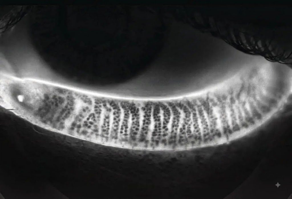
Meibum
A thick, oily fluid produced by the meibomian glands inside the eyelids
Associated structure: Tarsal plates/eyelid margin (Eye-tear oil)
Tarsal plate (tarsus)
provide structural support, shape, and rigidity to the upper and lower eyelids while housing oil-producing glands
Glands of Krause
Glands of Wolfring
ACCESSORY LACRIMAL GLANDS
Accessory lacrimal glands
They are located within the conjunctival tissue of the eyelids.
They produce additional watery tear secretion, so they help maintain the moisture of the eye
Glands of Krause
Small accessory lacrimal glands located in the conjunctiva of the eyelids that produce watery tear fluid.
Location: Mainly conjunctival fornices
Glands of Wolfring
Accessory lacrimal glands located mainly along the upper and lower borders of the tarsal plates of the eyelids that produce watery tear secretion.
Location: Along the tarsal plates
Sebum
A complex, waxy, oily substance made by your body's sebaceous glands.
Where: Skin and hair follicles, including eyelashes (Skin + hair oil)
Glands of Zeis
Small sebaceous (oil-producing) glands associated with the eyelash follicles. They secrete sebum, an oily substance that helps lubricate and protect the eyelashes and the surrounding eyelid margin.
Glands of Moll
Modified apocrine sweat glands located at the eyelid margin, usually associated with the eyelash follicles.
They produce a small amount of sweat-like secretion that contributes to the lubrication and protection of the eyelid margin.
Valve of Hasner
Opening of nasolacrimal duct in the inferior meatus