PTA 230 Nero Chapters 3-4
 Call Kai
 Learn
Learn Practice Test
 Spaced Repetition
 Match
Match Flashcards
 Knowt Play
1/73

There's no tags or description
Looks like no tags are added yet.
Name | Mastery | Learn | Test | Matching | Spaced | Call with Kai | Chat |
|---|
No analytics yet
Send a link to your students to track their progress
74 Terms
an area of damage of dysfunction
lesion
understanding the effects of lesions enables the therapist to
1. select appropriate therapy
2. predict therapeutic outcomes
3. recognize signs and symptoms that indicate the need to refer the patient to another health professional.
true or false.
where the lesion is located is more important than what caused it.
evidence a disease or an impairment that can be observed by someone other than the patient
sign
subjective experience of a patient
symptoms
limited to a single location
focal
limited to several nonsymmetric locations
multifocal
affects bilaterally symmetric structures but does not cross the midline as a single lesion
diffuse
the proportion of population that develops a new case of the disorder within in a defined time period. Exp. ( cavities in last 5 years)
incidence
current proportion of the population with the condition, including both old and new cases. Exp. (total number in room, ever had a cavity)
prevalence
is reported per 1000 people or as a % of people.
prevalence
tumor in spinal cord is an example of
focal lesion
tumor that has metastasized to several locations
multifocal
Alzheimer disease, a memory and cognitive disorder is
diffuse
Events that may affect the nervous system
. Trauma
. Vascular disorders
. Inflammation
. Degenerative disorders
. Developmental disorders
. Tumors
. Immunological disorders
. Toxic or metabolic disorders
Acute onset usually indicates a
vascular problem, trauma
subacute onset indicates an
inflammatory process, infection
chronic onset often suggests a
tumor or degenerative disease
indicating minutes or hours to maximal signs and symptoms
acute
progressing to maximal signs and symptoms over a few days
subacute
"Gradual worsening of signs and symptoms continuing for weeks or years"
chronic
questions used to obtain neurologic history
. What problems are you having
. when did the problem begin
. Did the problem start abruptly, over a few hours or days or gradually.
. How severe is the problem.
. Are symptoms constant or intermittent
. Have you had previous episodes
. Do you have any other symptoms
. Do you take medications or supplements
stroke, traumatic spinal cord injury is diagnostic example of
acute
improving or stable is a pattern of progression of
acute
multiple sclerosis, bacterial meningitis are diagnostic examples of
subacute
fluctuating and gradual improvement are pattern progressions of
subacute
meningioma, parkinson disease are diagnostic examples of
chronic
progressive and worsening are pattern progressions of
chronic
. x-rays pass through the body to the detector, to measure relative densities of tissue.
. used to see acute hemorrhage, abnormalities or bone fractures, calcified lesion, sinus disease
. scan time 5 minutes
. radiation exposure
Computed Tomography (CT)
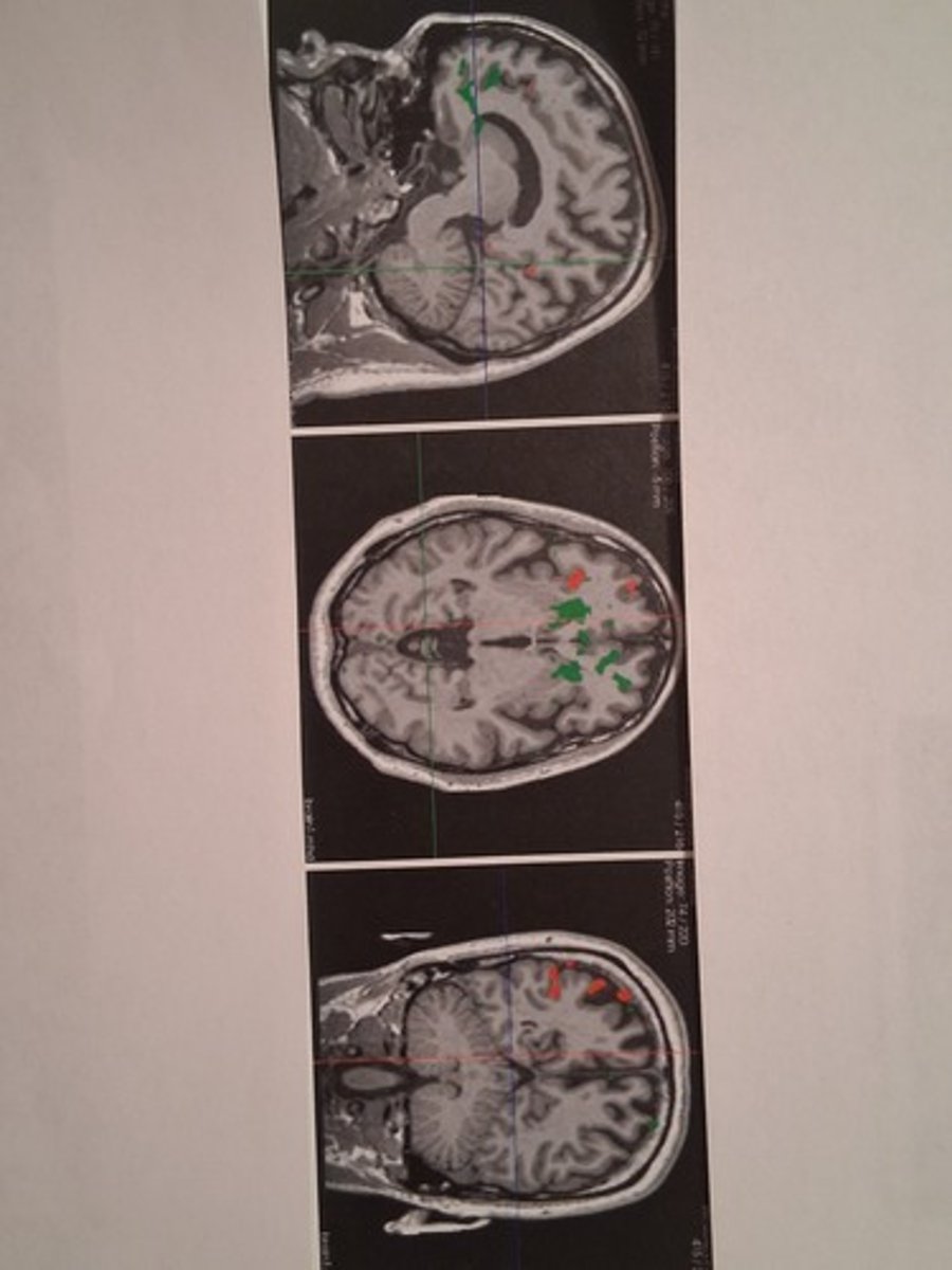
. measures changes in oxygenated blood flow
. used to detect neural activity in the brain by evaluating changes in blood flow
. scan time 60 minutes
. no radiation
Functional Magnetic Resonance Imaging (fMRI)
. magnetic fields and radio waves detect hydrogen ions
. used to detect: stroke, tumors, infection, multiple sclerosis
. scan time 30-120 minutes
. no radiation
Magnetic Resonance Imaging (MRI)
. magnetic fields and radio waves detect water diffusion along axons
. used for: detailed images of white matter tracts, surgical planning
. scan time 30 minutes
. no radiation
Diffusion Tensor Imaging (DTI)
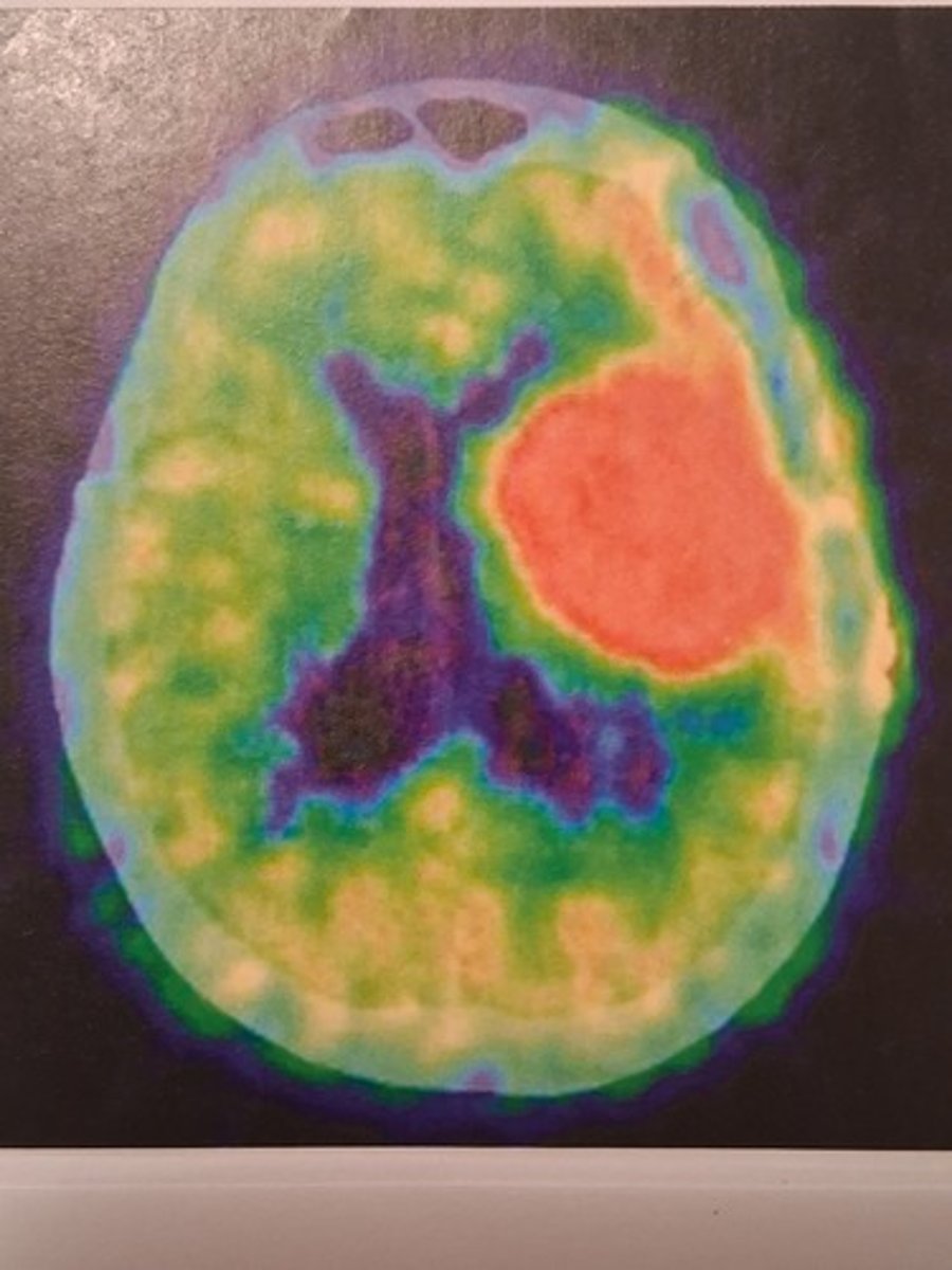
. detects radioactive isotopes as they travel through the blood
. use to: measure blood flow, glucose metabolism, and oxygen consumption
. scan time 30 minutes
. radiation exposure
Positron Emission Tomography (PET)
axial images are oriented
as if you are standing at the feet and looking toward the head of an inclined person
coronal images are oriented
as if you are standing in front of the person looking at him or her
what are the results in viewing axial and coronal images
the right side of the person body is on the left image, and the left side of the body is on the right side of the image
sagittal image is a
vertical slice that divides the body into right and left sides
horizontal slice that divides the body into caudal and rostral positions
axial cut
vertical slice that divides the body into posterior and anterior portions
coronal cut
tissues with higher water content appear____ on CT images
dark gray
metal and bone appear _____ on CT images, because they have increased density
white
substances with a high protein concentration will be_____ on CT images
light gray
air and cerebrospinal fluid appear ____ on CT images, because they are less dense structures
black
structures with similar intensity to the brain are called
isodense
CT should be avoided in
children and pregnant women
brain tissue has what color
light gray
term used to describe structures that are light gray
hyperdense
term used to describe structure that are dark gray
hypodense
the densest structure in the body
bone
indications for for contrast-enhanced CT
to detect increased angiogenesis ( sign of tumor), inflammation, infection
the intensity of MR images depends on
the presence of hydrogen protons
MRI is contraindicated for patients with
. metal fragments in the body
. pacemaker
. cochlear implants
since air and calcified bone lack water, they appear _________to brain tissue on MRIs
hypointense
fMRI is safer and noninvasive than _____ , as there is no need for injection of radioactive isotopes
PET
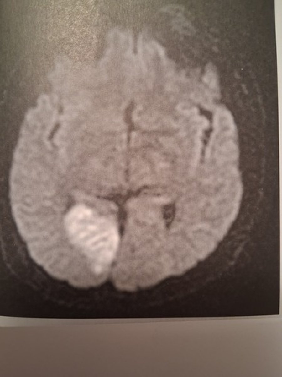
. scans useful when evaluating for ischemia
. helpful in differentiating various lesions
. highlights areas of reduced water movement
diffusion-weighted imaging (DWI)
. creates an image of the white matter tracts
. used to measure how water diffuses around bundles of axons
Diffusion tensor imaging (DTI)
. type of T2 image in which CSF and vitreous fluid signal is suppressed.
. useful when looking for plaques associated with multiple sclerosis, near ventricles
. useful look for areas of edema of neural tissue
(FLAIR) fluid-attenuated inversion recovery magnetic resonance imaging
what is helpful in screening for carotid stenosis, aneurysm, and abnormal connections between arteries and veins?
CT angiography or MRA
what is the difference between CTA and MRA?
CTA injects a contrast agent and MRA can be done with or without a contrast agent
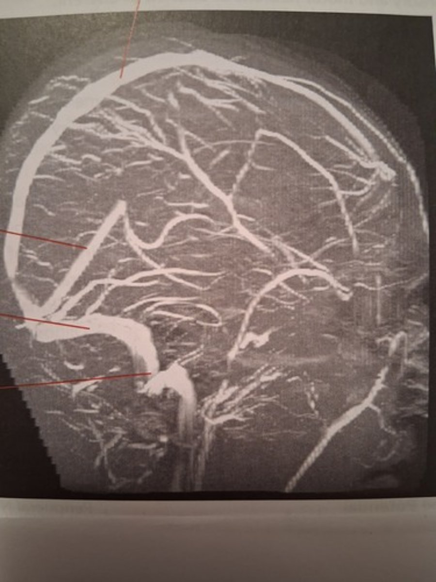
CT venography
MRV
MRI
T1, T2
which picture shows a Diffusion weighted imaging
which image shows a fMRI
which image shows a PET
which image shows an axial MRI
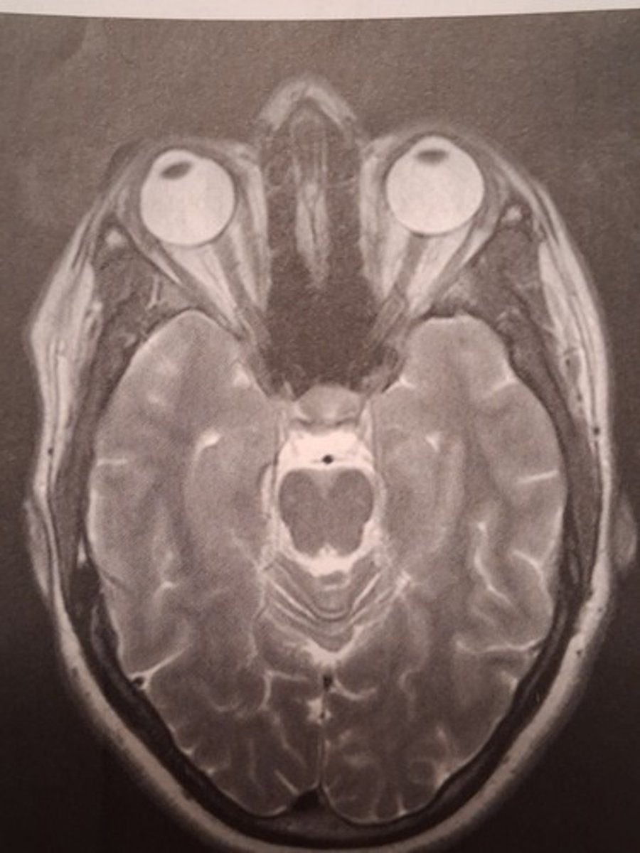
which image shows a Diffusion tensor imaging
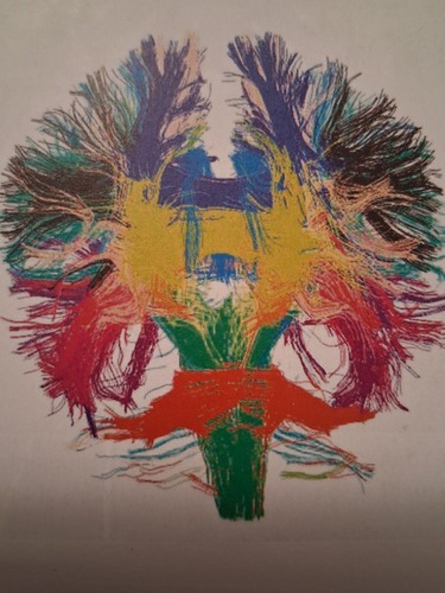
which image shows a Sagittal MRI
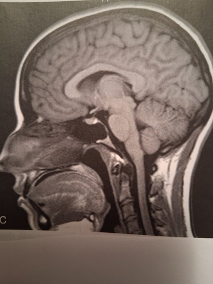
which image shows a CTA computed tomography angiography
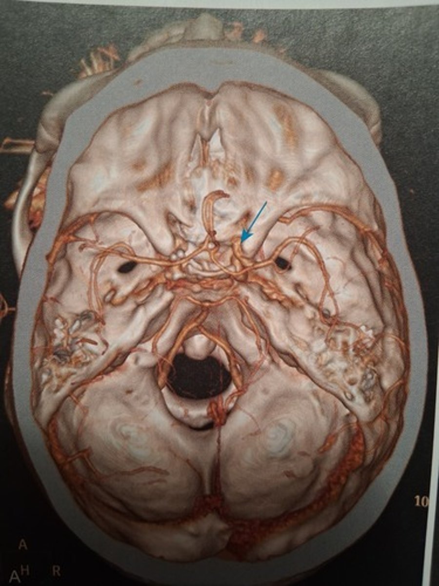
which image shows a Coronal MRI
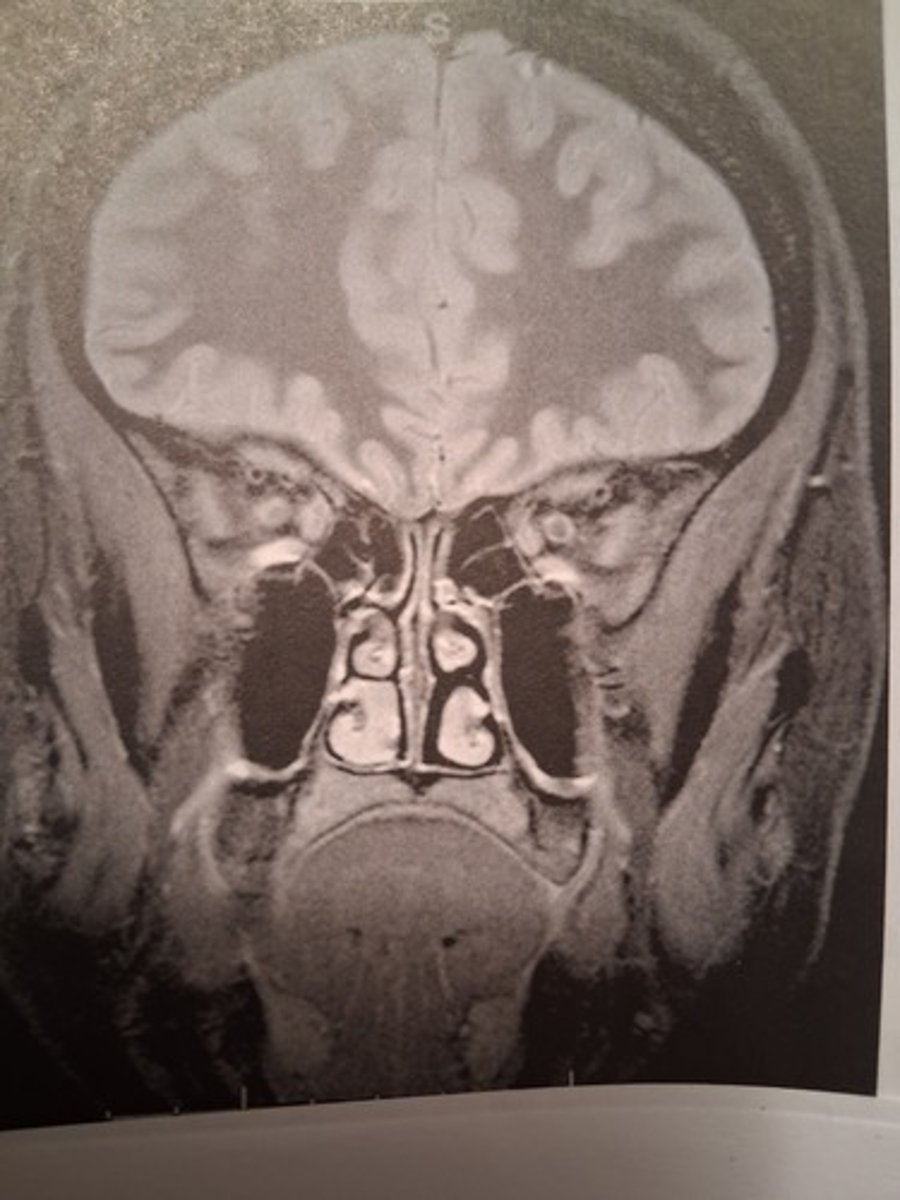
which image shows an Axial CT
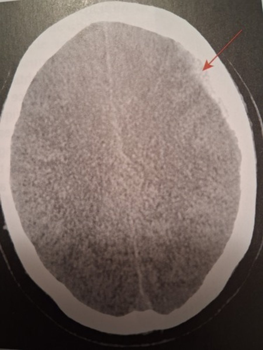
which image shows a MRA
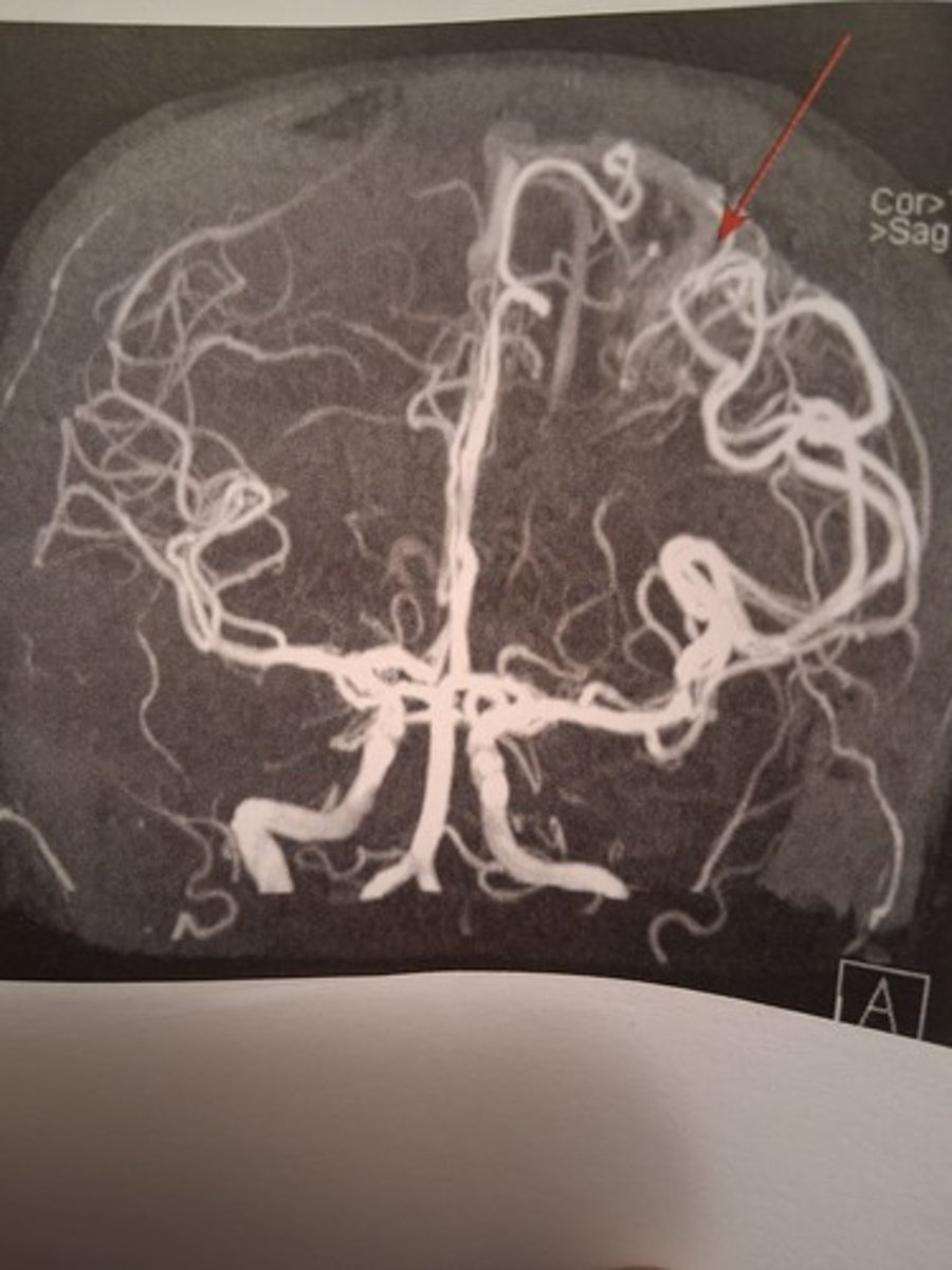
which images shows a MRV