Respiratory and Lymphatic system
 Call Kai
 Learn
Learn Practice Test
 Spaced Repetition
 Match
Match Flashcards
 Knowt Play
1/156

Earn XP
Description and Tags
For Bio 211/ Anatomy and Physiology 2, includes the anatomy of some parts as well as general information.
Name | Mastery | Learn | Test | Matching | Spaced | Call with Kai | Chat |
|---|
No analytics yet
Send a link to your students to track their progress
157 Terms
2 semi-independent parts of lymphatic system
A network of lymphatic vessels, and Lymphoid tissues and organs scattered throughout the body
function of lymphatic system
returns interstitial fluid and leaked plasma proteins back to the blood after cleansing them
lymph
the interstitial fluid once it has entered lymphatic vessels
how many major segments are in the lymphatic system
3 major parts
Lymph vessels, like arteries, contain valves.
False
The right lymphatic duct receives lymph from the:
upper right side of the body
Which of these organs atrophies as you age?
thymus
What is the purpose of the tonsils?
To offer initial immunity from pathogens entering the mouth
The thoracic duct receives lymph from ___________.
the upper left body and the lower body
Isolated areas of lymphoid follicles found in the intestinal mucosa are called _________________
peyer’s patches
Large clusters of lymph nodes are found near the body surface in all of the following areas EXCEPT the:
popliteal region
Once fluid enters the lymphatic system it is called ___________
Lymph
The thymus gland is covered by a thick...
connective tissue capsule
Lymphatic capillaries allow interstitial fluid into them because there are overlapping endothelial cells that open when pressure is higher outside those capillaries than inside.
True
Which is not something typically transported by the lymphatic system on purpose?
Carbohydrates
At what point is the fluid of the lymphatic system actually called lymph?
Once it has entered a lymphatic vessel
Of the 20L of fluid that is released from the blood into the interstitia fluid, 5.25L is immediately returned to the blood.
False
BALT stands for:
Bronchus associated Lymphatic Tissue
Which lymphatic duct is the largest, taking blood from the entire left side of the body and the lower left side?
Thoracic duct
The lining of the small intestine is called mucosa.
True
Check all the functions of the spleen that apply.
Resivoir of excess blood, Destruction of red blood cells
A specialized lymphatic capillary found in the digestive system is called a lacteal and they help the absorption of dietary proteins.
False
Which other system is the lymphatic system most closely related to?
Immune
Which tonsils are primarily responsible for inhaled pathogens?
pharyngeal
The diaphysis of long bones contain red bone marrow from birth until death.
False
Which of these is not a secondary structure?
Bone marrow
Which of these is responsible for the destruction of pathogens as they move through lymphatic vessels?
Lymph Nodes
When cancerous cells block lymphatic vessels, fluid builds up. This is called the...
lymphedema
The major function of the lymphatic system is to:
Drain fluids, filter them, and return them to the blood
What makes an organ a primary lymphoid organ?
They are a primary organ because lymphocytes develop/mature there under normal circumstances
______________ mature in the thymus gland.
T-lymphocytes
The two secondary structures of the lymphatic system are the bone marrow and thymus.
False
Which of these has a very weak capsule?
Spleen
Transportation within the lymphatic system
Once interstitial fluid enters lymphatics, it is called lymph and can transport molecules to other parts of the body
Defense within the lymphatic system
Together with lymphoid organs and tissues, provide the structural basis of the immune system
What are the 3 functions of the lymphatic system?
Fluid Balance and Drainage, Defense, and transportation
The Lymphatic system is a one-way system, meaning it will go throughout and re-enter the blood stream.
True
Lymphatic pathway
The system begins in Lymphatic capillaries, These drain into Lymphatic collecting vessels, These drain into collection locations called Lymphatic trunks, These all drain into two main large lymphatic duct. One-way system, lymph flows toward the heart, near where it will re-enter the blood stream at the bifurcation of the subclavian and internal jugular vein

Lymphatic Capillaries
Similar to blood capillaries, except: Very permeable (take up cell debris, pathogens, and cancer
cells, among other materials) Endothelial cells overlap to form one- way mini-valves, and
are anchored by collagen filaments, preventing the collapse of capillaries, Blunt, blind-endings

Lymphatic collecting vessels
Collecting vessels in the skin travel with superficial veins, Deep collecting vessels travel with arteries Nutrients are supplied from branching vasa vasorum, Distribution varies from person to person. Vessel walls resemble those of veins, including the presence of valves. The walls of lymphatics are thinner. The valves are closer together in lymphatics Lymphatics contain lymph nodes
Lymphatic Trunks
Before draining into one of two main ducts, lymphatic collecting vessels lead to a few major trunks
(collecting points) in the body, generally corresponding with the major body regions. These are the: jugular, subclavian, bronchomediastinal, lumbar, and intestinal

Lymphatic Duct
There are two lymphatic ducts into which all capillaries, vessels, and finally trunks, drain. The Right lymphatic duct is the smaller of the two and receives lymph from the right upper quadrant of the body. The Thoracic Duct is the larger of the two and receives lymph from the rest of the body
Drainage Area for Lymphatic Duct
The right lymphatic duct drains the right arm and the right side of the head and thorax. The thoracic duct arises from the cisterna chyli (which has drained the abdominal region) and drains the rest of the body
How does Lymph transport or move?
Lymph is propelled by: Pulsations of nearby arteries, Contractions of smooth muscle in the walls of the lymphatics, Skeletal Muscle contractions that squeeze the walls of the vessels, Breathing movements that change the volume and pressure of the thoracic cavity. It is not propelled by the pumping of the heart, unlike the circulatory system
What are the “main warriors” of the immune system and their varieties?
Lymphocytes in the form of T-cells and B-cells.
T-Cells
Manage the immune response and Attack and destroy foreign cells
B- Cells
Produce plasma cells, which secrete antibodies
Primary Lymphatic organs
Primary Lymphatic organs are the sites where lymphocytes are created, mature, and proliferate These are the bone marrow and thymus gland
Secondary Lymphatic Organs
Secondary Lymphatic organs are those where immune responses arise. When the body has need for
immunity, these organs release naïve lymphocytes (those that haven’t yet “seen” foreign pathogens)
Most secondary organs contain centers for dividing lymphocytes, fixed macrophages that function in
phagocytosis, and a heavy blood supply. These are the lymph nodes, spleen, and lymph nodules (tonsils, MALT, BALT)
What are the 2 primary lymphatic organs?
Thymus and bone marrow
Bone marrow
Primary lymphatic organ, Hematopoiesis for the formed elements, Site of B-lymphocyte maturation while T-lymphocytes are sent to the thymus gland for maturation. Loose collection of cells and reticular fibers (connective tissue)

Thymus
Primary Lymphatic Organ, Matures T-lymphocytes, Size changes with age. In infants, it is found in the inferior neck and extends into the mediastinum, where it partially overlies the heart, Increases in size and is most active in childhood. Stops growing in adolescence and atrophies, Differs from other lymphoid organs in important ways, Secretes a hormone called thymosin, Functions strictly in T-lymphocyte maturation, Does not directly fight antigens, Provides the environment in which T-lymphocytes become immunocompetent
The thymus fights antigens directly.
False

Lymph nodes
Secondary lymphatic organs, Lymphoid organs of the body that have the most overall effect. Embedded in connective tissue and found in clusters along lymphatic vessels, Strong connective tissue capsule covers/contains them. Many are near the body surface in inguinal, axillary, and cervical regions of the body. Functions: Filter lymph—macrophages destroy microorganisms
and debris, Immune system—lymphocytes are activated and mount an attack against antigens, Hematopoiesis if necessary
Where are lymph nodes usually found?
Many are near the body surface in inguinal, axillary, and cervical regions of the body

Spleen
Secondary lymphatic organ, Largest lymphoid organ, Located in the abdominal cavity, lateral to the stomach, Has a very weak, hardly protective capsule, Site of lymphocyte proliferation and immune surveillance and response, Defense by cleansing the blood of aged cells and platelets, and debris. Serves as a reservoir for excess blood formed elements, Under extreme conditions, can engage in
hematopoiesis – does it naturally in the fetus but this (normally ceases after birth) Contains lymphocytes, macrophages, and huge numbers of erythrocytes
Tonsils
Secondary lymphatic organs, Simplest contained lymphoid organs, First line of defense against inhaled and ingested pathogens, Epithelial tissue overlying tonsil masses invaginates, forming tonsillar crypts that trap and destroy matter, Form a ring of lymphatic tissue around the pharynx in
three locations: Pharyngeal tonsil—in posterior wall of the nasopharynx, Palatine tonsils—at posterior end of the oral cavity, Lingual tonsils—grouped at the base of the tongue
lymphoid nodules
Secondary lymphoid organ, Peyer’s patches, MALT, and BALT are patches of lymphoid tissue in particular areas of the body, They are clusters of lymphoid follicles, Peyer’s Patches, also called Mucosa Associated Lymphoid Tissue (MALT) are in the wall of the distal portion of the small intestine Bronchus Associated Lymphoid Tissue (BALT) is found in the bifurcation of the two primary bronchi
Similar structures are also found in the appendix, All of these destroy bacteria and prevent them from
entering the cavities of the body while protecting the digestive and respiratory systems from invasion They generate “memory” lymphocytes
3 major parts of respiratory tract
Upper tract, lower tract and accessory organs
Upper tract of respiratory system
Nose, Pharynx, and Larynx
Lower Tract
Trachea, Primary Bronchi, Secondary Bronchi, Tertiary Bronchi, Bronchioles Lungs
Accessory organs
Diaphragm, Thoracic Cavity, Oral Cavity
Functions of Respiratory System
Provide O2 for cellular respiration, Remove CO2 from cellular respiration, Maintain acid/base balance of the fluids of the body, Olfaction, Influence of speech, Straining when coughing, vomiting, childbirth
The Conducting Zone
Provide a route for incoming and outgoing air, Remove debris and pathogens from inhaled air. Warm and humidify incoming air, Allows sensing of odors via the Olfactory Nerve, Metabolize some airborne carcinogens, Extends from the Anterior nares to the terminal bronchioles
The Conducting Zones Structures
While the picture on the previous slide is general in nature, the structures of the conducting zone are even more specific, In order they are the: Nose (Nasal cavity), Pharynx (naso-, oro-, and laryngo-), Larynx, Trachea, Primary Bronchi, Secondary Bronchi, Tertiary Bronchi, Conducting Bronchioles, Terminal Bronchioles

The Nose and Naval Cavity
Structure – entrance and exit for the respiratory system with an internal and external structure, Openings – anterior nares vs. posterior nares, Dividing septum with conchae and meati, Epithelium –
pseudostratified ciliated columnar epithelium with nerve endings and bloodvessels
Functions of the nose
entry and exit of air, conduction of air, warming and humidifying air (due to conchae, meati, and blood vessels), contributing to speech, protection against inhaled solids (physical barrier), Immune function (the mucus of the epithelium contains lysozymes and defensins which are antibacterial)
Nasal Septum 3 parts
1- septal cartilage, 2- vomer bone, 3- perpendicular plate of ethmoid bone


What sinus is labeled with green?
Frontal Sinus

What sinus is labeled in purple?
Ethmoid Sinus

What sinus is labeled in red?
Sphenoid sinus

What sinus is labeled in blue?
Maxillary Sinus

pharynx
Structure of skeletal muscle lined with epithelium (pseudostratified in the superior third, stratified squamous in the inferior two-thirds), Functions- Passageway for air food, liquids, Aids with speech Three regions- Nasopharynx, Oropharynx, Laryngopharynx
The conducting zone of the respiratory system conducts gas.
False
Which of the 2 systems (circulatory and lymphatic) is considered a “high speed train”?
Circulatory system
Which of the 2 systems (circulatory and lymphatic) is considered a calculated taxi system?
Lymphatic system

What is the blue circle laying on?
The hyoid bone
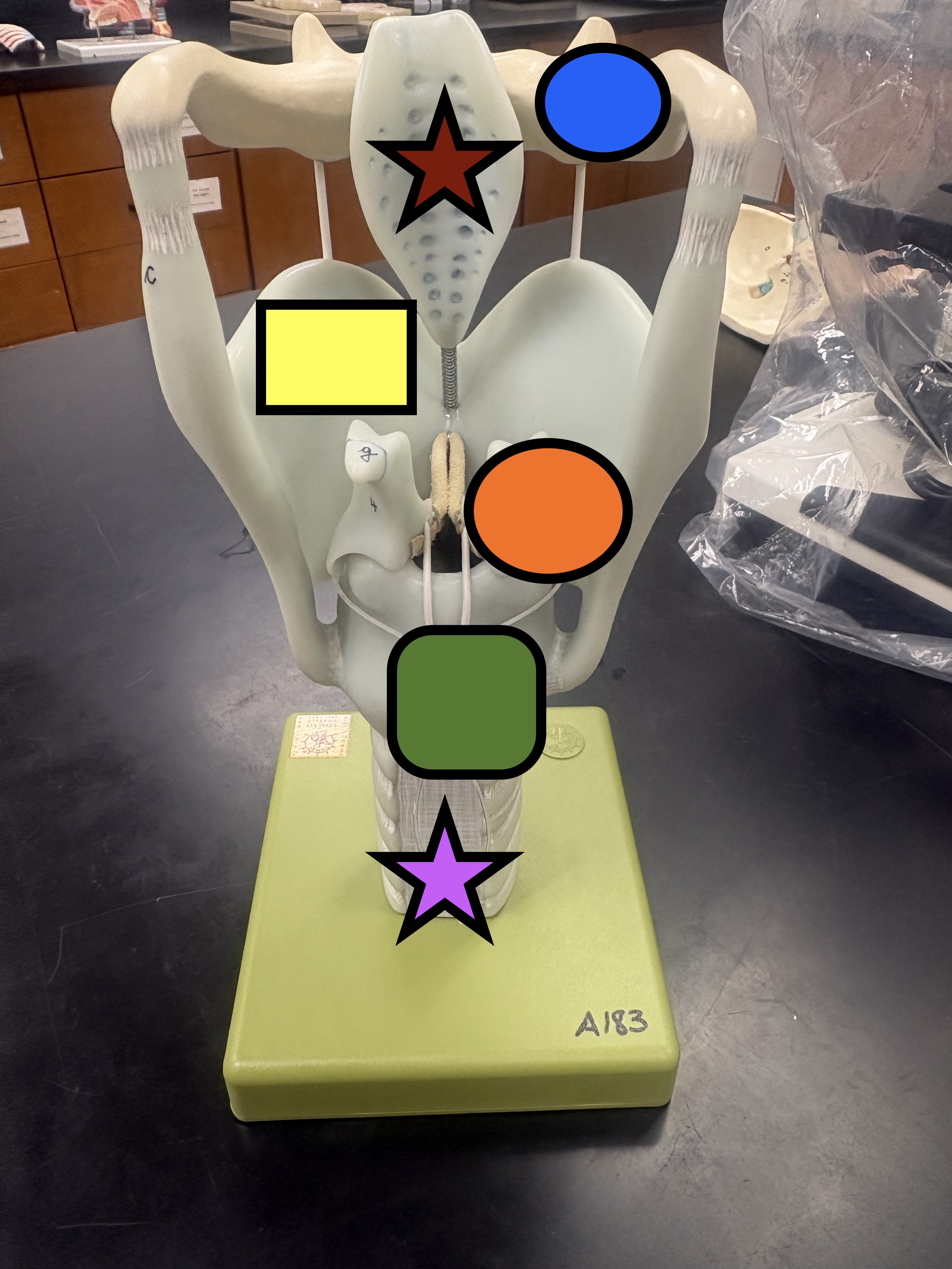
What is the red star representing?
The epiglottis

What is the yellow rectangle representing?
The thyroid cartilage (front and back of model)

What is the orange circle representing?
arytenoid cartilage

What is the green square representing?
Cricoid Cartilage

What is the purple star representing?
Trachea

What is the orange square representing?
Pharynegeal tonsils

What is the green circle?
Lingual tonsils

What sinus is 25 representing?
Frontal Sinus

what sinus is 26 representing?
Maxillary Sinus

What sinus is 27 representing?
Ethmoid Sinus

What sinus is 28 representing?
Sphenoid Sinus

What is 18 representing?
External Nose

What is 19 representing?
Anterior Nare
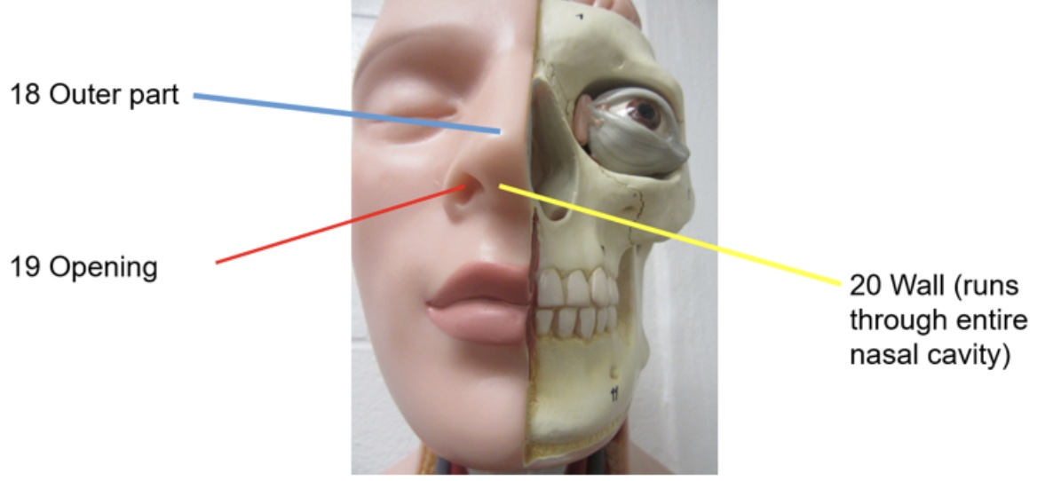
What is 20 representing?
Nasal Septum

What did 14 represent?
Olfactory Epithilium

What does 15 represent?
Posterior nare

What does 16 represent?
Anterior Nare

What does 17 represent?
Eustachian tube opening

What does 9 represent?
Superior Turbinate

What does 10 represent?
Middle Turbinate

What does 11 represent?
Inferior Turbinate

What does 3 represent?
Superior Meatus

What does 4 represent?
Middle Meatus