Respiratory System
 Call Kai
 Learn
Learn Practice Test
 Spaced Repetition
 Match
Match Flashcards
 Knowt Play
1/31

There's no tags or description
Looks like no tags are added yet.
Name | Mastery | Learn | Test | Matching | Spaced | Call with Kai | Chat |
|---|
No analytics yet
Send a link to your students to track their progress
32 Terms

Nasal cavity

Oral cavity
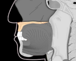
Hard palate (made of bone)

Soft palate (made of soft tissue)

Pharynx, the 3 sections are: Nasopharynx, Oropharynx, and Laryngopharynx

Larynx, here is another view

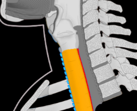
Trachea, here is another view

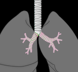
Bronchial tree
How many lobes in each lung and why?
3 in right, 2 in left because of heart asymmetry (it has cardiac notch)

What are the layers of a typical respiratory tube?
1. Mucosal layer: Ciliated pseudostratified columnar epithelium, submucosal layer of loose connective tissue
2. Fibromusculocartilaginous layer: Large moons of hyaline cartilage connected with muscle, glued together with connective tissue
3. Adventitial layer: Dense irregular connective tissue

Thyroid cartilage

Cricoid cartilage

What is this, and how is it connected to the cricoid cartilage?
Arytenoid cartilage, synovial joint
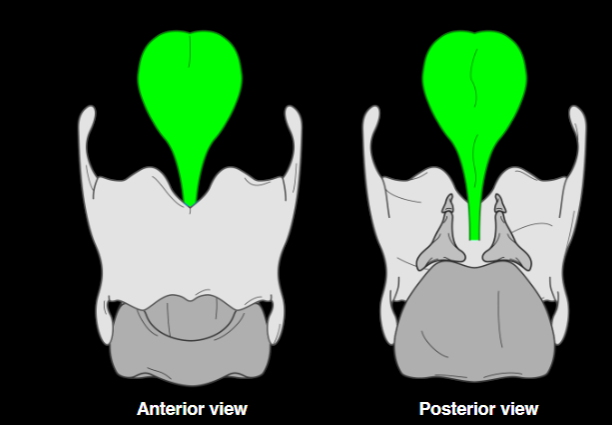
Epiglottis
What part of the larynx is muscle attached to? What type of muscle is it?
Attached to arytenoids, it is skeletal muscle (voluntary)

Describe the surface tissue of all these zones
External body: keratinized stratified squamous epithelium. Oral cavity: nonkeratinized stratified squamous epithelium. Nasal cavity, Larynx, Trachea: Ciliated pseudostratified columnar epithelium (as expected for respiratory tubes)
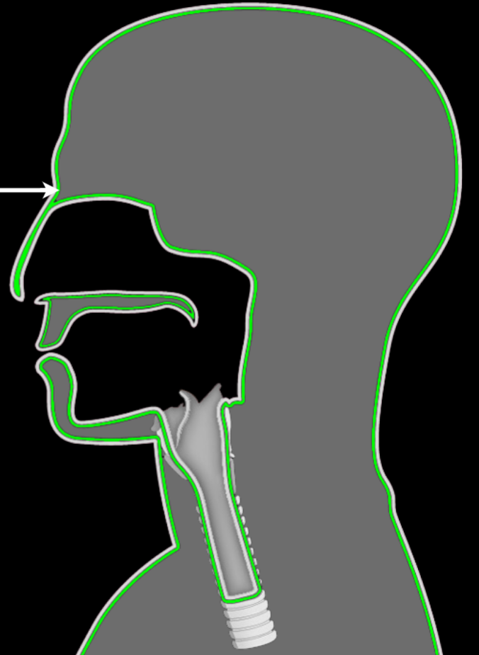
Describe the tissue just deep of the surface for all these zones
External body: Dermis made of papillary layer and reticular layer. Cavities: No reticular layer, just loose connective tissue

What do you call the cavity of the larynx?
Glottis
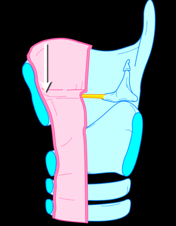
What is being pointed at, and its structure?
Vocal folds. A flap of the mucosal layer.

What is being pointed at, and what structures is it connected to?
Vocal ligaments, connect arytenoids to the thyroid cartilage

What do you call the space between the vocal folds?
Rima glottidis
Describe sound production and variation in the larynx
Air rushes past vocal folds, causing vibration and sound. The skeletal muscles move the arytenoids to change tension, changing the pitch.

What is this, and its structure:
Mucosa of trachea: Ciliated pseudostratified columnar epithelium and loose connective tissue
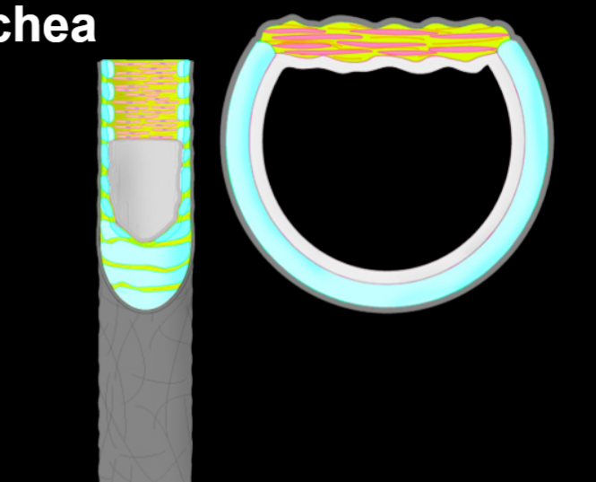
What is this, and its structure?
Fibromusculocartilaginous layer of trachea: Incomplete rings of tracheal cartilage (hyaline), trachealis muscle (smooth), and anular ligaments orbiting the trachea connecting the tracheal cartilages and surrounding muscle cells

Adventital layer of trachea

Large bronchus
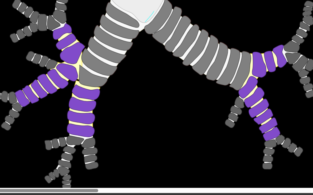
Lobar bronchus (one for each lobe)

Segmental bronchus
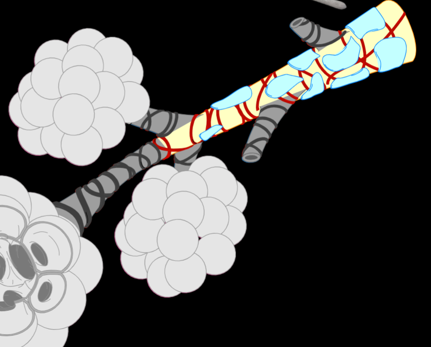
What is this, how is its structure different from the larger bronchi?
Bronchiole. Cartilage is now plates, muscle runs around tube, epithelium turns to simple columnar/cuboidal, adventitia and walls get thinner

What is this, how is it different from larger bronchi?
Alveolar duct. There is not fibromusculocartilagenous layer. It is very thin (near gas exchange)
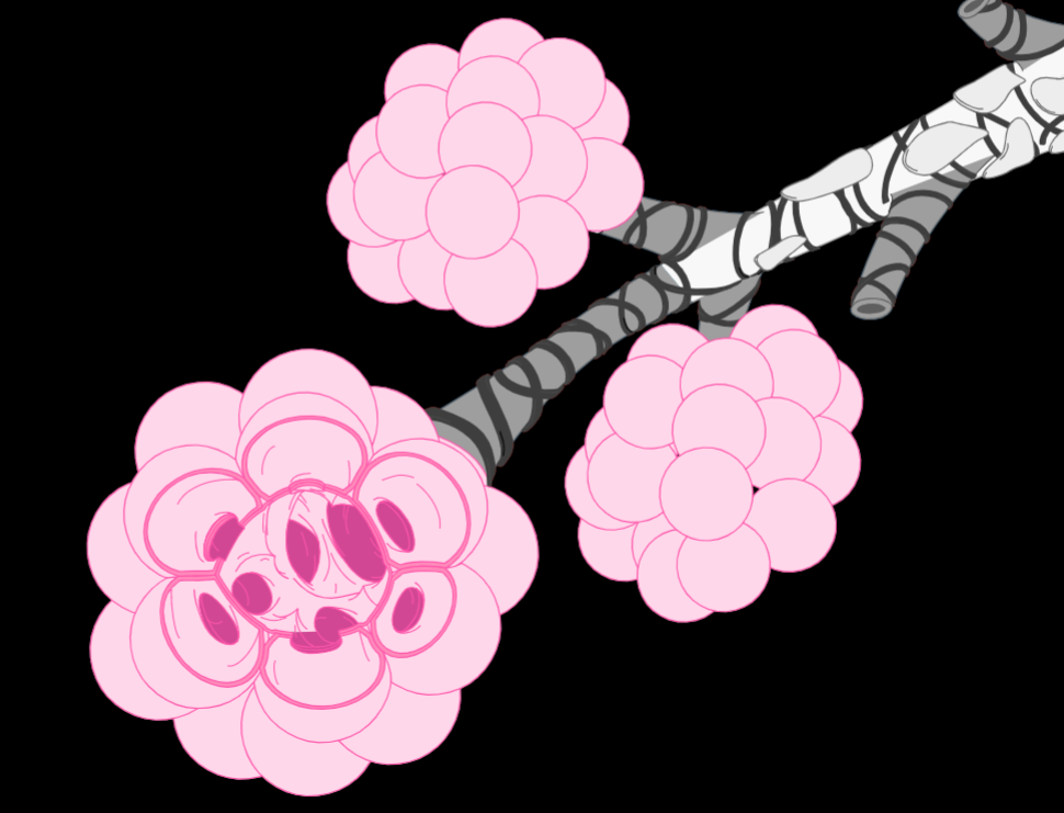
Alveolar saccule, it is hollow

What is this, and its structure
Pulmonary alveolus. Each sphere is simple squamous epithelium for exchange with capillaries.