BIO162 - Structure of Brain and Skull
1/48
There's no tags or description
Looks like no tags are added yet.
Name | Mastery | Learn | Test | Matching | Spaced |
|---|
No study sessions yet.
49 Terms
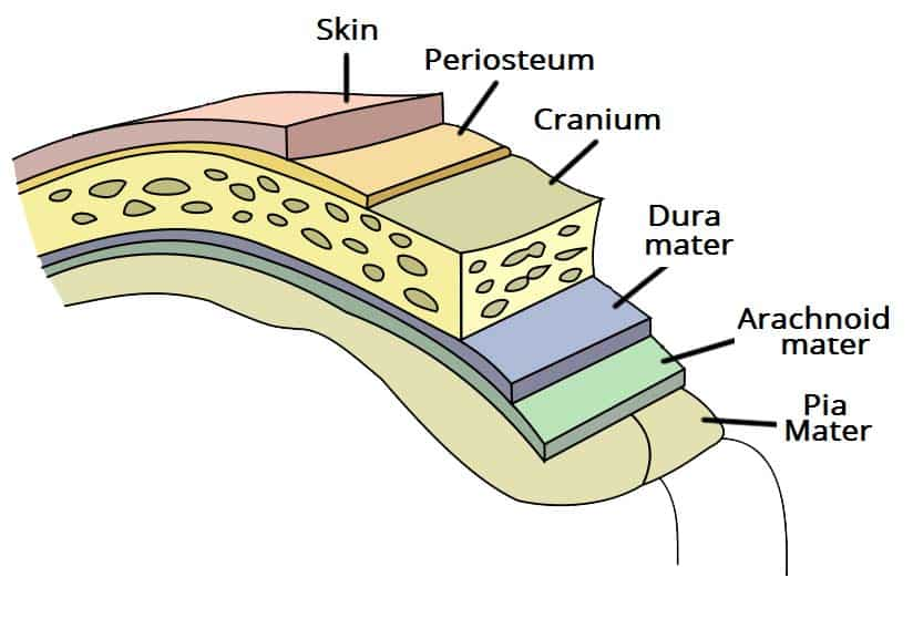
dura mater
thick, tough layer of CT that separates the meninges and brain from the skull

arachnoid mater
loose tissue between the dura and pia mater
pia mater
thin delicate sticks to the surface of the brain
subarachnoid space
space between the pia and arachnoid maters filled with CSF
cerebrum (cerebral cortex)
consists of 2 hemispheres and 4 lobes
gyri
ridges on the surface of the cerebrum
sulci
valleys between the gyri
longitudinal fissure
divides the cerebrum into 2 hemispheres
corpus callosum
provides communication highway between the hemispheres (white matter)
lateral ventricles
chambers within the hemispheres bw the corpus callosum and the fornix
choroid plexus
produce the CSF in the lateral ventricles
thalamus
routes info into the cerebrum (role in memory and basic emotion)
third ventricle
forms ring around the thalamus, CSf flows through it
hypothalamus
involved in hormone secretion, circadian rhythm, and memory
pituitary gland
secretes endocrine hormones
midbrain
top of brain stem, relays auditory and visual stimuli
cerebral aqueduct
tube between 3rd and 4th ventricle
pons
relays info to cerebrum, medulla and cerebellum
cerebellum
deals with muscle memory and learned reflexes; has arbor vitae and folia on the interior
fourth ventricle
sends CSF to the spinal cord
medulla oblongata
connects brain to the spinal cord
cranium
8 bones that encase the brain
sutures
ridged joints that connect the bones of the cranium
frontal bone
bone that makes up most of the forehead
nasal bones
form the bridge of the nose
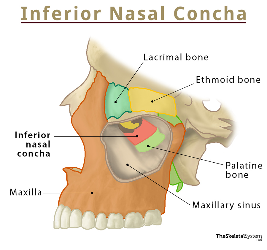
maxilla
forms the upper part of the jaw
zygomatic bone
forms cheekbones and part of the orbit
lacrimal bone
medial wall of the orbit
ethmoid bone
posterior to the lacrimal bone, makes up superior part of septum
sphenoid bone
the posterior part of the orbit, posterior to the zygomatic and inferior to the frontal
vomer
inferior portion of the septum
inferior nasal conchae
bumps in the lower part of the nasal cavity

parietal bone
covers most of the top of the skull
sagittal suture
connects the two parietal bones
coronal suture
connects the parietal bones to the frontal bone
temporal bone
forms most of the sides of the cranium
squamous suture
joins parietal and temporal bones
external acoustic meatus
the opening to the ear
mastoid process
bump behind the earlobe
occipital bone
forms most of the back of the skull
lambdoid suture
joins occipital and parietal bones
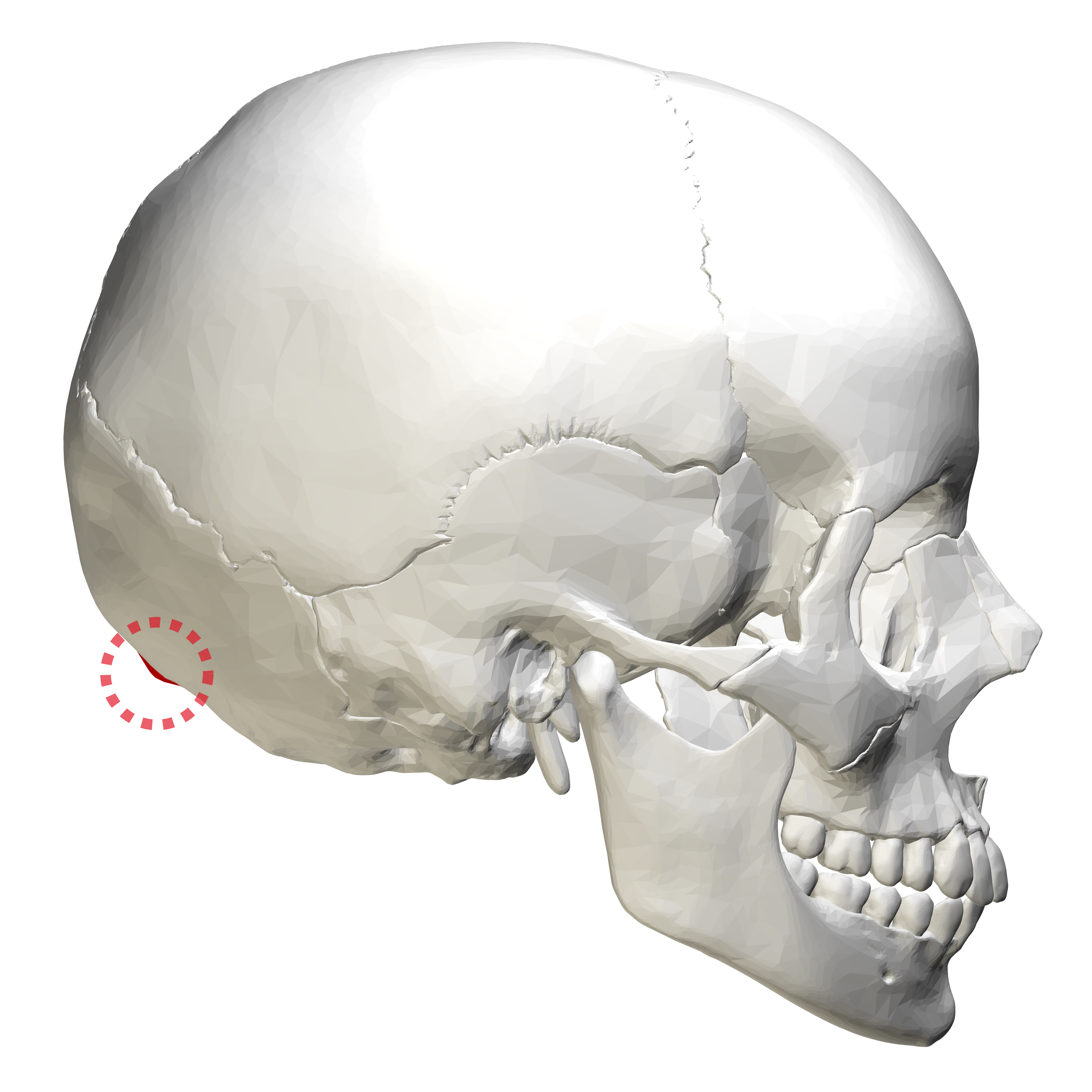
external occipital protuberance
bump in the middle of the occipital bone

nuchal lines
extend laterally from the external occipital protuberance
occipital condyles
articulate with the C1 vertebra
palatine bone
form the posterior edge of the hard palate with the maxilla bone
anterior cranial fossa
made of frontal and ethmoid bones
middle cranial fossa
made of temporal and sphenoid
posterior cranial fossa
surrounds foramen magnum
hypophyseal fossa
of sphenoid bone