Pitt Anatomy Lab Quiz 3 - cell types
1/71
Earn XP
Description and Tags
Name | Mastery | Learn | Test | Matching | Spaced |
|---|
No study sessions yet.
72 Terms
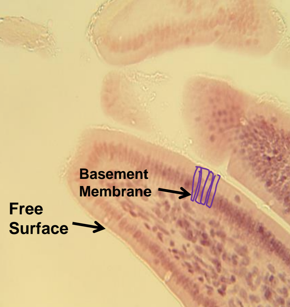
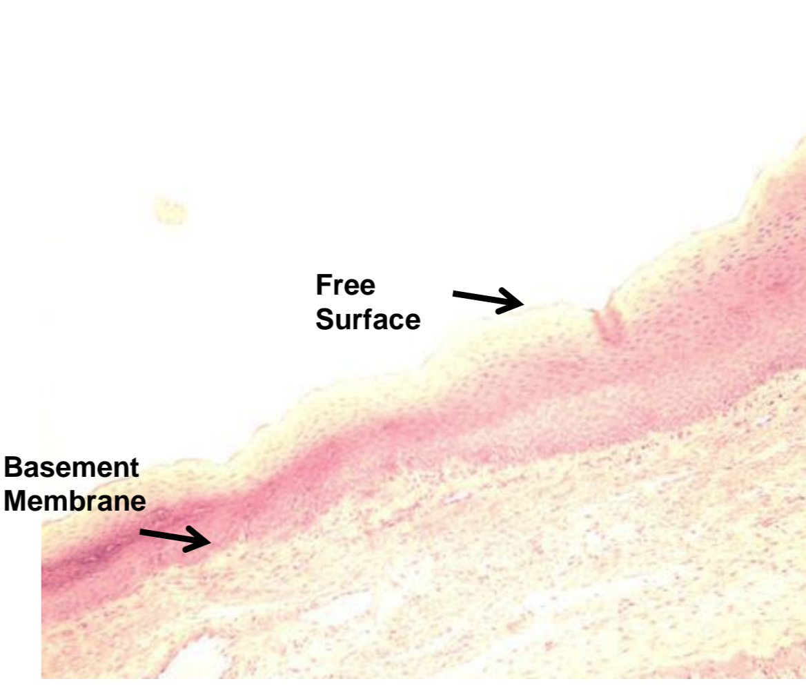
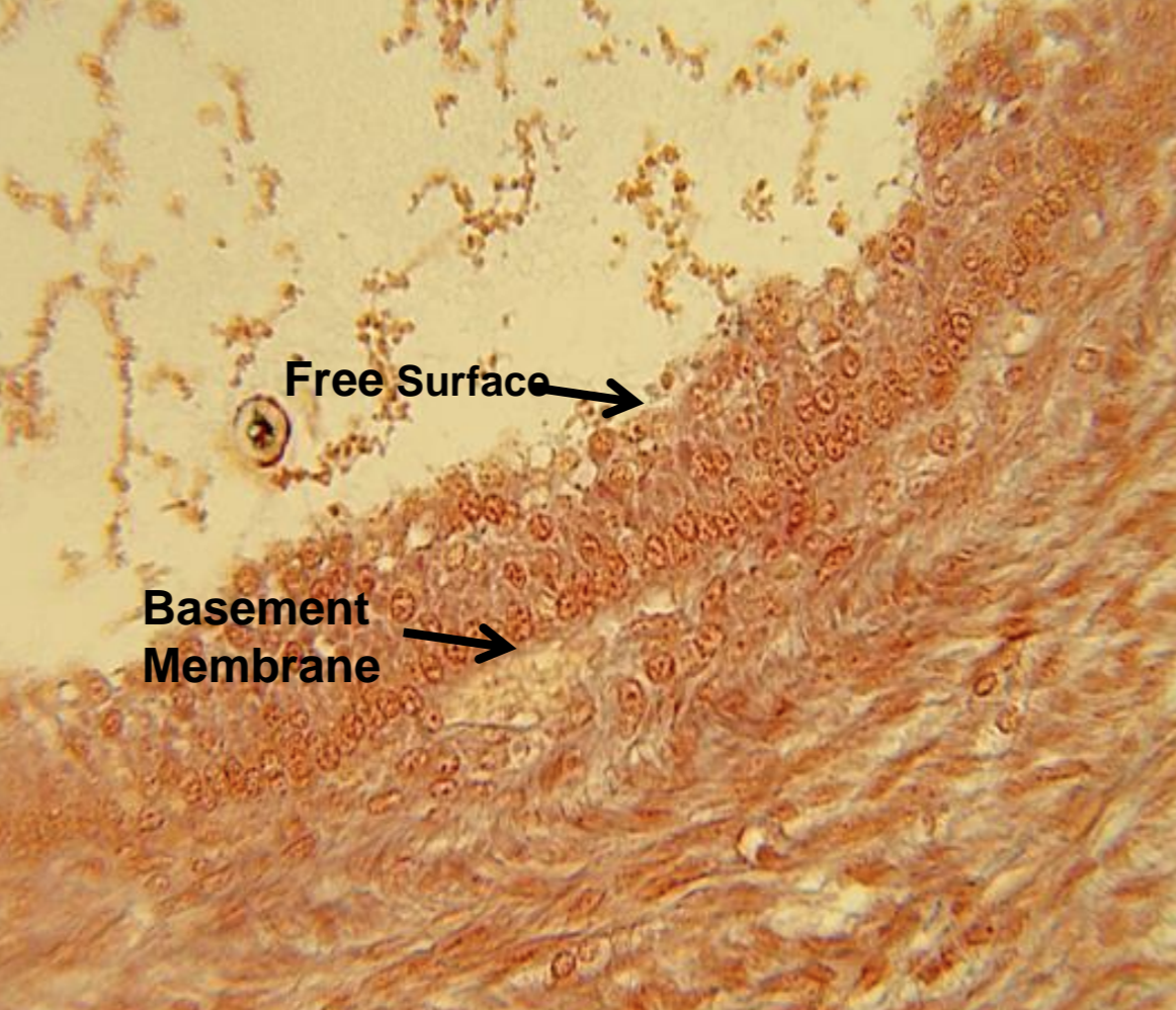
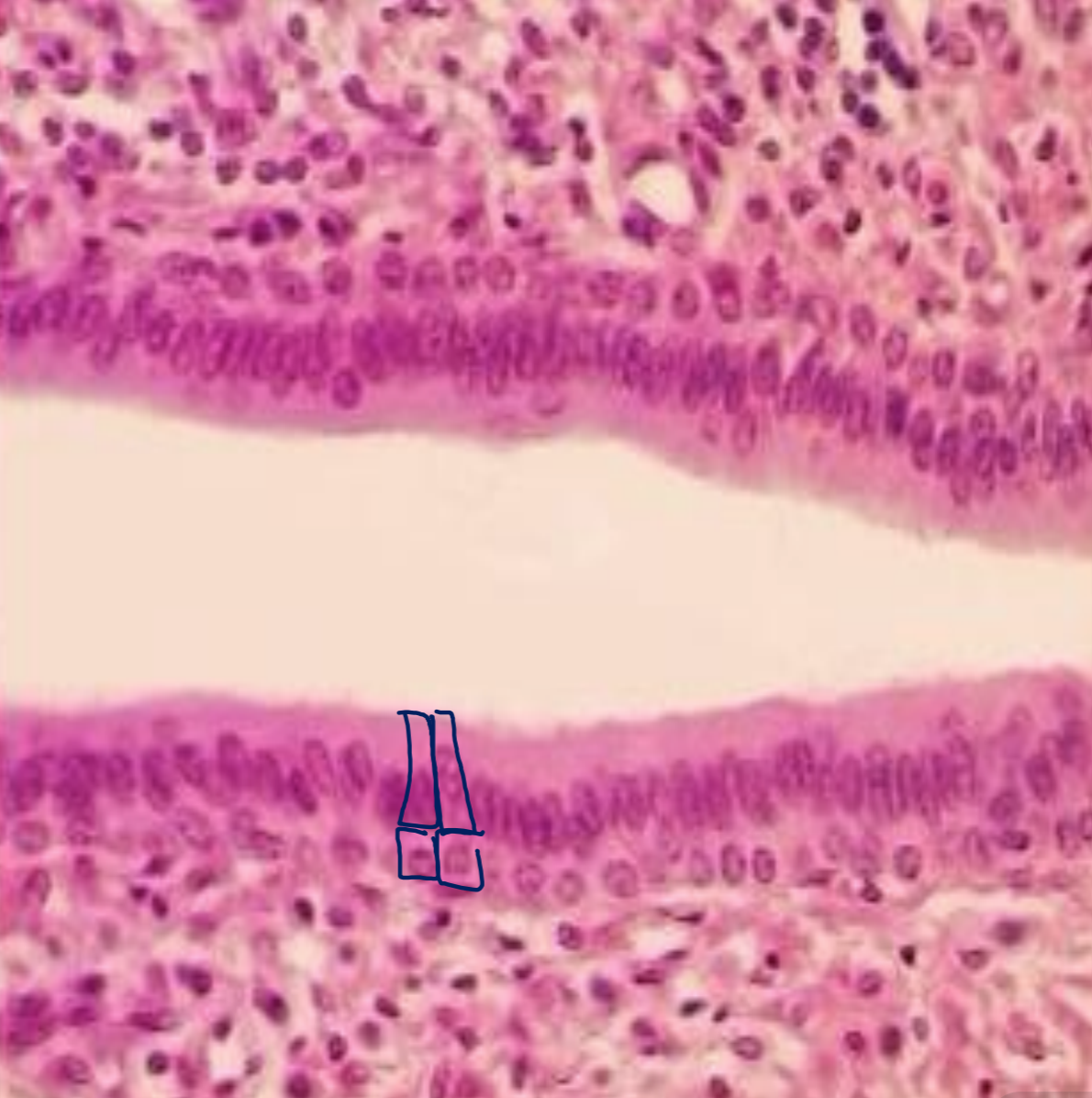
simple columnar
single layer of column shaped cells with round nucleus, contains goblet cells
simple columnar - function
secretion of mucous, absorption
simple columnar - location
lines digestive tract and portions of the uterus and uterine tubes
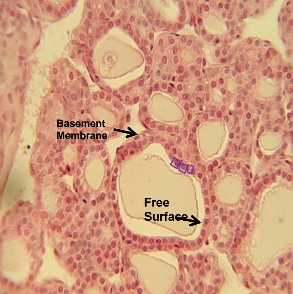
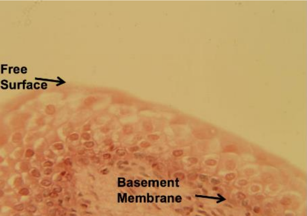
simple cuboidal
single layer of cubed shaped cells, large and round with central nuclei
simple cuboidal - function
absorption, secretion, active transport
simple cuboidal - location
thyroid, ovaries, kidney duct
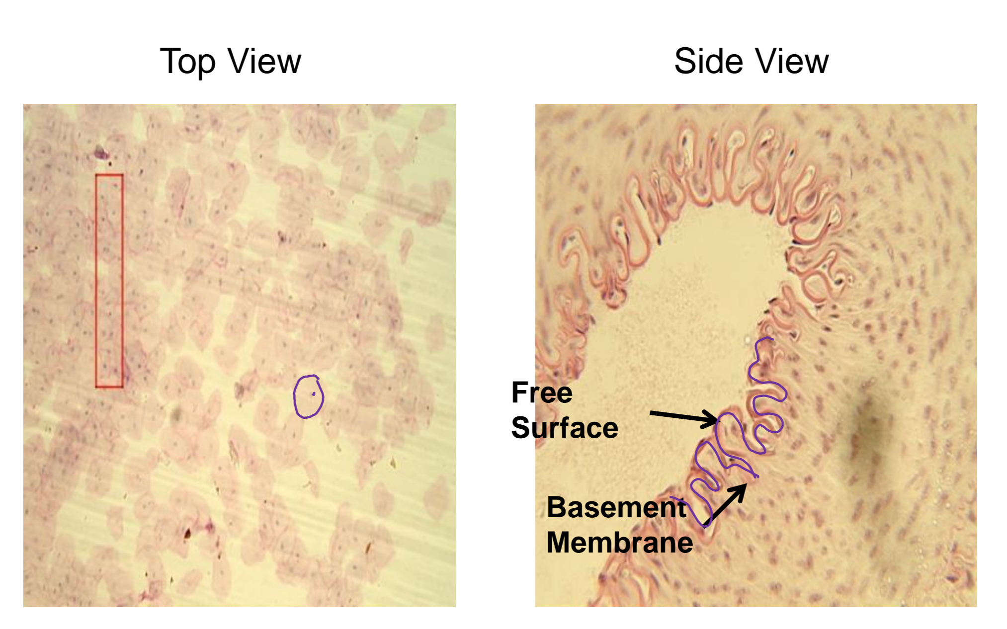
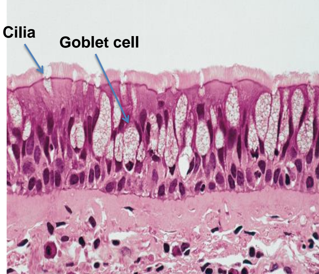
pseudostratified
single layer of cells with differing nuclei, nuclei located at different heights, all cells touch base membrane, not all touch apical surface, contain cilia
pseudostratified - function
goblet cells release mucous that coats passage ways
pseudostratified - location
respiratory tract, male urethra
simple squamous
single layer thick, flat or oval with a central nucleus
simple squamous - function
diffusion, secretion, filtration
simple squamous - location
capillaries, endothelium, alveoli
stratified squamous
multiple layers of cells with nuclei throughout
stratified squamous - function
protection against abrasion
stratified squamous - location
esophagus, anus, mouth, vagina, epidermis of skin
stratified cuboidal
many layers of cube cells
stratified cuboidal - function
protection, secretion
stratified cuboidal - location
sweat glands, ovary follicles, mammary glands
transitional
tissue changes shape, relaxed = cuboidal, stretched = squamous
transitional - function
allows stretching as it fills
transitional - location
bladder, urethra, ureters
stratified columnar
apical layer is composed of columnar cells, basal is cuboidal
stratified columnar - function
protection, secretion
stratified columnar - location
large ducts and some glands
what tissue is depicted in the image
simple squamous
what tissue is depicted in the image
simple cuboidal
what tissue is depicted in the image
simple columnar
what tissue is depicted in the image
pseudostratified
what tissue is depicted in the image
stratified squamous
what tissue is depicted in the image
stratified cuboidal
what tissue is depicted in the image
stratified columnar
what tissue is depicted in the image
transitional
what are the parts of the extracellular matrix (areolar) aka protein fibers
collagen, reticular fibers, elastin fibers
collagen
rope-like and strong but inflexible
reticular fibers
tiny collagen fibers that act like web between cells
elastin fibers
can be stretched and returned to its original shape
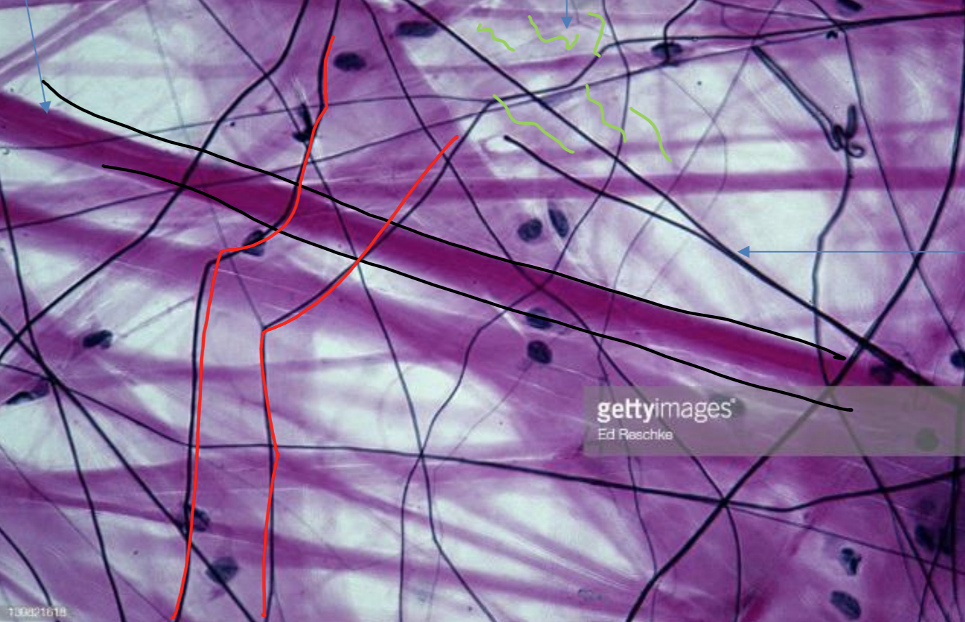
areolar (ECM)
matrix contains all three fibers collagen, elastic, reticular held loosely together
areolar (ECM) - function
anchors skin to underlying tissues, anchors tissues together, surrounds capillaries and tissue, around organs
areolar (ECM) - location
everywhere in body
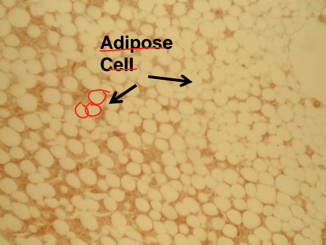
adipose
fat cells, a drop of fat is put in each cell and this pushes the nucleus to the edge of the cell, nucleus is highly vascular
adipose - function
cushioning, insulation, energy storage
adipose - location
center of femur, under skin, around kidney, eye balls
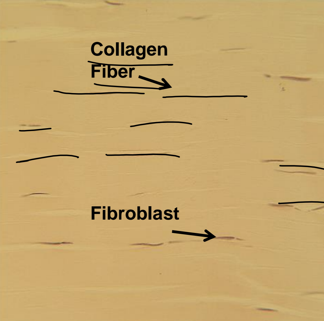
dense regular
unidirectional, nuclei are aligned parallel, densely packed fibers
dense regular - function
attaches muscles to bones (tendons), attaches bones to bones (ligaments)
dense regular - location
tendons, ligaments, aponeuroses
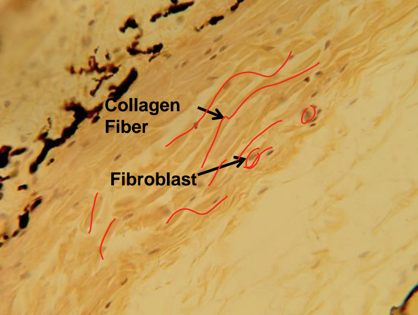
dense irregular
random assignment, multiple directions
dense irregular - function
strong in all directions
dense irregular - location
dermis of skin, capsules around organs and joints
what tissue is depicted in the image
areolar (ECM)
what tissue is depicted in the image
adipose
what tissue is depicted in the image
dense regular
what tissue is depicted in the image
dense irregular
what tissue is depicted in the image
elastic
what tissue is depicted in the image
hyaline
what tissue is depicted in the image
fibrocartilage
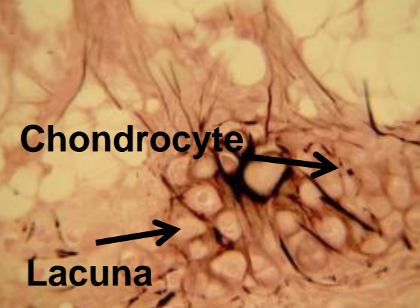
hyaline cartilage
articular surfaces and joints, has lacuna and chondrocyte
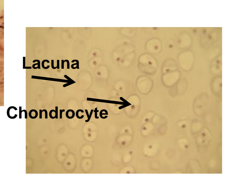
elastic cartilage
larynx, nose, ear, very flexible, has lacuna and chondrocyte
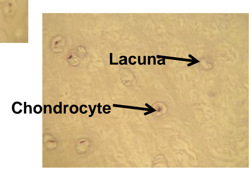
fibrocartilage
absorbs shock, intervertebral disks, has lacuna and chondrocyte
what tissue is depicted in the image
bone
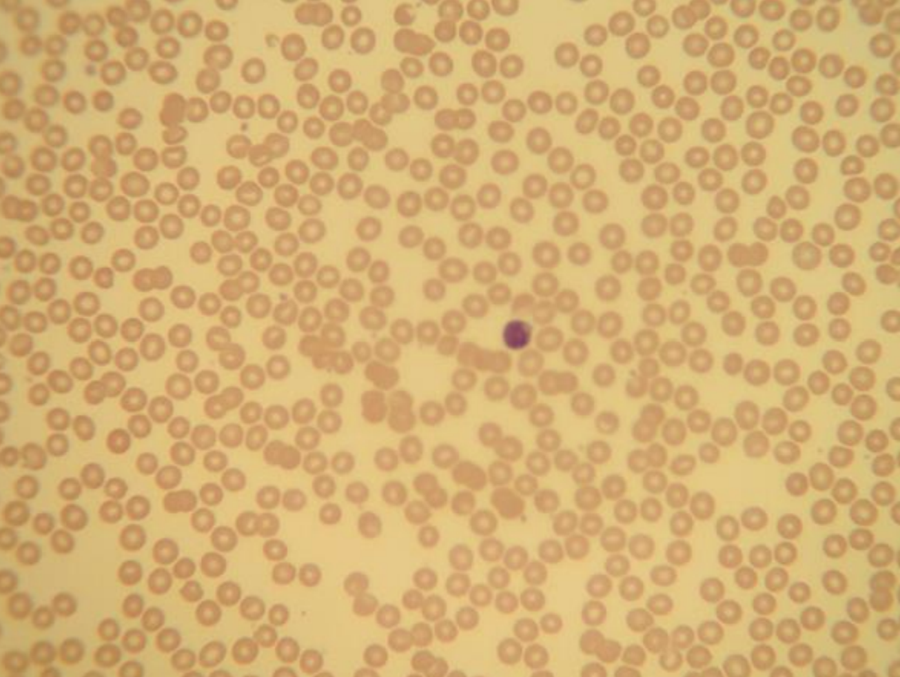
what tissue is depicted in the image
blood
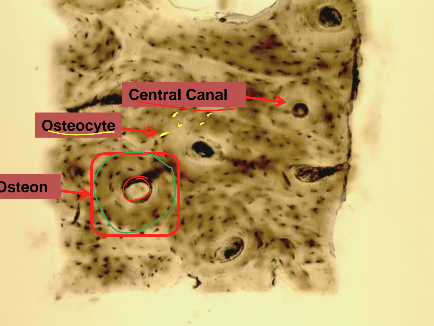
bone
consists of mineralized ECM, lacunae is holes in the matrix where osteocytes reside
blood cells
suspended in fluid matrix, classified as connective tissue because it derives from mesenchyme
nervous tissue - function
specialized to carry electrical signals called action potentials
nervous tissue
dendrites, cell body, axon
nervous tissue - location
brain, spinal cord, peripheral nerves
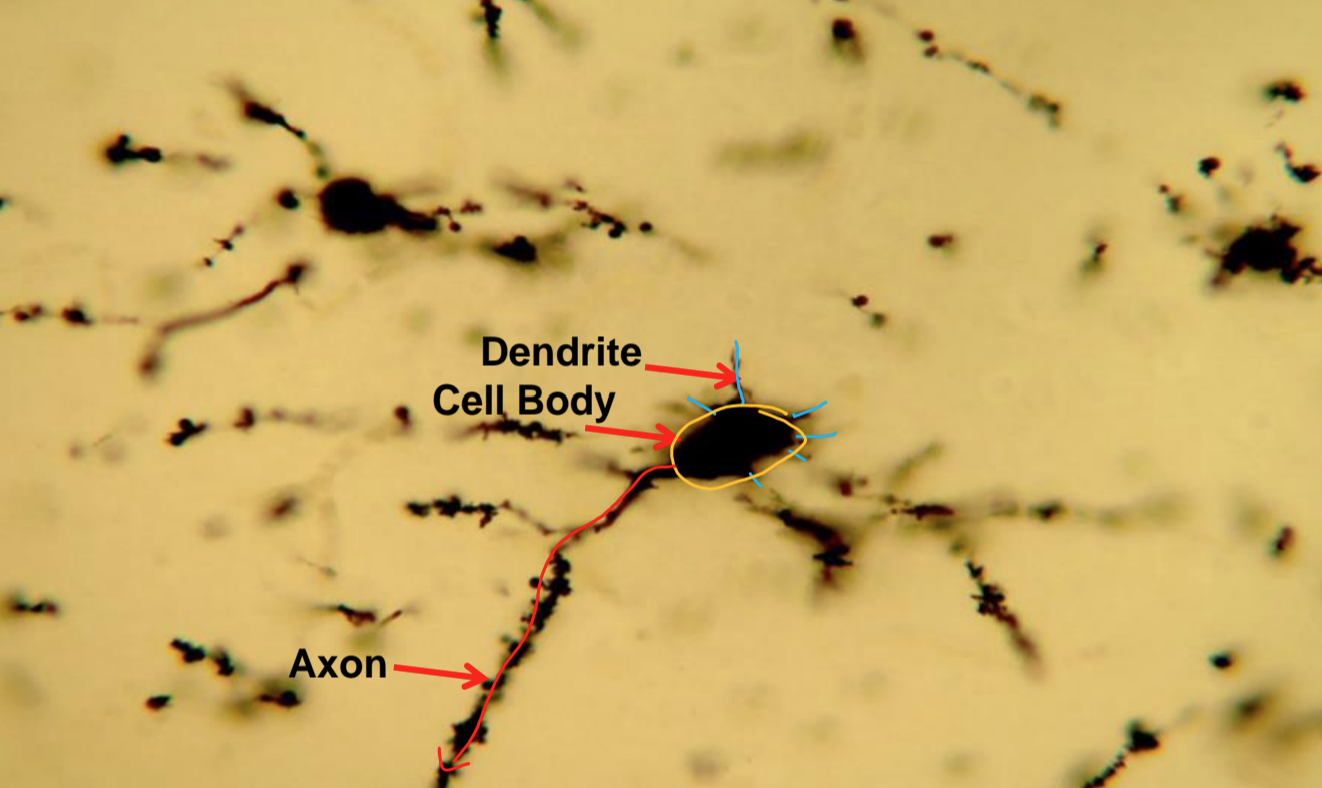
what tissue is depicted in the image
nervous tissue
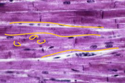
skeletal muscle tissue
striated, voluntary, multinucleated
cardiac muscle tissue
striated, involuntary, uninucleated
smooth muscle tissue
nonstriated, involuntary, uninucleated
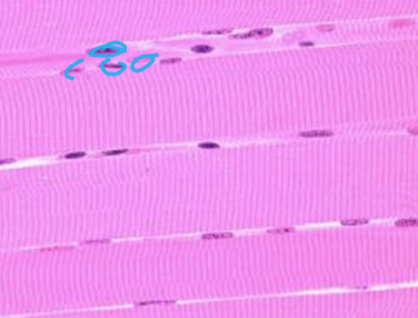
what tissue is depicted in the image
skeletal muscle tissue
what tissue is depicted in the image
cardiac muscle tissue
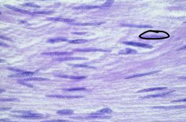
what tissue is depicted in the image
smooth muscle tissue