aqa a level biology topic 3
1/161
There's no tags or description
Looks like no tags are added yet.
Name | Mastery | Learn | Test | Matching | Spaced | Call with Kai |
|---|
No analytics yet
Send a link to your students to track their progress
162 Terms
what is the relationship between a small organism and its surface area?
small organism have large surface areas
these substances (glucose and oxygen) can diffuse directly into (or out of) the cell across the cell-surface membrane. The diffusion rate is quick because of the small distances the substances have to travel
what is the relationship between a large organism and its surface area
large organsims have small surface area
In multicellular animals, diffusion across the outer membrane is too slow, for two reasons:
• Some cells are deep within the body — there's a big distance between them and the outside environment.
environment.
• Larger animals have a low surface area to volume ratio — it's difficult to exchange enough substances to supply a large volume of animal through a relatively small outer surface.
how does body size and shape affect heat exchange?
The rate of heat loss from an organism depends on its surface area . If an organism has a large volume, its surface area is relatively small. This makes it harder for it to lose heat from its body.
how does body size and shape affect heat exchange? Part 2
If an organism is small, its relative surface area is large, so heat is lost more easily. This means smaller organisms need a relatively high metabolic rate, in order to generate enough heat to stay warm and larger oganisms need a relatively small metabolic rate.
how does body shape affect heat exchange?
1) Animals with a compact shape have a small surface area relative to their volume — minimising heat loss from their surface.

how does body shape affect heat exchange?
2) Animals with a less compact shape (those that are a bit gangly or have sticky outy bits) have a larger surface are a relative to their volume — this increases heat loss from their surface.
how does body shape affect heat exchange?
3) Whether an animal is compact or not depends on the temperature of its environment.
name all the adaptations to increase surface area?
villi and microvilli (absorption of digested food)
alveoli and bronchioles (gas exchange)
spiracles and tracheoles (gas exchange)
gill filaments and lamellae (gas exchange)
thin wide leaves (gas exchange)
many capillaries (capillary network)
what are the terms related to lung disease?
1) Tidal volume is the volume of air in each breath — usually between 0.4 dm and 0.5 dm for adults.
2) Ventilation rate is the number of breaths per minute.
what are the terms related to lung disease?
3) Forced expiratory volume (FEV1) is the maximum volume of air that can be breathed out in 1 second.
4) Forced vital capacity (FVC) is the maximum volume of air it is possible to breathe forcefully out of the lungs after a really deep breath in.
how does the lung diseases TB, fibrosis asthma and emphysema decrease rate of gas exchange?
TB, fibrosis, asthma and emphysema all reduce the rate of gas exchange in the alveoli. Less oxygen is able to diffuse into the bloodstream, the body cells receive less oxygen and the rate of aerobic respiration is reduced. This means less energy is released and sufferers often feel tired and weak.
how does tuberculosis affect gas exchange?
gaseous exchange surface is damaged, so tidal volume is decreased.
Tuberculosis also causes fibrosis , which further reduces the tidal volume.
A reduced tidal volume means less air can be inhaled with each breath.
Pulmonary tuberculosis is a disease of the lungs. Describe the transmission and course of infection of pulmonary tuberculosis.
(exam question) (5 marks)
1 (Bacteria transmitted in) droplets / aerosol;
2 (Bacteria) engulfed / ingested by phagocytes / macrophages;
3 (Bacteria) encased in named structure e.g. wall / tubercle / granuloma / nodule;
4 (Bacteria) are dormant / not active / not replicating;
5 If immunosuppressed, bacteria activate / replicate / released;
6 Bacteria destroy alveoli / capillary / epithelial cells;
7 (Leads to) fibrosis / scar tissue / cavities /calcification;
8 (Damage) leads to less diffusion /less surface area / increases diffusion distance;
9 (Activation / damage allows bacteria) to enter blood / spreads (to other organs);
how does fibrosis affect gas exchange?
formation of scar tissue in the lungs.
Scar tissue is thicker and less elastic than normal lung tissue.
how does fibrosis affect gas exchange? Part 2
This means that the lungs are less able to expand and so can't hold as much air as normal — tidal volume is reduced, and so is FVC
There's a reduction in the rate of gaseous exchange — diffusion is slower across a thicker scarred membrane.
how to dissect fish gills in bony fish?
1) Again, make sure you're wearing an apron or lab coat.
2) Place your chosen fish (something like a perch or salmon works well) in a dissection tray or on a cutting board.
3) Gills are located on either side of the fish's head. They're protected on each side by a bony flap called an operculum and supported by gill arches.
how to dissect fish gills in bony fish?
4) To remove gills, push back the operculum and use scissors to carefully remove the gills. Cut each hill arch through the bone at the top and bottom.
5) If you look closely, you should be able to fill filamentes
You Can Dissect the Gaseous Exchange System in Insects too
how?
1)First fix the insect to a dissecting board. You can put dissecting pins through its legs to hold it in place.
2) To examine the tracheae, you'll need to carefully cut and remove a piece of exoskeleton (the insect's hard outer shell) from along the length of the insect's abdomen.
You Can Dissect the Gaseous Exchange System in Insects too
how?
3)Use a syringe to fill the abdomen with saline solution. You should be able to see network of very thin, silvery-grey tubes- these are tracheae. They look silver because they're filled with air.
You Can Dissect the Gaseous Exchange System in Insects too
how?
4) You can examine the tracheae under and optical microscope using a temporary mouth slide. A gain, the tracheae well appear silver of grey. You should be able to she rings of chitin I'm the walls of the tracheae. The we are there for support (like the sings of cartilage in a human tracheae)
describe the process when you breath in air
1) As you breathe in, air enters the trachea.
2) The trachea splits into two bronchi — one bronchus leading to each lung.
3) Each bronchus then branches off into smaller tubes called bronchioles.
describe the process when you breath in air
4) The bronchioles end in small 'air sacs' called alveoli
5) The ribcage, intercostal muscles and diaphragm all work together to move air in and out
what happens during inspiration?
1) The external intercostal and diaphragm muscles contract.
2) This causes the ribcage to move upwards and outwards and the diaphragm to flatten, increasing the volume of the thoracic cavity
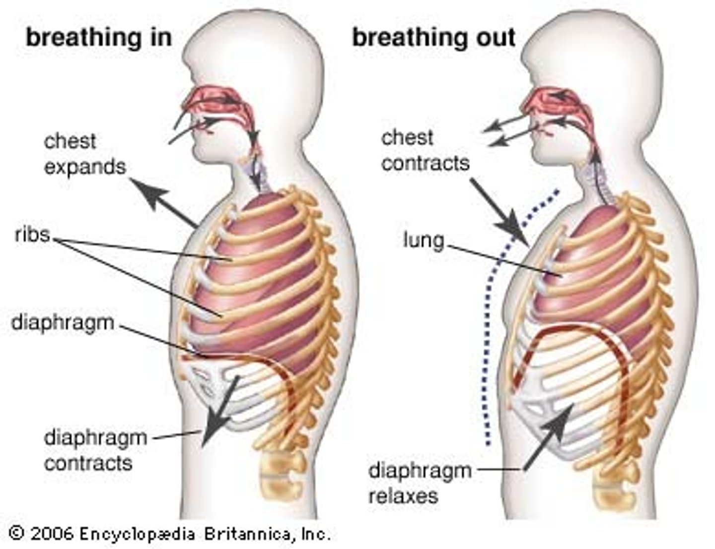
what happens during inspiration?
3) As the volume of the thoracic cavity increases, the lung pressure decreases
4) Air will always flow from an area of higher pressure to an area of lower pressure so air flows down the trachea and into the lungs.
what happens during inspiration?
5) Inspiration is an active process — it requires energy.
what happens during expiration?
1) The external intercostal and diaphragm muscles relax.
2) The ribcage moves downwards and inwards and the diaphragm becomes curved again.
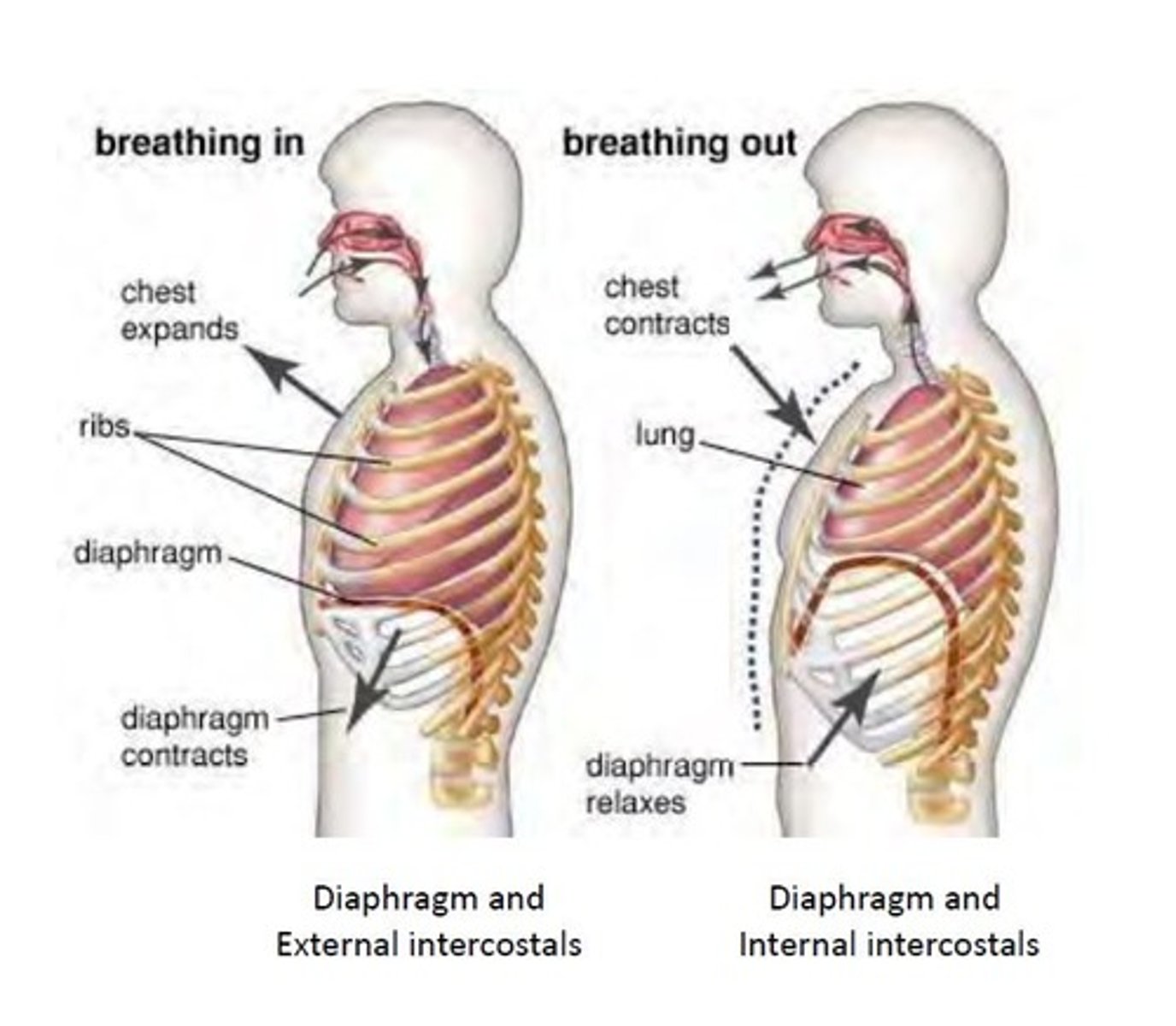
what happens during expiration?
3) The volume of the thoracic cavity decreases, causing the air pressure to increase
4) Air is forced down the pressure gradient and out of the lungs.
what happens during expiration?
5)Normal expiration is a passive process — it doesn't require energy.
6) Expiration can be forced though
what happens during expiration?
7) During forced expiration, the external intercostal muscles relax and internal intercostal muscles contract, pulling the ribcage further down and in. During this time, the movement of the two sets of intercostal muscles is said to be antagonistic (opposing).
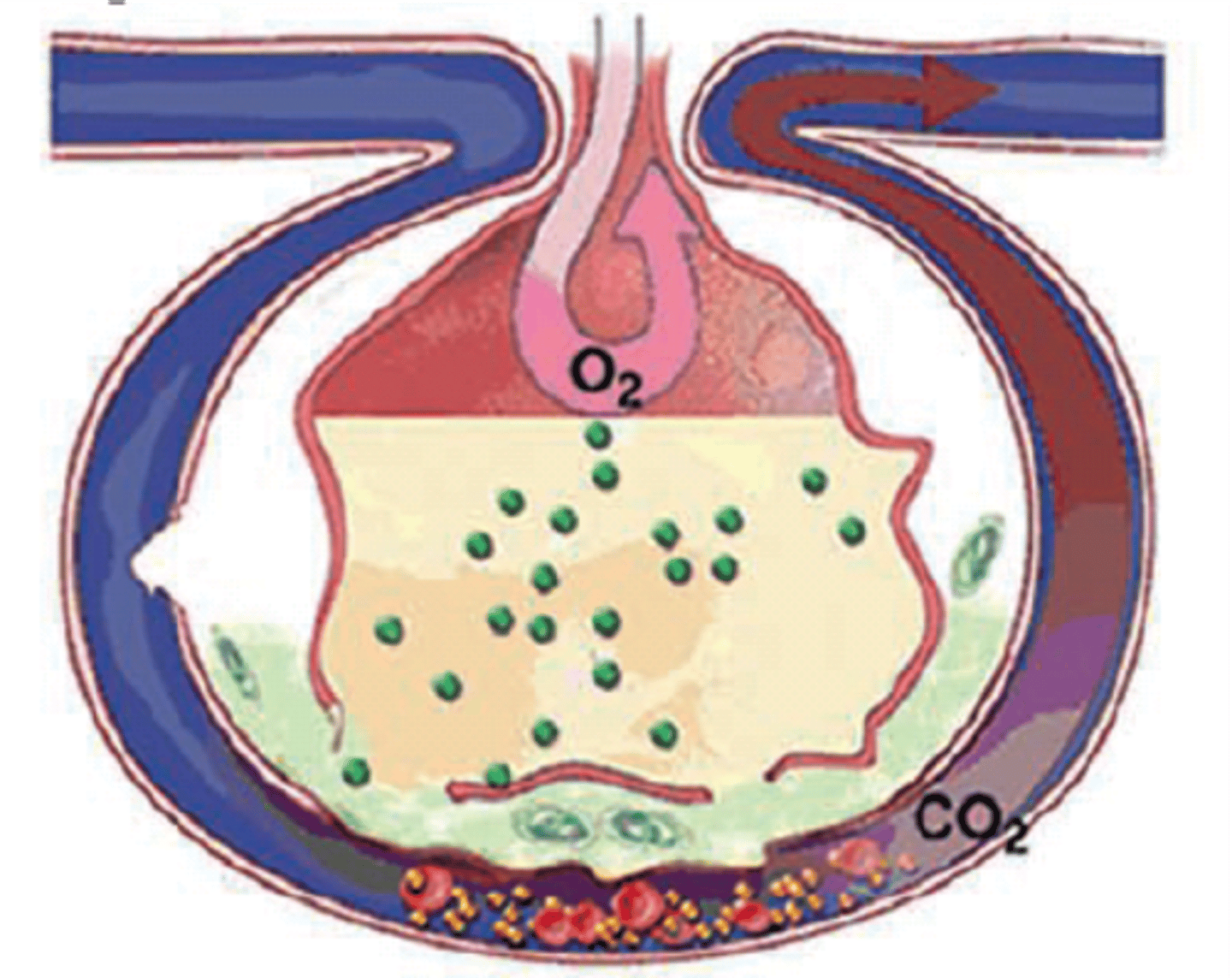
describe the process of gas exchange in the alveoli
1) There's a huge number of alveoli in the lungs, which means there's a big surface area for exchanging oxygen and carbon dioxide
2) The alveoli are surrounded by a network of capillaries.
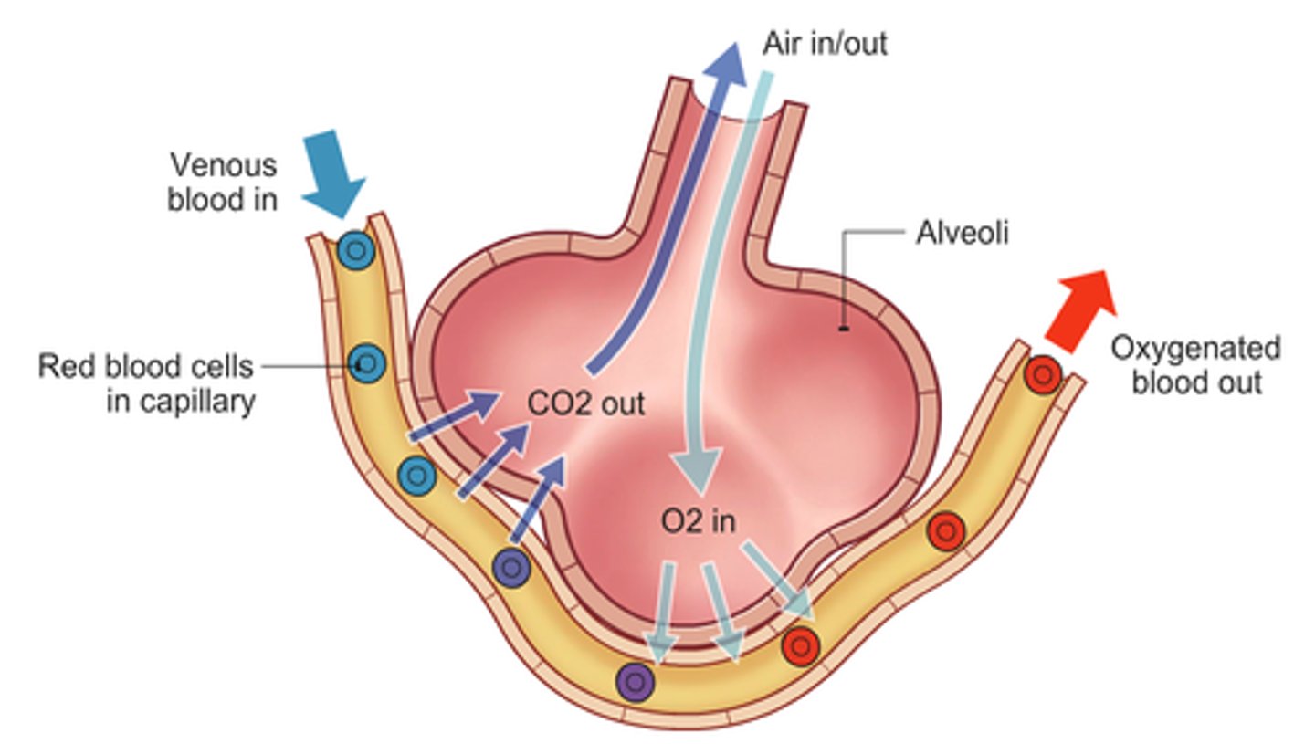
describe the process of gas exchange in the alveoli
3) oxygen diffuses out of the alveoli, across the alveolar epithelium and the capillary endothelium and into haemoglobin in the blood.
4) C 0 2 diffuses into the alveoli from the blood, and is breathed out.
describe the process of gas exchange in the alveoli
IN SUMMARY
oxygen from the air moves down the trachea, bronchi and bronchioles into the alveoli. This movement happens down a pressure gradient. Once in the alveoli, the oxygen diffuses across the alveolar epithelium, then the capillary endothelium, ending up in the capillary itself. This movement happens down a diffusion gradient.
what are the adaptations of gas exchange in alveoli?
1) A thin exchange surface — the alveolar epithelium is only one cell thick. This means there's a short diffusion pathway
2) A large surface area — the large number of alveoli means there's a large surface area for gas exchange.
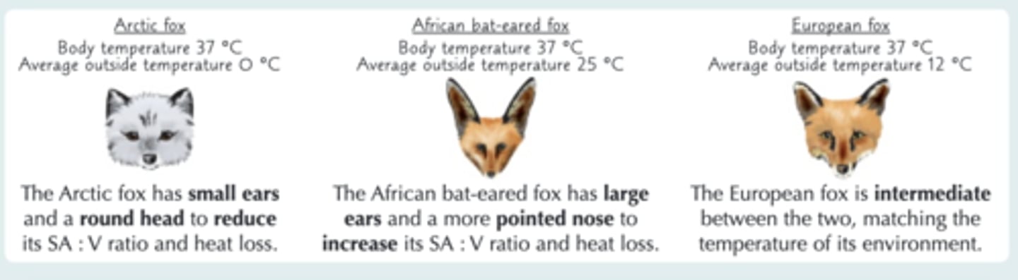
how does behavioural and physiological adaptations Aid Exchange
1) Animals with a high SA : volume ratio tend to lose more water as it evaporates from their surface. Some small desert mammals have kidney structure adaptations so that they produce less urine to compensate.
how does behavioural and physiological adaptations Aid Exchange
2) To support their high metabolic rates, small mammals living in cold regions need to eat large amounts of high energy
3) Smaller mammals may have thick layers of fur or hibernate when the weather gets really cold.
how does behavioural and physiological adaptations Aid Exchange
4) Larger organisms living in hot regions, find it hard to keep cool as their heat loss is relatively slow. Elephants have developed large flat ears to increase their surface area, allowing them to lose more heat. Hippos spend much of the day in the water — a behavioural adaptation to help them lose heat.
describe the structure of a terrestrial insect
insects have an exoskeleton made of hard fibrous material for protection and a lipid layer to prevent water loss
has a tracheal system
what are the adaptations of insects to prevent water loss?
small surface are to volume ratio where water can evaporate from
has a water proof exoskeleton
has spiracle, where gases enter and water can evaporate from, which can open and close to reduce water loss
what is the insect tracheal system?
1) Insects have a tracheae which they use for gas exchange. 2) Air moves into the tracheae through pores on the surface called spiracles
3) Oxygen travels down the concentration gradient towards the cells.
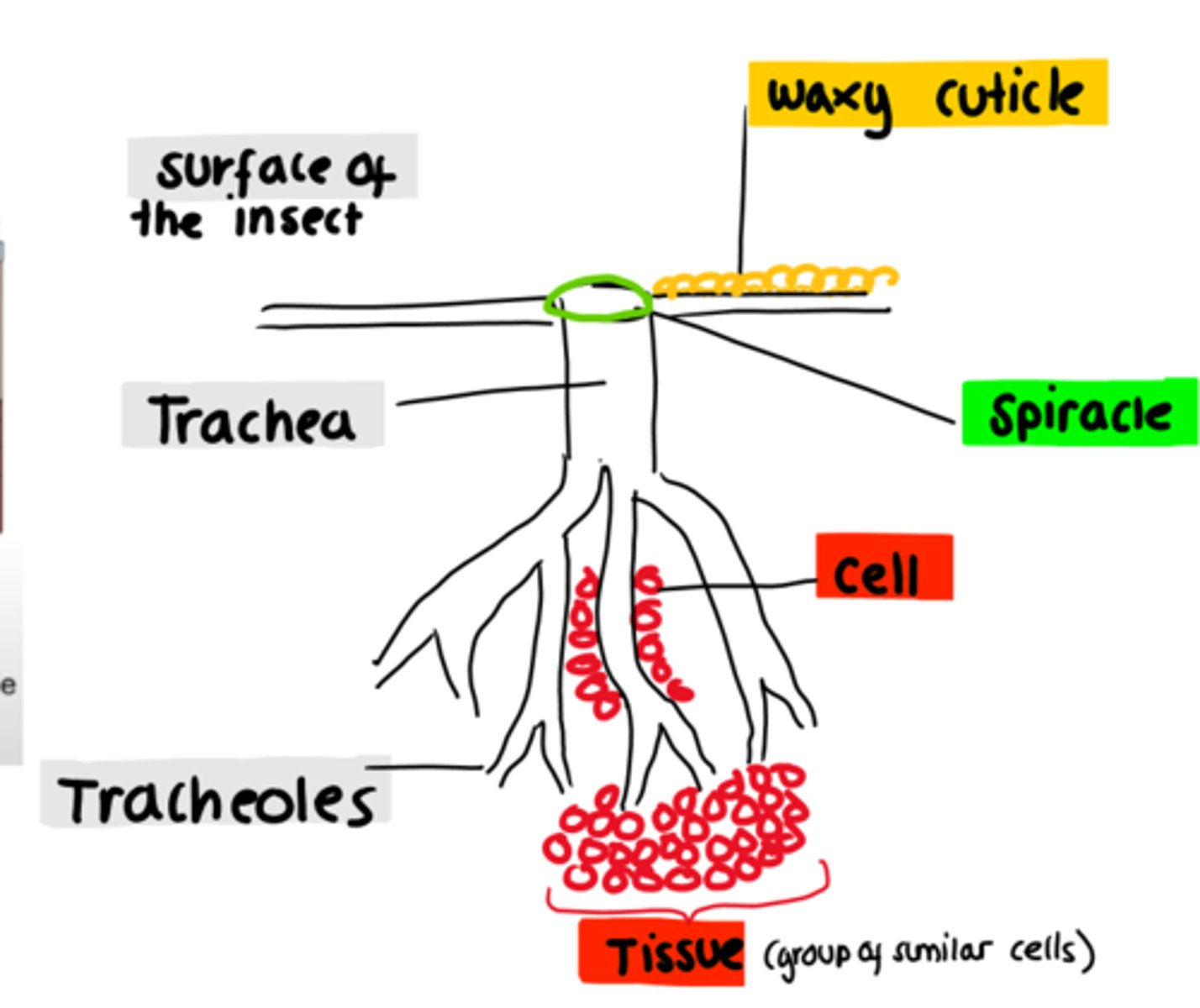
what is the insect tracheal system?
4) The tracheae branch off into smaller tracheoles which have thin, permeable walls and go to individual cells. This means that oxygen diffuses directly into the respiring cells — the insect's circulatory system doesn't transport O2
what is the insect tracheal system?
5) Carbon dioxide from the cells moves down its own concentration gradient towards the spiracles to be released into the atmosphere.
6) Insects use rhythmic abdominal movements to move air in and out of the spiracles
what are three ways of moving gases in the tracheal system?
1)gases can exchange by diffusion as when cells respire they use oxygen and produce carbon dioxide creating a concentration gradient from the trachealoes to the atmosphere
2)mass transport- insects contract and relax their abdominal muscles to move gases on mass
what are the adaptations for efficient gas exchange?
large number of tracheoles (provides large surface area)
walls of the tracheoles are thin and has a short distance from the spiracles (short diffusion pathway)
use of oxygen and production of carbon dioxide sets up steep diffusion gradient
what are the gas exchange surface feature?
1) They have a large surface area.
2) They're thin (often just one layer of epithelial cells) — this provides a short diffusion pathway across the gas exchange surface
The organism also maintains a steep concentration gradient of gases across the exchange surface.
describe fish gill anatomy:
- four layers of gill on both sides of the head
- each gill filament is covered in gill lamellae which are at a right angle to gill filaments. this allows for a large surface area
when fish open their mouth water rushes in and over the gills and through a hole in the sides of their head
what are the adaptations of the fish gas exchage?
Each gill is made of many gill filaments, which give a big surface area for exchange of gases.
The gill filaments are covered in lots of lamellae, which increase the surface area even more.
The lamellae have lots of blood capillaries and a thin surface layer of cells to speed up diffusion.
what is the counter current system?
Blood flows through the lamellae in one direction and water flows over in the opposite direction.
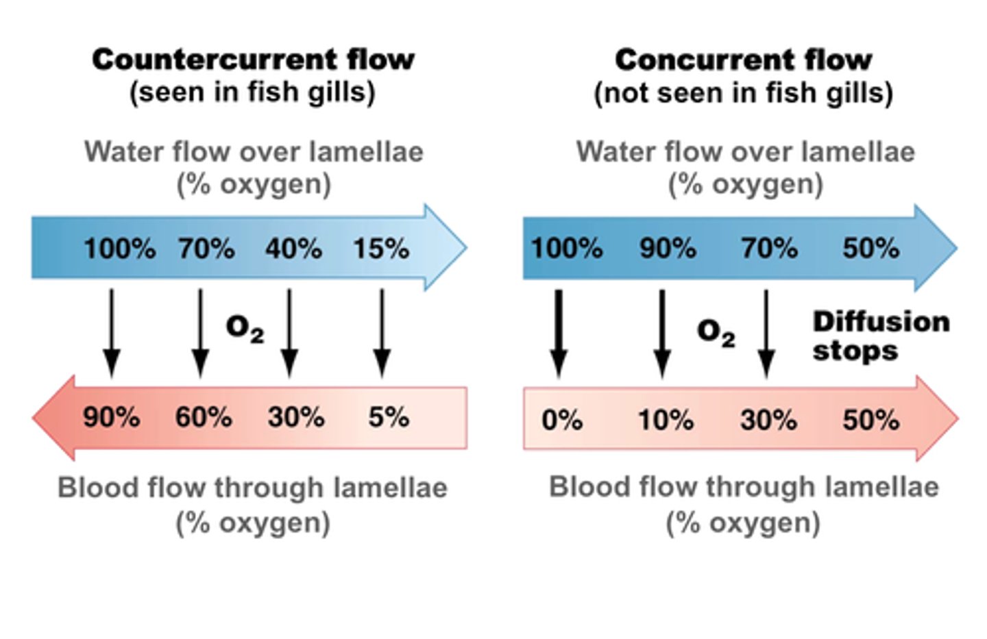
It maintains a large concentration gradient between the water and the blood. The concentration of oxygen in the water is always higher than that in the blood, so as much oxygen as possible diffuses from the water into the blood.
what is the counter current system?
counter current flow ensures equilibrium isnt reached
this ensures that a diffusion gradient is maintained across the entire length of the gill lamellae.
what happens at the gas exchange of a stomata?
The main gas exchange surface is the surface of the mesophyll cells in the leaf. They're well adapted for their function — they have a large surface area.
The mesophyll cells are inside the leaf.

what happens at the gas exchange of a stomata?
Gases move in and out through special pores in the epidermis called stomata The stomata can open to allow exchange of gases, and close if the plant is losing too much water. Guard cells control the opening and closing of stomata.
what happens at the gas exchange of a stomata?
3)Use a syringe to fill the abdomen with saline solution. You should be able to see a network of very thin, silvery-grey tubes — these are the tracheae. They look silver because they're filled with air.
how do insects and plants control water loss?
1)If insects are losing too much water, they close their spiracles using muscles. They also have a waterproof, waxy cuticle all over their body and tiny hairs around their spiracles, both of which reduce evaporation.
how do insects and plants control water loss?
2) Plants' stomata are usually kept open during the day to allow gaseous exchange. Water enters the guard cells, making them turgid, which opens the stomatal pore. If the plant starts to get dehydrated, the guard cells lose water and become flaccid, which closes the pore.
how do insects and plants control water loss?
3) Some plants are specially adapted for life in warm, dry or windy habitats, where water loss is a problem. These plants are called xerophytes.
how do insects and plants control water loss?
4)You can examine the tracheae under an optical microscope using a temporary mount slide. Again, the tracheae will appear silver or grey. You should also be able to see rings of chitin in the walls of the tracheae — these are there for support (like the rings of cartilage in a human trachea).
what are adaptations of xerophytes?
• Stomata sunk in pits that trap moist air, reducing the concentration gradient of water between the leaf and the air. This reduces the amount of water diffusing out of the leaf and evaporating away.
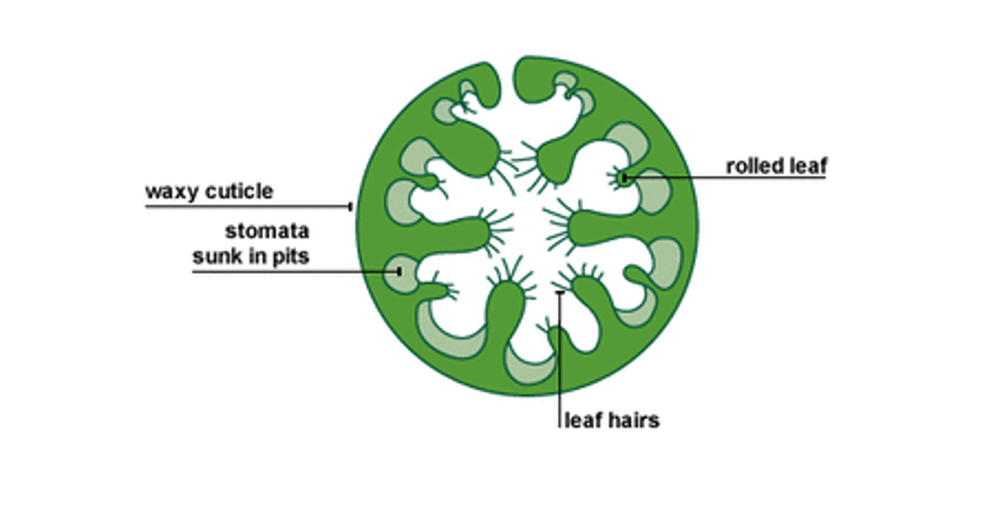
what are adaptations of xerophytes?
• A layer of 'hairs' on the epidermis — again to trap moist air round the stomata
• Curled leaves with the stomata inside, protecting them from wind (windy conditions increase the rate of diffusion and evaporation).
what are adaptations of xerophytes?
•A reduced number of stomata, so there are fewer places for water to escape
• Waxy, waterproof cuticles on leaves and stems to reduce evaporation.
what happens during digestion?
During digestion, large biological molecules are hydrolysed to smaller molecules that can be absorbed across cell membranes.
what are the enzymes used in digestion in mammals
amylase and membrane-bound disaccharidases (for carbohydrates)
lipase (for lipids)
how are carbohydrates broken down by amylase and membrane-bound disaccharidases?
1) Amylase is a digestive enzyme that catalyses the conversion of starch into the smaller sugar maltose. This involves the hydrolysis of the glycosidic bonds in starch.
2) Amylase is produced by the salivary glands and also by the pancreas.
how are carbohydrates broken down by amylase and membrane-bound disaccharidases?
3) Membrane-bound disaccharidases are enzymes (lactase and sucrase) that are attached to the cell membranes of epithelial cells lining the ileum. They help to break down disaccharides (e.g. maltose, sucrose and lactose) into monosaccharides (e.g. glucose, fructose and galactose). Again, this involves the hydrolysis of glycosidic bonds
how are proteins broken down?
Endopeptidases act to hydrolyse peptide bonds between amino acids in the middle of the polymer chain.
Exopeptidases act to hydrolyse peptide bonds between amino acids at the end of a polymer chain
membrane-bound dipeptidases hydrolyses peptide bonds between two amino acids
how are lipids digested?
1) Lipase enzymes catalyse the breakd own of lipids into monoglycerides and fatty acids. This involves the hydrolysis of the ester bonds in lipids.
2) Lipases are made in the pancreas. They work in the small intestine.
how are lipids digested?
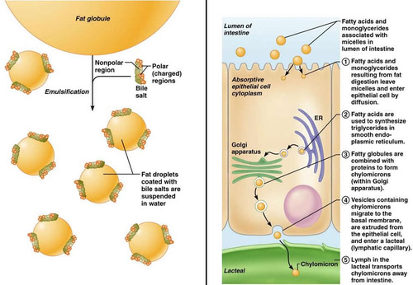
3) Bile salts are produced by the liver and emulsify lipids — this means they cause the lipids to form small droplets.
how are lipids digested?
4) Bile salts are really important in the process of lipid digestion. Several small lipid droplets have a bigger surface area than a single large droplet. So the formation of small droplets greatly increases the surface area of lipid that's available for lipases to work on.
how are lipids digested?
5) Once the lipid has been broken down, the monoglycerides and fatty acids stick with the bile salts to form tiny structures called micelles
what are micelles and outline their function?
micelles are water soluble vesicles formed of the fatty acids, glycerol and bile salts
Micelles help to move monoglycerides and fatty acids towards the epithelium.
Because micelles constantly break up and reform they can 'release' monoglycerides and fatty acids, allowing them to be absorbed — whole micelles are not taken up across the epithelium. Monoglycerides and fatty acids are lipid-soluble, so can diffuse directly across the epithelial cell membrane.
what are the features of the ileum that maximise absorption?
the walls are covered in villi which have thin walls surrounded by a network of capillaries and epithelial cells which have even smaller microvilli
this increases the surface area , decreases the diffusion gradient and maintaining a concentration gradient.
how are monosaccharides absorbed across the cell membrane?
Glucose is absorbed by active transport with sodium ions via a co-transporter protein.
Galactose is absorbed in the same way using the same co-transporter protein.
Fructose is absorbed via facilitated diffusion through a different transporter protein.
how are amino acids absorbed across the cell membrane?
Amino acids are absorbed in a similar way to glucose and galactose. Sodium ions are actively transported out of the epithelial cells into the ileum itself. They then diffuse back into the cells through sodium-dependent transporter proteins in the epithelial cell membranes, carrying the amino acids with them.
what happens during lipid absorption?
lipids are digested into monoglycerides and fatty acids by the action of lipase and bile salts.
these form tiny structures called micelles
when these micelles encounter the ileum epithelial cells,
due to the non polar nature of the fatty acids and monoglycerides, they can simply diffuse across the cell surface membrane to enter the epithelial cells
once in the cells these will be modified back into triglycerides inside the endoplasmic reticulum and Golgi body
What is haemoglobin?
A protein with a quaternary structure that carries oxygen
how is oxygen carried by haemoglobin?
1) Red blood cells contain haemoglobin .
2) Haemoglobin is a large protein with a quaternary structure
3) Each chain has a haem group, which contains an iron ion and gives haemoglobin its red colour
how is oxygen carried by haemoglobin?
4) Haemoglobin has a high affinity for oxygen — each molecule can carry four oxygen molecules.
5) In the lungs, oxygen joins to haemoglobin in red blood cells to form oxyhaemoglobin.
how is oxygen carried by haemoglobin?
6) This is a reversible reaction — when oxygen leaves oxyhaemoglobin (dissociates from it) near the body cells, it turns back to haemoglobin.
9 (a) Explain how oxygen is loaded, transported and unloaded in the blood
exam question
1. Haemoglobin carries oxygen / has a high affinity for oxygen / oxyhaemoglobin;
2. In red blood cells;
3. Loading/uptake/association in lungs;
4. at high p.O2;
5. Unloads/ dissociates / releases to respiring cells/tissues;
6. at low p.O2;
7. Unloading linked to higher carbon dioxide (concentration);
how does Haemoglobin Saturation Depends on the Partial Pressure of Oxygen?
1) The partial pressure of oxygen is a measure of oxygen concentration. The greater the concentration of dissolved oxygen in cells, the higher the partial pressure.
2) Similarly, the partial pressure of carbon dioxide is a measure of the concentration of CO2 , in a cell.
how does Haemoglobin Saturation Depends on the Partial Pressure of Oxygen?
3) Haemoglobin's affinity for oxygen varies depending on the partial pressure of oxygen: Oxygen loads onto haemoglobin to form oxyhaemoglobin where there's a high partial pressure of oxygen, Oxyhaemoglobin unloads its oxygen where there's a lower partial pressure of oxygen.
how does Haemoglobin Saturation Depends on the Partial Pressure of Oxygen?
4) Oxygen enters blood capillaries at the alveoli in the lungs. Alveoli have a high partial pressure of oxygen so oxygen loads onto haemoglobin to form oxyhaemoglobin.
how does Haemoglobin Saturation Depends on the Partial Pressure of Oxygen?
5) When cells respire, they use up oxygen — this lowers the partial pressure of oxygen. Red blood cells deliver oxyhaemoglobin to respiring tissues, where it unloads its oxygen
6) The haemoglobin then returns to the lungs to pick up more oxygen
The relationship between loading, transport and unloading of oxygen in relation to the oxyhaemoglobin dissociation curve.
Where partial pressure of oxygen is high (e.g. in the lungs), haemoglobin has a high affinity for oxygen (i.e . it will readily combine with oxygen), so it has a high saturation of oxygen. Where partial pressure of oxygen is low (e.g. in respiring tissues), haemoglobin has a low affinity for oxygen, which means it releases oxygen rather than combines with it. That's why it has a low saturation of oxygen.
what is cooperative binding?
when haemoglobin combines with the first oxygen molecule, its shape alters in a way that makes it easier for other oxygen molecules to bind too.
What is the Bohr effect?
when a high carbon dioxide concentration causes the oxyhaemoglobin curve to shift to the right. The affinity for oxygen decreases because the acidic carbon dioxide changes the shape of haemoglobin slightly.
What happens at lower partial pressure of carbon dioxide in the alveoli?
curve shifts to the left
affinity for oxygen increases and therefore haemoglobin uploads more oxygen
What happens at higher partial pressure of carbon dioxide at respiring tissues?
curve shifts to the right
affinity for oxygen decreases and therefore haemoglobin unloads more oxygen
How is haemoglobin different in different organisms?
1) Organisms that live in environments with a low concentration of oxygen have haemoglobin with a higher affinity for oxygen than human haemoglobin — the dissociation curve is to the left of ours.
2) Organisms that are very active and have a high oxygen demand have haemoglobin with a lower affinity for oxygen than human haemoglobin — the curve is to the right of the human one.
What is the circulatory system?
A mass transport system
1) Multicellular organisms, like mammals, have a low surface area to volume ratio, so they need a specialised transport system to carry raw materials from specialised exchange organs to their body cells — this is the circulatory system.
What is the circulatory system?
2) The circulatory system is made up of the heart and blood vessels.
3) The heart pumps blood through blood vessels (arteries, arterioles, veins and capillaries) to reach different parts of the body.
What is the circulatory system?
4) Blood transports respiratory gases, products of digestion, metabolic wastes and hormones round the body. There are two circuits.
One circuit takes blood from the heart to the lungs, then back to the heart. The other loop takes blood around the rest of the body. The heart has its own blood supply — the left and right coronary arteries.
role of managing pressure blood flow in the circulatory system?
The blood flows through the lung at a lower pressure. This prevents damage to the capillaries in the alveoli and also reduces the speed at which blood flows, enabling more time for gas exchange.
The oxygenated blood from the lungs then goes back through the heart to be pumped out at a higher pressure to the rest of the body. This is important to ensure that the blood reaches all the respiring cells in the body.
What are the key blood vessels?
coronary arteries
heart ( vena cava, aorta, pulmonary artery, and pulmonary vein)
lungs (pulmonary artery and pulmonary vein)
kidneys (renal artery and renal vein)
These major blood vessels are connected within the circulatory system via the arteries, arterioles, capillaries and veins
what is the function of coronary arteries?
Supply oxygenated blood to the heart muscle
function of left ventricle?
1) The left ventricle of the heart has thicker, more muscular walls than the right ventricle, because it needs to contract powerfully to pump blood all the way round the body. The right side only needs to get blood to the lungs, which are nearby
function of right ventricle?
pumps blood to the lungs. This needs to be at a lower pressure to prevent damage to the capillaries in the lungs and blood flows slowly to allow time for gas exchange, so it has thinner walls in comparison to the left ventricle.