ANSC 2202 - Appendicular Skeleton
1/50
There's no tags or description
Looks like no tags are added yet.
Name | Mastery | Learn | Test | Matching | Spaced |
|---|
No study sessions yet.
51 Terms
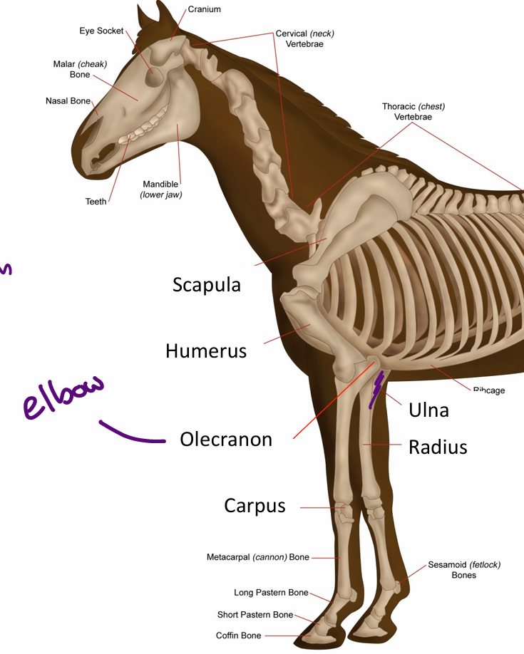
The Thoracic Limb
Divided into two parts:
Antebrachium - Forearm
Scapula
Humerus
Radius
Ulna
Manus - Distal portion (hand)
Carpus
Metacarpus: between carpus and digit(s)
Digit(s)
Olecranon
Elbow
Equine Thoracic Limb Diagram
The Ulna and Radius are sometimes fused in livestock
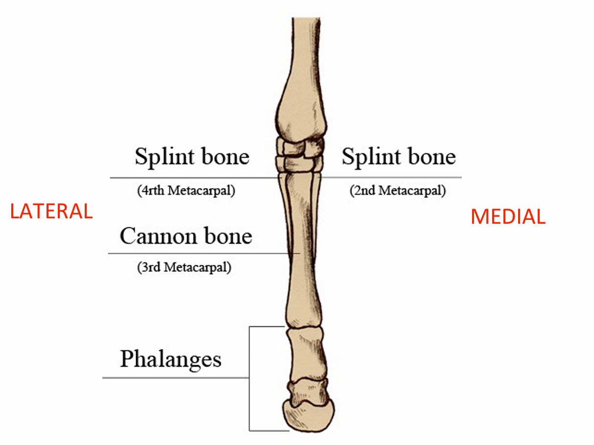
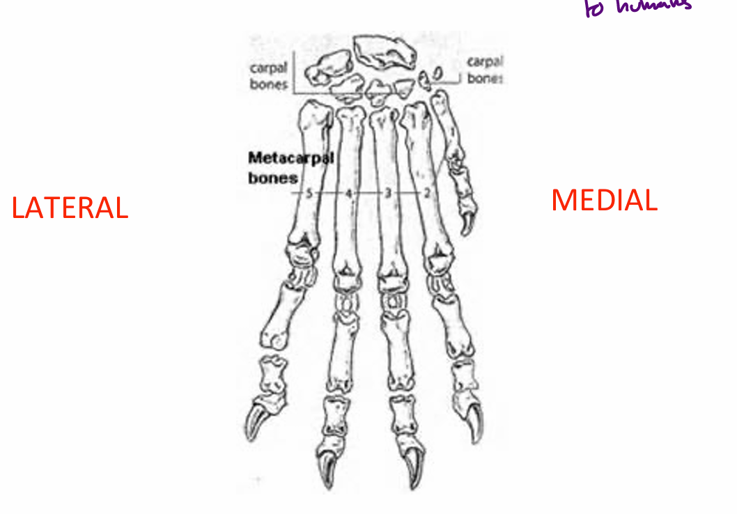
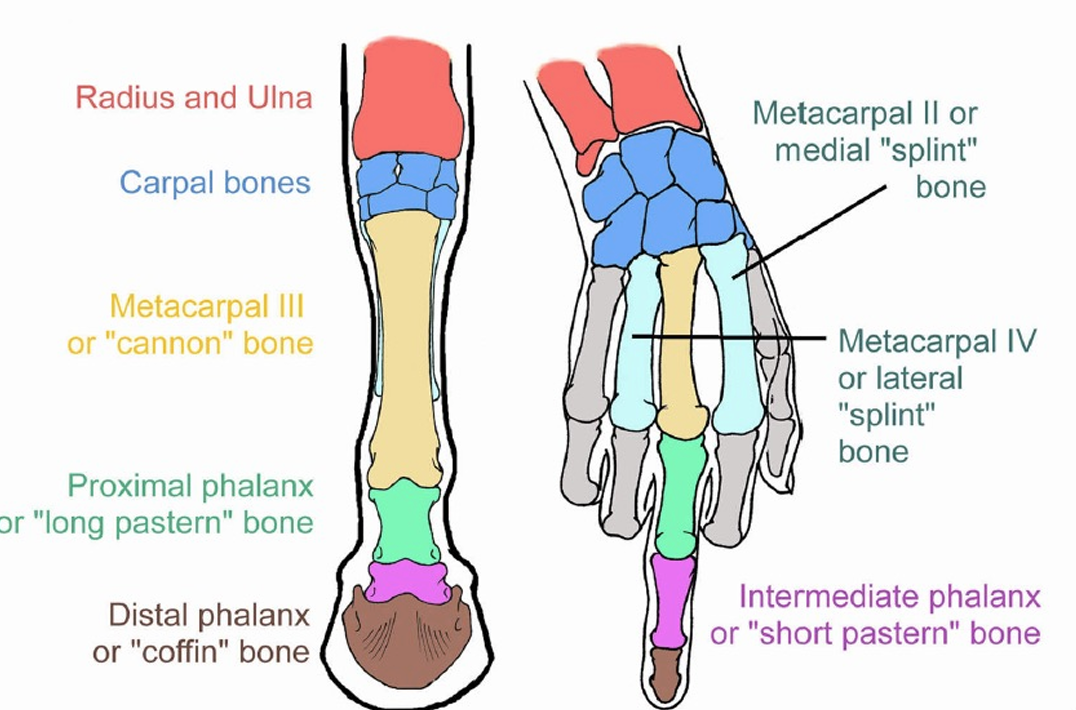
Equine: Metacarpus
Metacarpal bones II-IV (medial to lateral)
Mc II and IV are splint bones
Mc III is the cannon bone
Mc I is absent
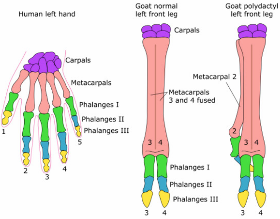
Ruminant: Metacarpus
Mc III and IV are fused to create the cannon bone
Mc V is a small metacarpal bone
Mc I and II are absent

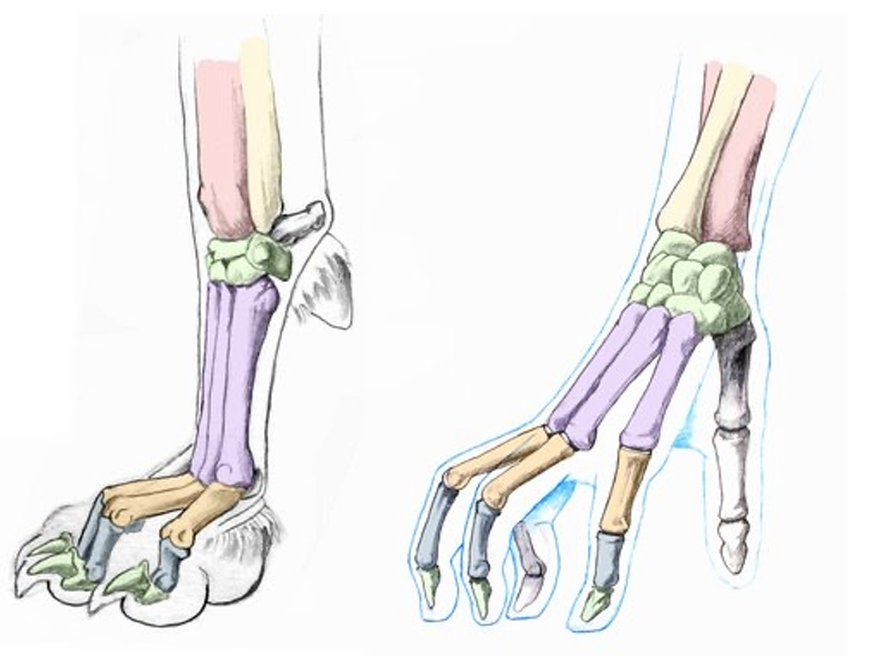
Carnivore: Metacarpus
Mc I is small and non-weight bearing (dewclaw)
Mc II-V are all present
Very similar to humans
Digits
Correspond to fingers and toes in humans; variable in number depending on the species
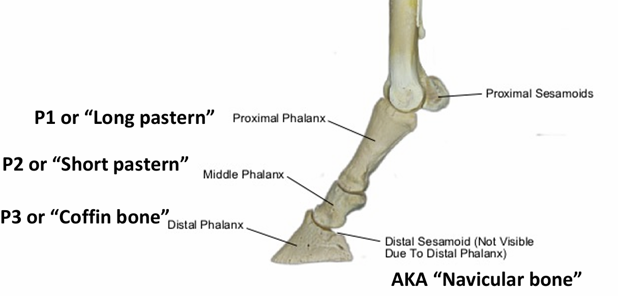
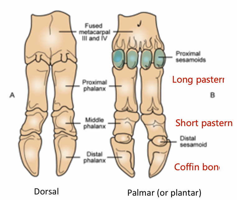
Proximal phalanx, P1 (long pastern* bone)
Middle phalanx, P2 (short pastern* bone)
Distal phalanx, P3 (coffin* bone)
Sesamoids (proximal and distal)
*Common names used in horses and ruminants
Note: Phalanges is plural for phalanx
Equine: Digits
Proximal phalanx (long pastern): one per foot
Middle phalanx (short pastern): one per foot
Distal phalanx (coffin bone): one per foot
Sesamoid bones: one proximal sesamoid (two per foot) at the fetlock joint and one distal sesamoid (one per foot) at the coffin joint
Distal also known as the navicular bone
Equine: Digits vs Humans
Compared to humans
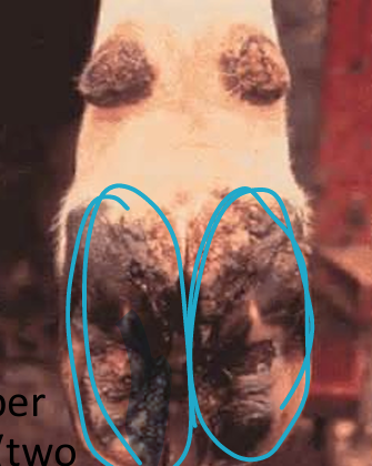
Ruminant: Digits
Two weight bearing (3rd and 4th, circled)
Two non-weight bearing (2nd and 5th)
Manifested as horny dewclaws on the palmar surface of the fetlock joint
1st digit is missing
Ruminant: Digits 2
Proximal phalanx (long pastern): two per foot.
Middle phalanx (short pastern): two per foot.
Distal phalanx (coffin bone): two per foot
Sesamoid bones: two proximal sesamoids (four per foot) at the fetlock joint and one distal sesamoid (two per foot) at the coffin joint.
Ruminant: Digits vs Humans
Compared to humans
Carnivore: Digits 2
Four weight bearing digits (II-V)
Non-weight bearing dewclaw (I)
May not exist
Carnivore: Digits 2
Proximal phalanx
Middle phalanx
Distal phalanx
Ungual Process
Horny Claw
Sesamoid bones: two sesamoid bones on the palmar/plantar surface of each metatarsophalangeal joint of each main digit (distal sesamoid bones are cartilaginous
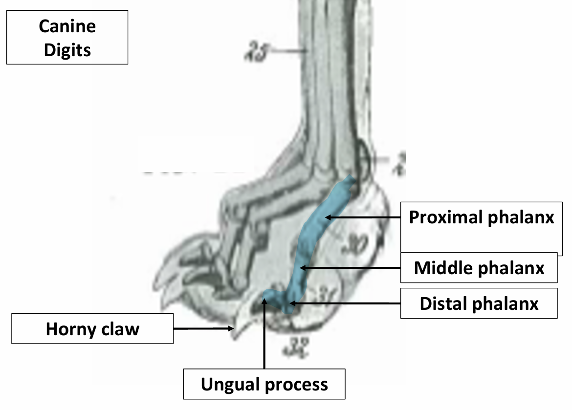
Ungual Process
a curved, cone-like extension of the distal phalanx that is covered by the horny claw
Horny Claw
a curved, fingernail-like projection covering and protecting the ungual process (this is the toenail)
Carnivore: Digits vs Humans
Compared to humans
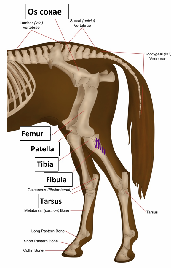
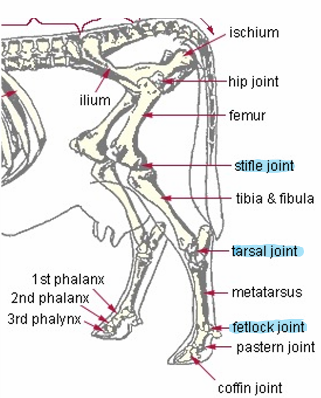
Pelvic Limb
Proximal portion
Os Coxae
Femur
Patella
Tibia
Fibula
Pes - hindpaw/distal portion of the pelvic limb (foot)
Tarsus
Metatarsus (Mt): between tarsus and digit(s)
Digit(s): phalanges and sesamoid bones
Equine Pelvic Limb Diagram
The fibula is fibbing, so it hides behind the tibia
Tibia and fibula are sometimes fused in livestock
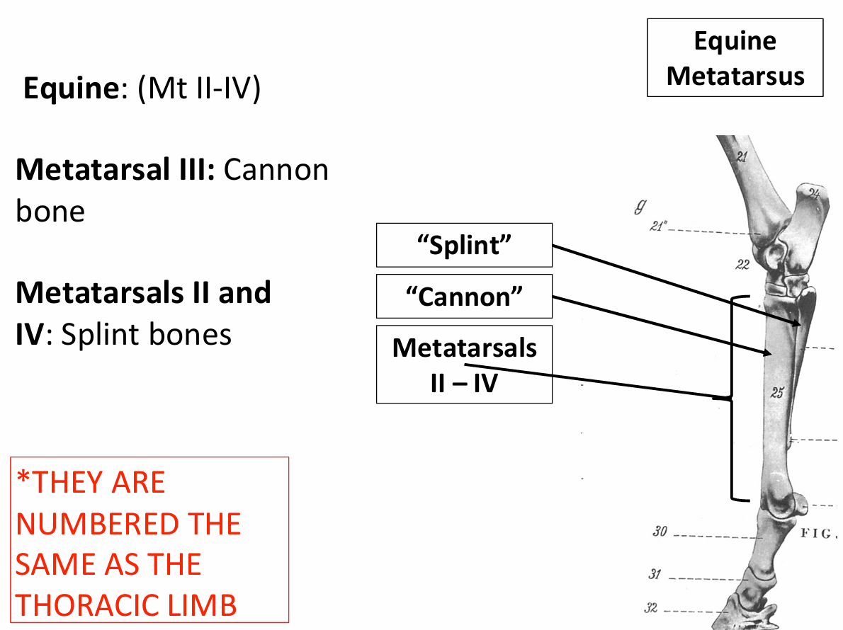
Equine: Metatarsals
Mt II-IV
Mt III is the cannon bone
Mt II-IV are splint bones
They are numbered the same as the thoracic limb
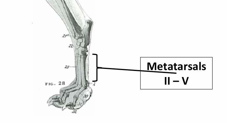
Ruminant: Metatarsals
Mt III and IV are fused to create the cannon bone
Mt II is a small metatarsal bone
Mt I is absent
Canine: Metatarsals
Mt I is small and non-weight bearing
Dewclaw — often absent
Mt II-V are all present
Do the pelvic limb digits differ from the thoracic limb digits?
No, they are the same.
Anthrology
The study of joints
Articulation
Two or more bones are united or joined to create a joint
How are joints classified? What are the classifications?
Classified based on the number of bones that articulate with one another
Simple joints: two bones that articulate with one another
Vertebrae, shoulder
Compound joints: more than two bones articulating with one another
Carpus, tarsus, fetlock
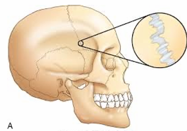
Fibrous joint
Often temporary
Little or no movement
Skull
Young animals
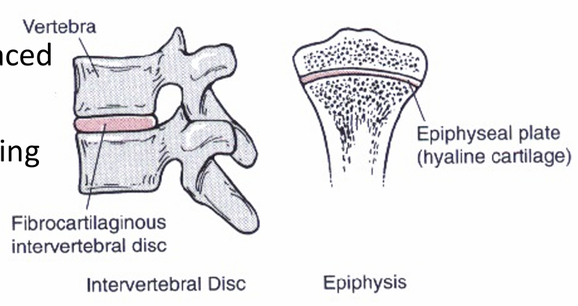
Cartilaginous joint
United by fibrocartilage or hyaline cartilage
Limited movement, such as compression or stretching (vertebrae)
Cartilage is replaced by bone when the animal stops growing
Not in vertebrae, sometimes in epiphyseal plate
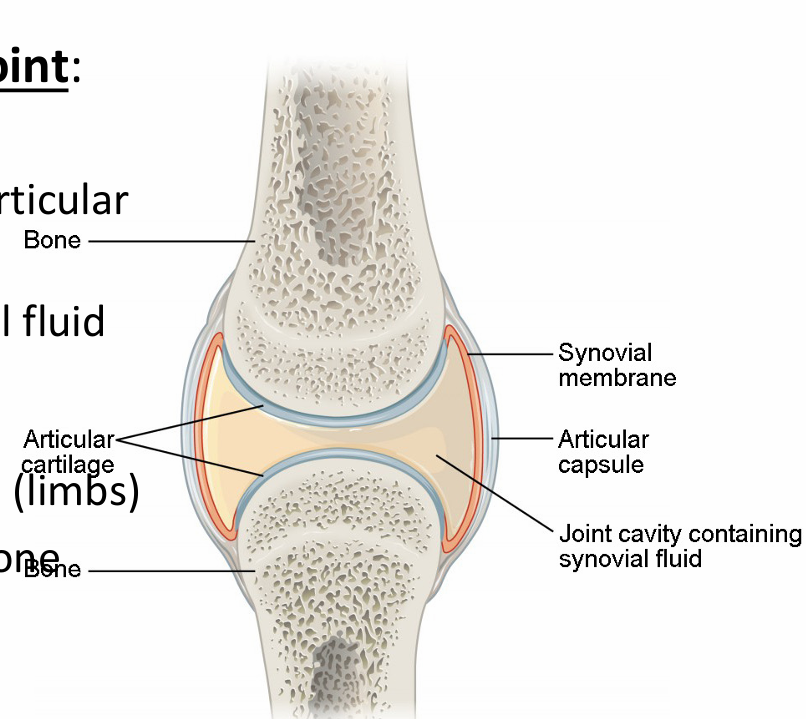
Synovial joint
“True joint”
Encasulated by articular capsule
Filled with synovial fluid
Made by synovial membrane
High motion joints (limbs)
Cartilage covers bone ends
Scapulohumeral/shoulder joint
Scapula and humerus

Cubital/elbow joint
Humerus, radius, and ulna
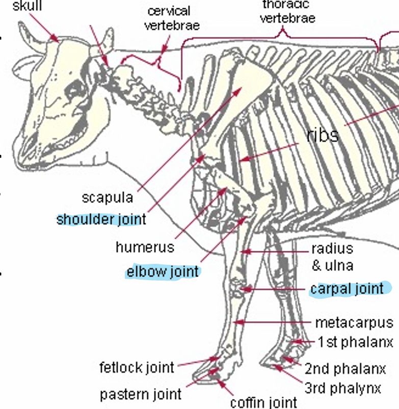
Carpus/carpal joint
Between radius/ulna and metacarpal(s)

Metacarpophalangeal/fetlock* joint
Metacarpal(s), proximal phalanx and proximal sesamoid bones
*Equine/Ruminant term, not used for carnivores
Proximal interphalangeal/pastern* joint
Proximal and middle phalanx
*Equine/Ruminant term, not used for carnivores
Distal interphalangeal/coffin* joint
Middle and distal phalanx
*Equine/Ruminant term, not used for carnivores
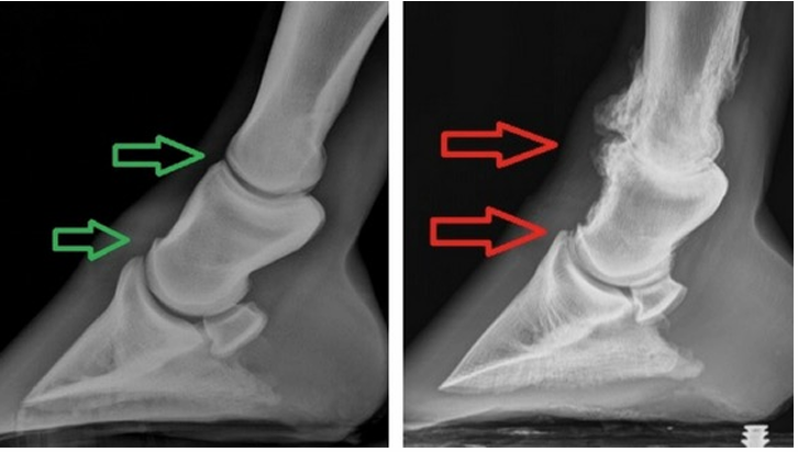
Ringbone (arthritis)
Periosteal bone deposition in the distal limb
High ringbone: deposition at the pastern joint
Low ringbone: deposition at the coffin joint
Onychectomy
Declawing
Removal of the distal phalanx by disarticulating the distal interphalangeal joint
Onych = nail
Sacroiliac joint
Sacrum and ilium
White arrow
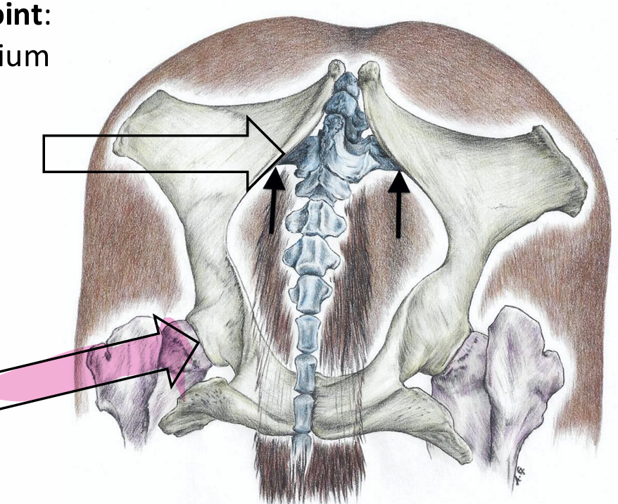
Coxofemoral/hip joint
Acetabulum and femur
Pink arrow

Stifle joint
Femur, patella, tibia (and fibula)cetabulum and femur
Tarsus/Tarsal/Hock joint
Between tibia and metatarsal bone(s)

Metatarsophalangeal/fetlock joint
Metatarsal(s), 1st phalanx and proximal sesamoid bones

Are the joints on the digit(s) of the pelvic limb the same as the thoracic limb?
Yes, they are the same.
Tendons
Story energy
Support weight on skinny legs
Allow animal to sleep standing
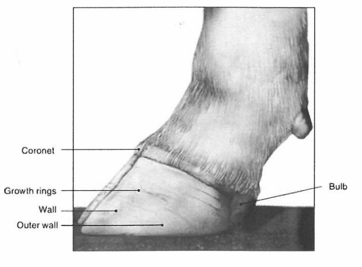
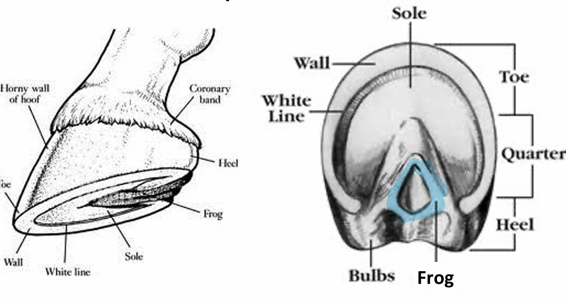
The Hoof
Wall: visible outer portion of the hoof
Bulbs of the heels: soft expanded part on caudal aspect of hoof
Coronet/coronary band: where hair meets hoof; hoof grows downward from here (like human cuticles)
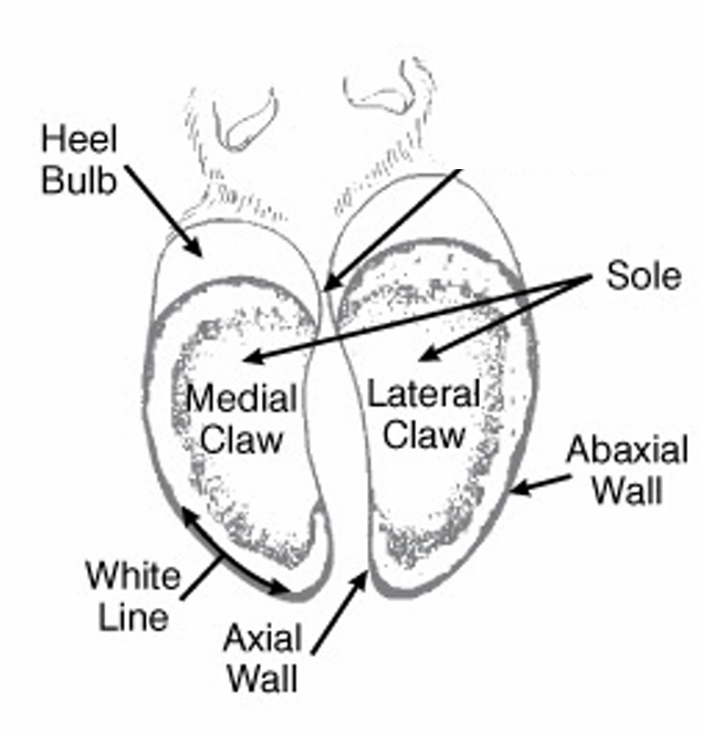
Ruminant Hoof
Sole: concave portion of the hoof visible on the ground surface
White line: Junction between wall and sole
Insensitive - where nails go in horse shoes
Equine Hoof and the Frog
Frog: V-shaped structure that allows expansion of the foot at the heel (only in horses)
Equine Hoof and the Digital Cushion
Wedge-shaped mass of fibrous and fatty tissue that is located INSIDE the hoof and functions to absorb concussive forces
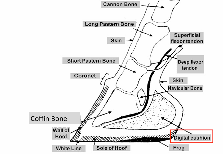
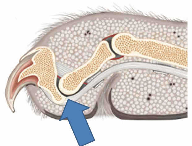
Laminae
Connects the hoof wall to the distal phalanx (coffin bone)
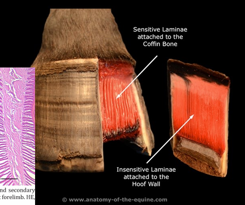
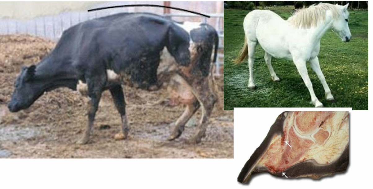
Laminitis
“Founder”
Degeneration or failure of the attachments between P3 and inner hoof wall.
May result in the rotation of the coffin bone
Lameness and even puncturing of the sensitive laminae