Introduction to Embryology - Basic Science Core
 Call Kai
 Learn
Learn Practice Test
 Spaced Repetition
 Match
Match Flashcards
 Knowt Play
1/112

Earn XP
Description and Tags
Embryology
Name | Mastery | Learn | Test | Matching | Spaced | Call with Kai | Chat |
|---|
No analytics yet
Send a link to your students to track their progress
113 Terms
Define Embryology
Is the branch of Biology studying the prenatal development of gametes, fertilization and the development of the embryos and fetuses
Three Anatomical Planes
1) Sagittal Plane
2. Coronal Plane
3. Transverse Plane
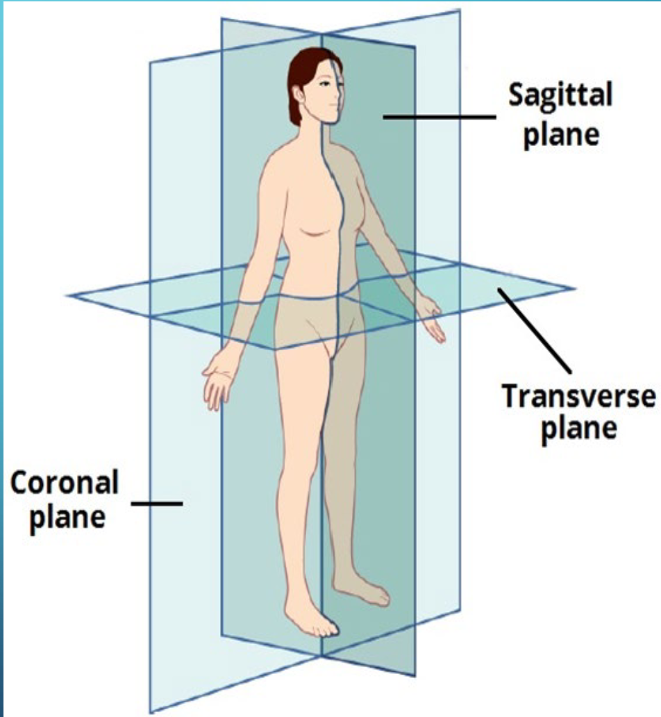
Sagittal Plane
Vertical plane that divides the body into left and right
Coronal Plane
Vertical plane that divides the body into front(anterior) and back(posterior)
Transverse Plane
Horizontal plane that divides the body into upper (superior) and lower(inferior)
Chromosomes in the human Genome
Human genome consists of 46 chromosomes total
46 chromosomes are arranged in 23 pairs (23 inherited from maternal parent & 23 inherited from paternal parent)
22 pairs are autosomes
1 pair are sex chromosomes
Chromosomes are Composed of?
DNA complexed with proteins
A means of packaging a large amount of information in a small space
Genes in the human genome
~23,000 genes in the human genome
• 1 gene → many different proteins
• Function of various splicing/editing mechanisms
• Proteins regulate gene expression and act as signaling molecule
Gene
Genes are segments of DNA that act as the instruction manual for your body on how to make proteins
Induction
One group of cells or tissues causes another to change their fate
How cells communicate to try to change
Inducer in cells
Produces a signal
Cells do this to communicate
Responder
Reacts to the signal
Cells do this to communicate
Ability for cells to respond to signals is called?
Competence
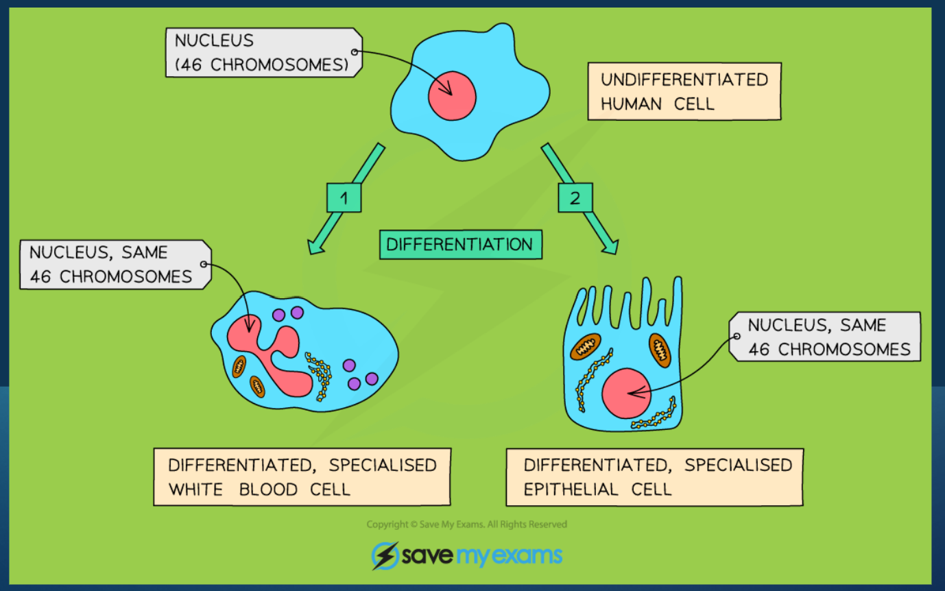
Differentiation (Cells)
A process where unspecialized stem cells or progenitor cells develop into mature, specialized cell types with unique structures and function
INDUCTION & ORGAN FORMATION
In Cells Signaling errors may occur (2)
Either genetic or environmental factors or BOTH can result in signaling errors
2 of many examples:
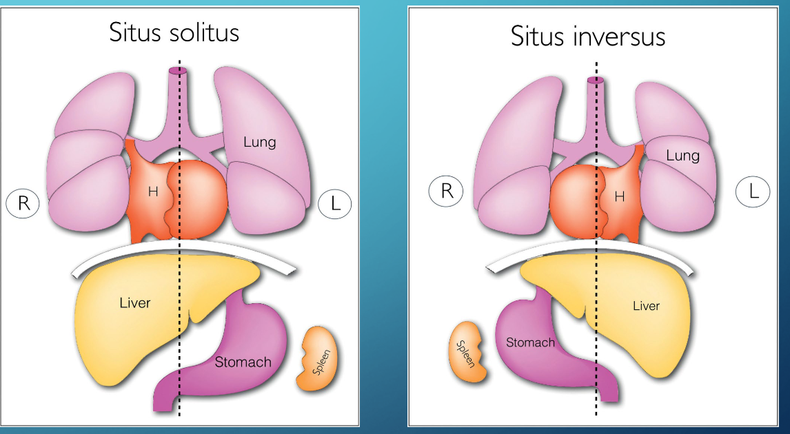
1) Situs Inversus
2) Heterotaxy
Situs Inversus:
condition where the positioning of
all organs is reversed in a mirror image
arrangement
Heterotaxy
one or more organs is abnormally
placed
Gametogenesis
The process by which gametes (germ cells) are produced in an organism
Gametes are derived from primordial germ cells (PGCs)
in the second week of fetal development
• PGCs arrive in the developing gonads of the genetically
female or male embryo by 5 th week
Female Gametogenesis
Female gamete (ova/egg cell) forms through process
called oogenesis.
Male Gametogenesis
Male gamete (sperm) forms through spermatogenesis
PGCs
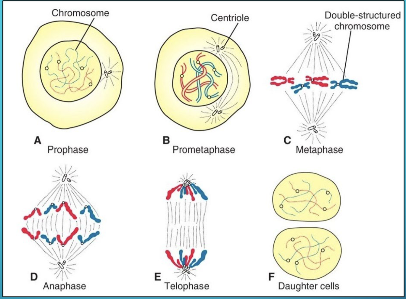
Mitosis (Cell Division)
germ cells (future mature gametes) first undergo mitosis
Cell division → 2 daughter cells that are genetically identical
Each cell receives the complete complement of 46 chromosomes
Meiosis
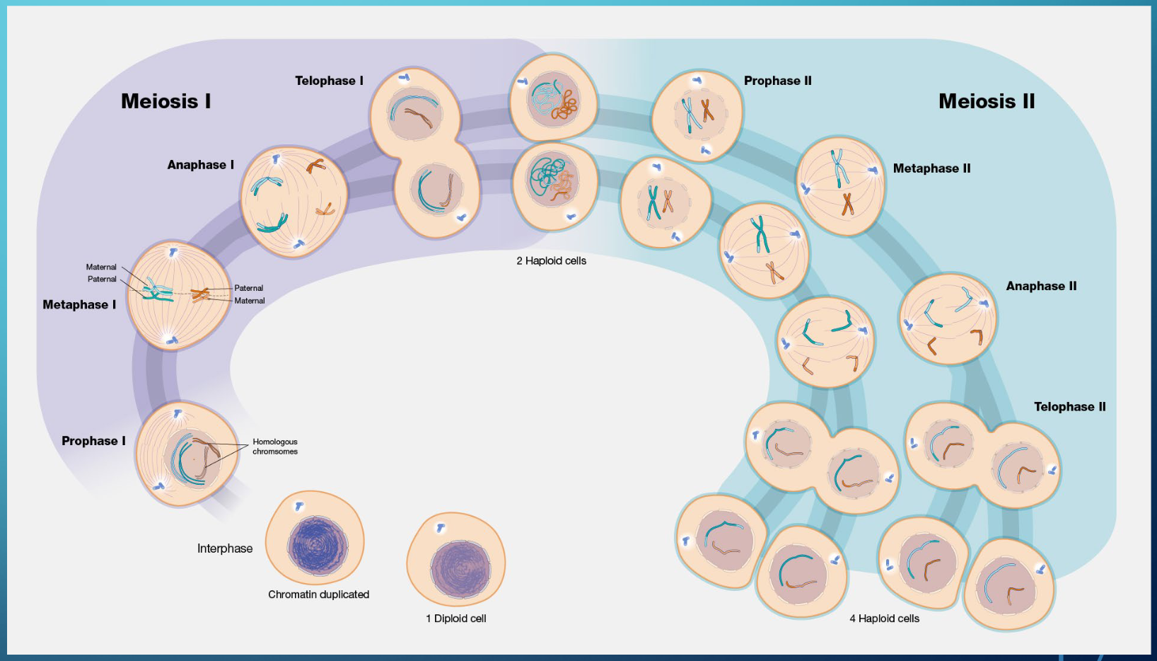
Future mature gametes then must undergo Meiosis
to reduce chromosome number (this process
completes at different times in males and
females)
• 2 successive meiotic divisions: Meiosis I and
Meiosis II
• Meiosis I reduces chromosome number to
23
• Meiosis II resembles mitosis
• Results in reduction of chromosomes from diploid
(46) to haploid (23)
Diploid
a cell or organism that has paired
chromosomes, one set from each parent
Haploid
Cell or organism that has a single set of chromosomes
Oogenesis
THE PROCESS BY WHICH OOGONIA BECOME MATURE OOCYTES
PGCs undergo mitosis, enter the female gonad(5th week), and
differentiate to oogonia
• @ 3 months: some oogonia cells synchronously arrest
division in prophase of meiosis 1 primary oocytes
• All primary oocytes are formed before birth
• Primary oocytes arrested in prophase 1 remain arrested
until puberty
• First meiotic division is completed at puberty
• Each ovarian cycle: Meiosis I Meiosis II, cell arrests
in metaphase and only completes meiosis II if fertilized
• 1 primary oocyte only produces 1 mature gamete
Oogenesis Extra, (she is flying make this latter)
Meiosis I completes when female starts ovulation
• Meiosis II starts but is only completed if secondary oocyte fertilization occurs
• If fertilization occurs secondary oocyte completes meiosis II and form mature oocyte +
second polar body
• OVERALL: If fertilization occurs
• 1 primary oocyte 1 mature oocyte and polar bodies
• Polar bodies are not fertilized and degenerate
• Ovarian follicles are simultaneously undergoing maturation and provide the support
for oocyte growth and maturation
• Corpus luteum secretes hormones to prepare the uterus for pregnancy
• If fertilization does not occur corpus luteum degenerates to corpus albicans
Spermatogenesis
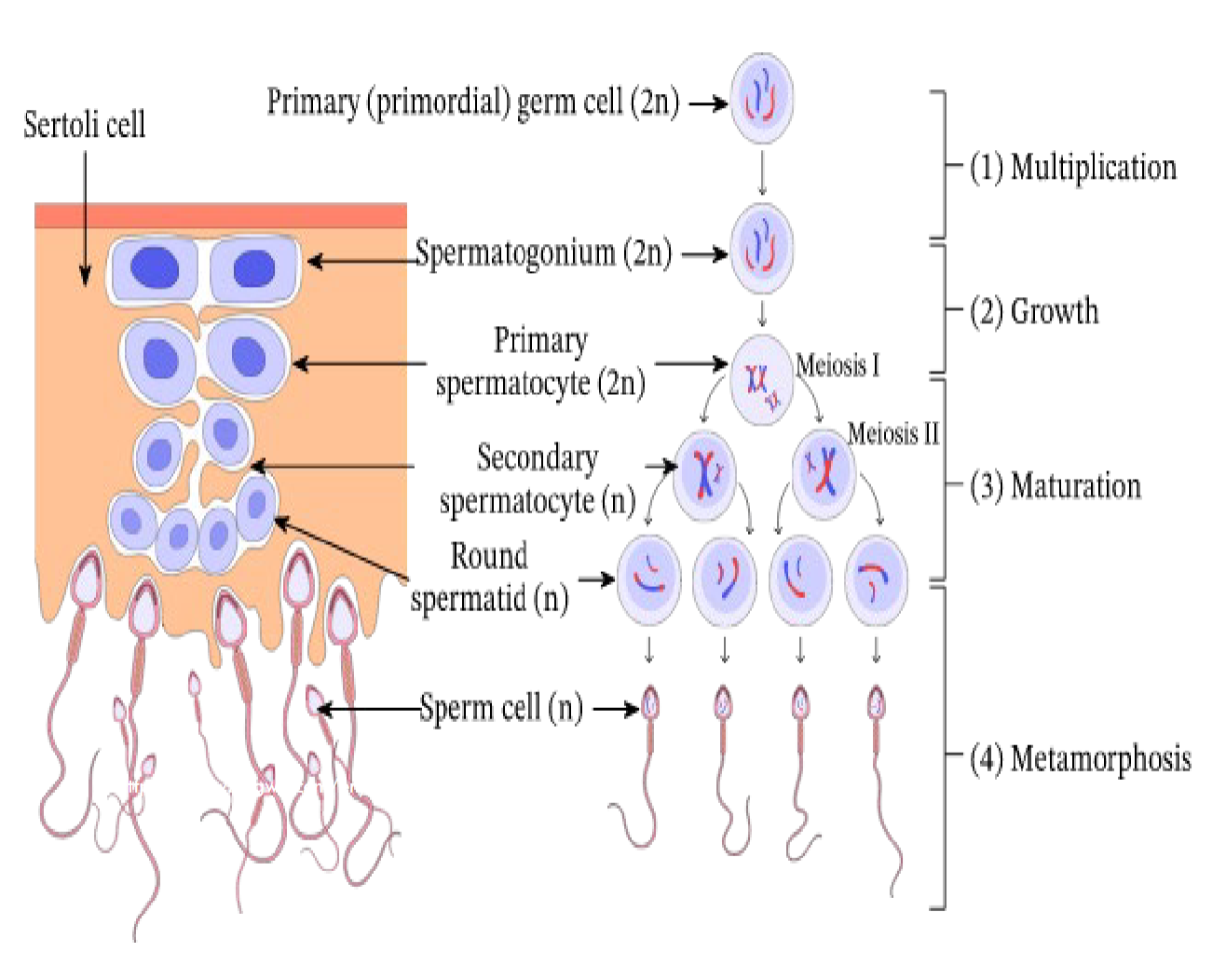
THE PROCESS OF SPERM CELL DEVELOPMENT
PGCs arrive in the genetically male gonad
(now called spermatogonia), undergo several
cycles of mitosis, and then enter mitotic arrest
• Spermatogonia remain dormant until puberty
when spermatogenesis begins
• Spermatogenesis: Spermatogonia (2n)
spermatocyte stages (2n n) spermatids (n)
mature spermatozoa (n)
• Process lasts about 74 days
• No formation of polar bodies
• 1 primary spermatocyte 4
mature spermatozoa
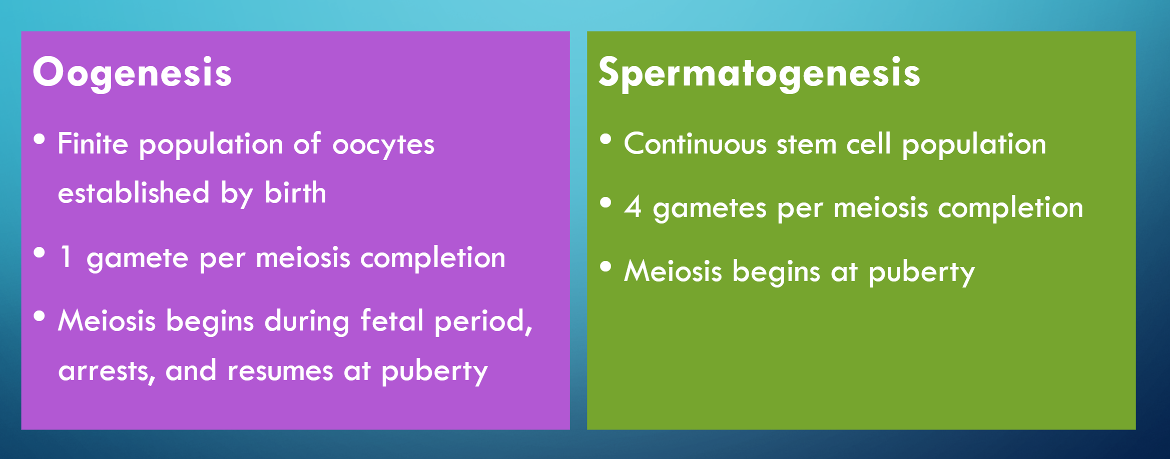
Major Difierences between Oogenesis vs Spermatogenesis
Finite population of oocytes established by birth vs Continuous stem cell population
How many pregnancies today result in a miscarriage?
As many as 50% of pregnancies end in miscarriage (spontaneous abortion)
EXTRA CHROMOSOME(S) in kids
More common then fewer chromosomes
Examples: Trisomy (3 copies of a chromosome), Klinefelter syndrome XXY
FEWER CHROMOSOMES
(absence of a chromosome from a pair); Turner syndrome XO
Death
Trisomy 21
Down Syndrome
Abnormality is due to the presence of an extra copy of
chromosome 21
Clinical features:
• Varying degrees of intellectual disability
• Craniofacial abnormalities, including upward slanting eyes, epicanthal folds
(extra skin folds at the medial corners of the eyes), flat facies, and small
ears
• Cardiac defects
• Hypotonia
• Increased risk of: leukemia, infections, thyroid
dysfunction, and premature aging
• Head and neck manifestations: macroglossia,
gingivitis, premature periodontitis
Cytogenetic analysis (She said she is not going to ask about it much)
Examination of the number of chromosomes (ploidy) and
their integrity
2. Microarrays
Small sequences of DNA on chips act as
probes to detect mutations
3. Exome sequencing
Coding regions only of DNA are sequenced
and assessed for abnormalities
FERTILIZATION AND IMPLANTATION
1) Development begins with fertilization (usually occurs in ampulla of fallopian tube)
2) Haploid (1n) gametes (sperm and oocyte) fuse to form diploid (2n) zygote
• Oocyte from female: 22 autosomal chromosomes + 1 sex chromosome (X)
• Sperm from male: 22 autosomal chromosomes + 1 sex chromosome (X or Y)
• Genetic male zygote: XY
• Y chromosome carries SRY gene that dictates testis differentiation
• Genetic female zygote: XX
• Absence of SRY gene ovary developme
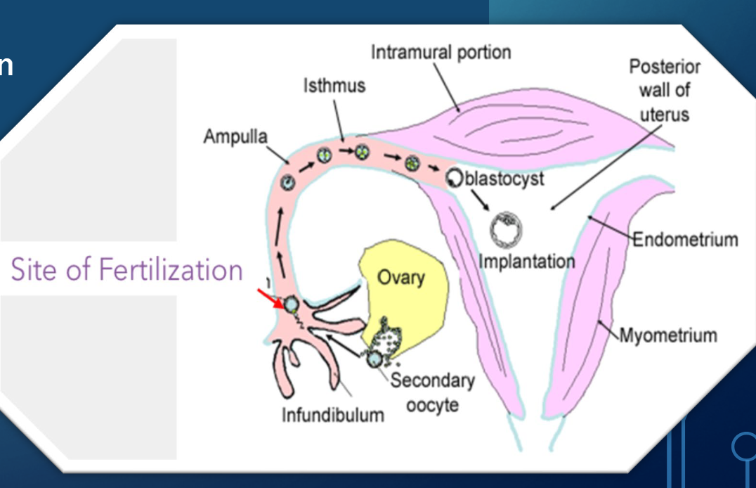
Once a spearm meets an egg what is it now considered?
A Zygote
At this point the male cell will determine the sex of the child
Time Line of Human Development
Birth typically in 38 weeks
First Trimester is a big no/no. Do not do dental procedures at this time
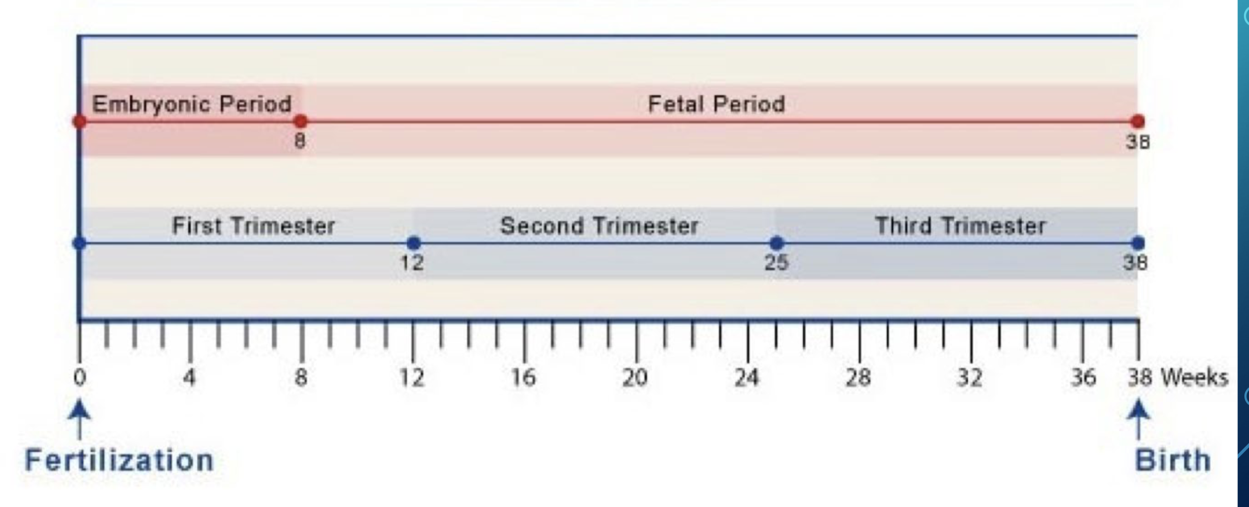
The Embryonic Period Timeline
1) 0 to 8 weeks
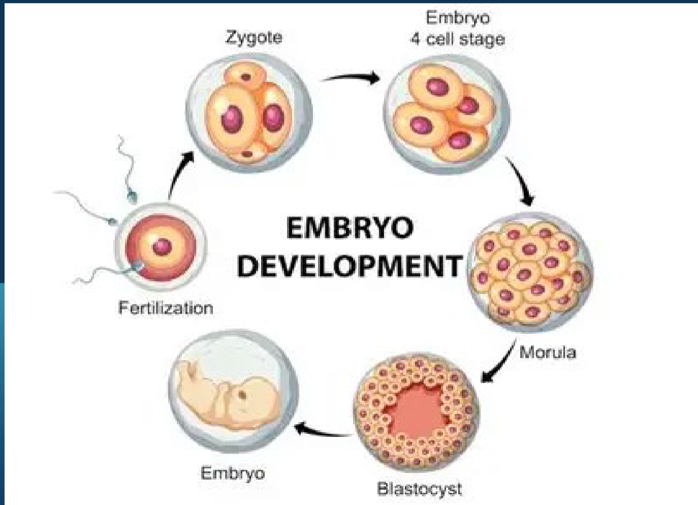
The Embryonic Period ZYGOTE Division (3)
@ 16 cells (approx. 3 days) is called a morula
Begins to separate into inner cell mass and outer cell mass
1) two cell stage
2) four cell stage
3) Morula
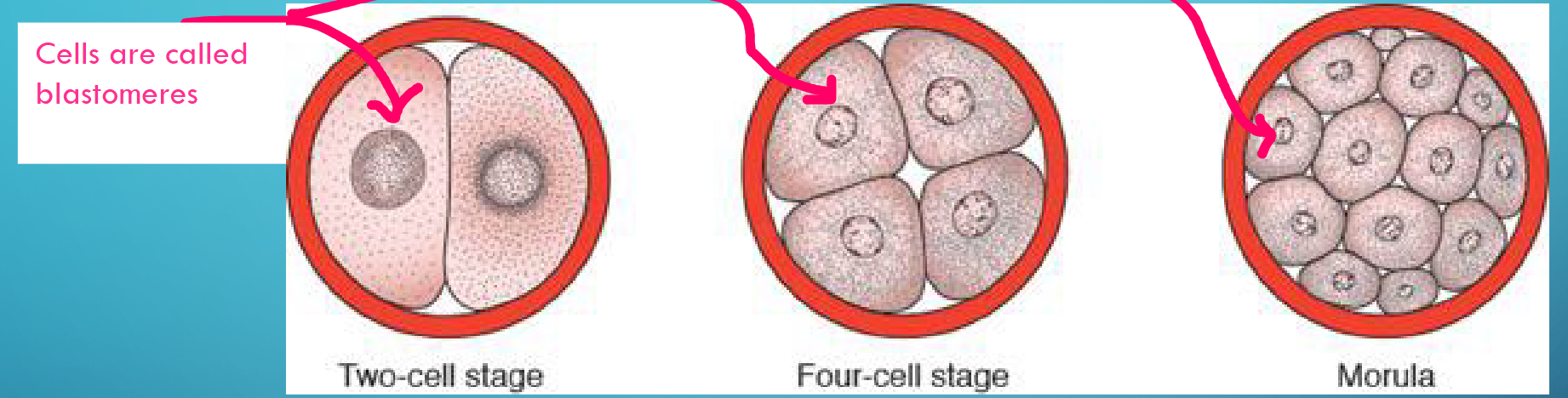
Blastomere
Cells in Zygotic Division
Blastocyst Formation
Morula travels to uterine cavity
• Fluid begins to penetrate and forms a single
cavity called the blastocele
• Embryo at this stage is called the
blastocyst
• Blastocyst implants in the endometrium
along the anterior or posterior wall
Blastocyst parts (2)
1) Inner cell mass → embryoblast
2) Outer cell mass → trophoblas
What happens by the first week of The Embryonic Period?
By the end of the first week the zygote morula blastocyst begins implantation in the uterine mucosa
Pluripotent
able to give rise to many different cell types
ABNORMAL IMPLANTATION
Implantation outside of the uterus can take place and results in extrauterine pregnancy → Ectopic pregnancy
Ectopic pregnancy
Can occur any place in abdominal
cavity, ovary, uterine tube
• 95% occur in the fallopian tube
• If left to grow can cause adjacent
organ damage or life-threatening
blood loss
MATERNAL IMMUNITY
Women, in order to not reject the embryo (which is ½ “foreign”), need to undergo an immune system shift
Shift from cell-mediated immunity to humoral (antibody-mediated) immunity
Not fully understood but may increase mothers' risk of infections
Change in immune system may alter symptoms of a mother’s autoimmune disease
• Ex. Multiple sclerosis, Rheumatoid arthritis are cell-mediated and show improvement
during pregnancy
• Ex. Systemic lupus erythematosus is predominantly antibody mediated and may
worsen during pregnancy
SECOND WEEK OF DEVELOPMENT
By second week human chorionic gonadotropic (hCG) hormone is in
significant quantities to be detected and are used for pregnancy testing
The week of the 2’s
Trophoblast(outer cell mass) differentiates into 2 layers:
cytotrophoblast and syncytiotrophoblast
* Embryoblast(inner cell mass) forms 2 layers: epiblast and hypoblast
• 2 cavities form: the amniotic and yolk sac cavities
• Extraembryonic mesoderm splits into two layers
Trophoblast cells during Week 1 & 2
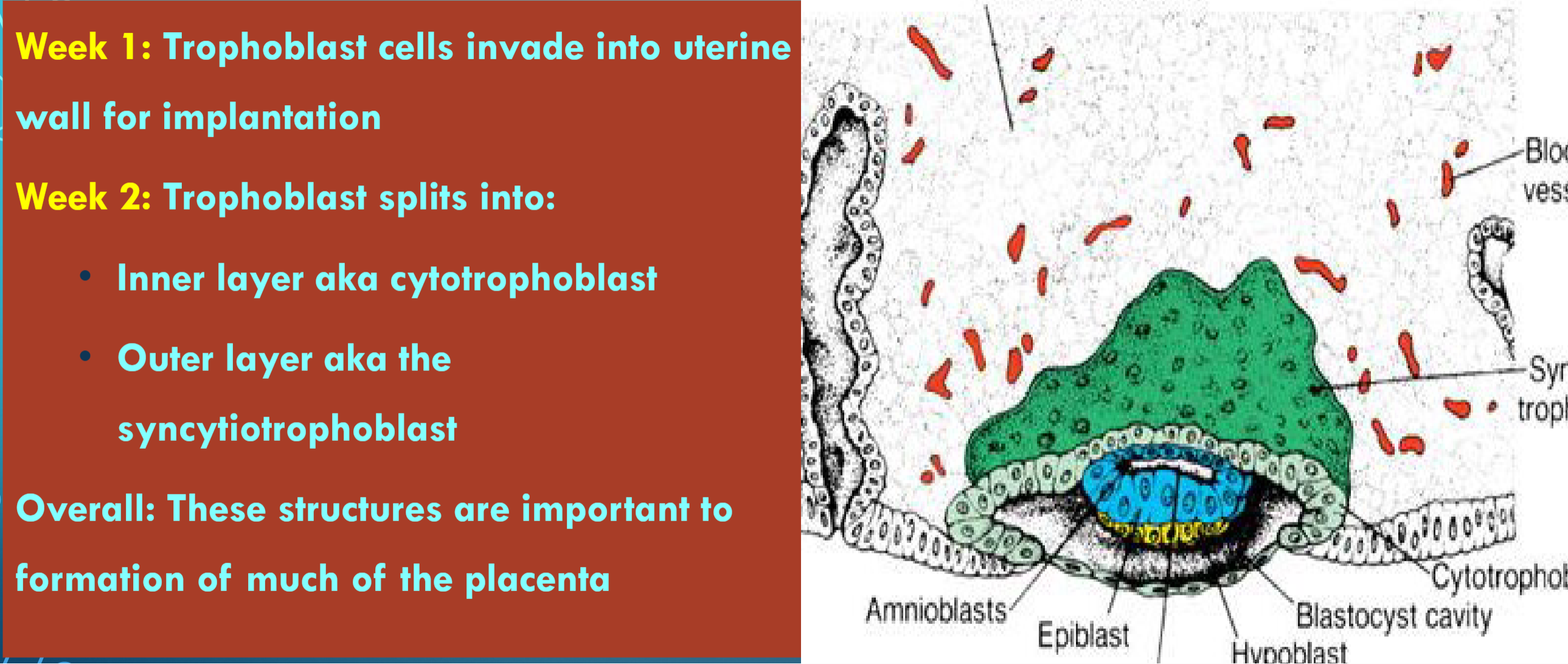
Embryoblast (Inner Cell Mass)
Embryoblast (inner cell mass) differentiates into epiblast and hypoblast
Epiblast
Definition
Epiblast: One of the two cell layers formed from the embryoblast; it becomes the dorsal layer of the bilaminar disc.
Answer
Embryoblast (inner cell mass) differentiates into epiblast and hypoblast.
These cells migrate and orient such that epiblast is dorsal.
Together with the hypoblast, it forms the bilaminar disc.
Hypoblast
Definition
Hypoblast: One of the two cell layers formed from the embryoblast; it becomes the ventral layer of the bilaminar disc.
Answer
Embryoblast (inner cell mass) differentiates into epiblast and hypoblast.
These cells migrate and orient such that hypoblast is ventral.
Together with the epiblast, it forms the bilaminar disc.
Some cells of the hypoblast differentiate into the anterior visceral endoderm (AVE).
Bilaminar Disc
Definition
Bilaminar disc: A two-layered embryonic disc formed by the epiblast and hypoblast.
Answer
Both of these layers together form the bilaminar disc.
The epiblast is the dorsal layer.
The hypoblast is the ventral layer
Anterior Visceral Endoderm (AVE)
Definition
Anterior visceral endoderm (AVE): Specialized cells that develop from the hypoblast and are important for early embryo patterning.
Answer
Some cells of the hypoblast differentiate into the anterior visceral endoderm (AVE).
AVE cells migrate to the future cranial site of the embryo.
AVE cells are important for signaling body axes
Amniotic cavity
Small cavity appears within the epiblast
Primitive yolk sac
Cavity forms between hypoblast and exocoelomic membrane (formed from cells of hypoblast)
THE YOLK SAC: SECOND WEEK Main Key Functions
Early Nutrition
• Gas exchange
• Hematopoiesis
• Germ cell Development
• Contributing to organ formation
• Early Immune and Metabolic Support
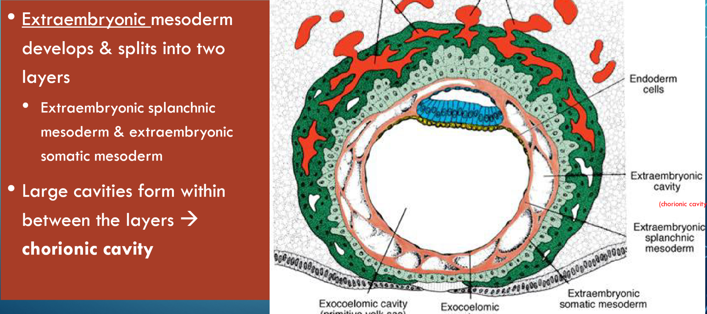
Amnion
refers to a membranous structure
which covers and protects the embryo
• Forms inside the chorion
Chorion
Chorion is a double-layered membrane formed by the trophoblast and the extra-
embryonic mesoderm, which eventually will give rise to the fetal part of the
placenta
Chorion & Amnion
These two membrane structures fuse later in development
THE TRILAMINAR DISC
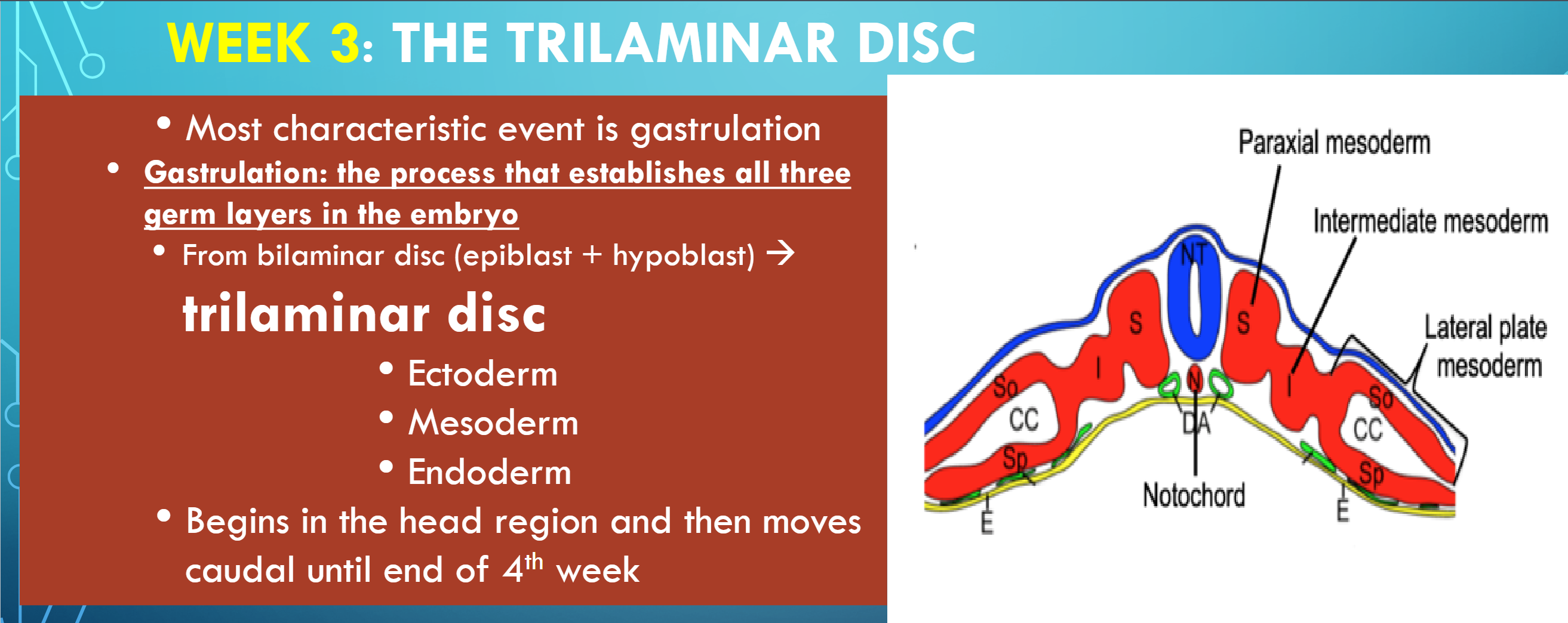
GASTRULATION
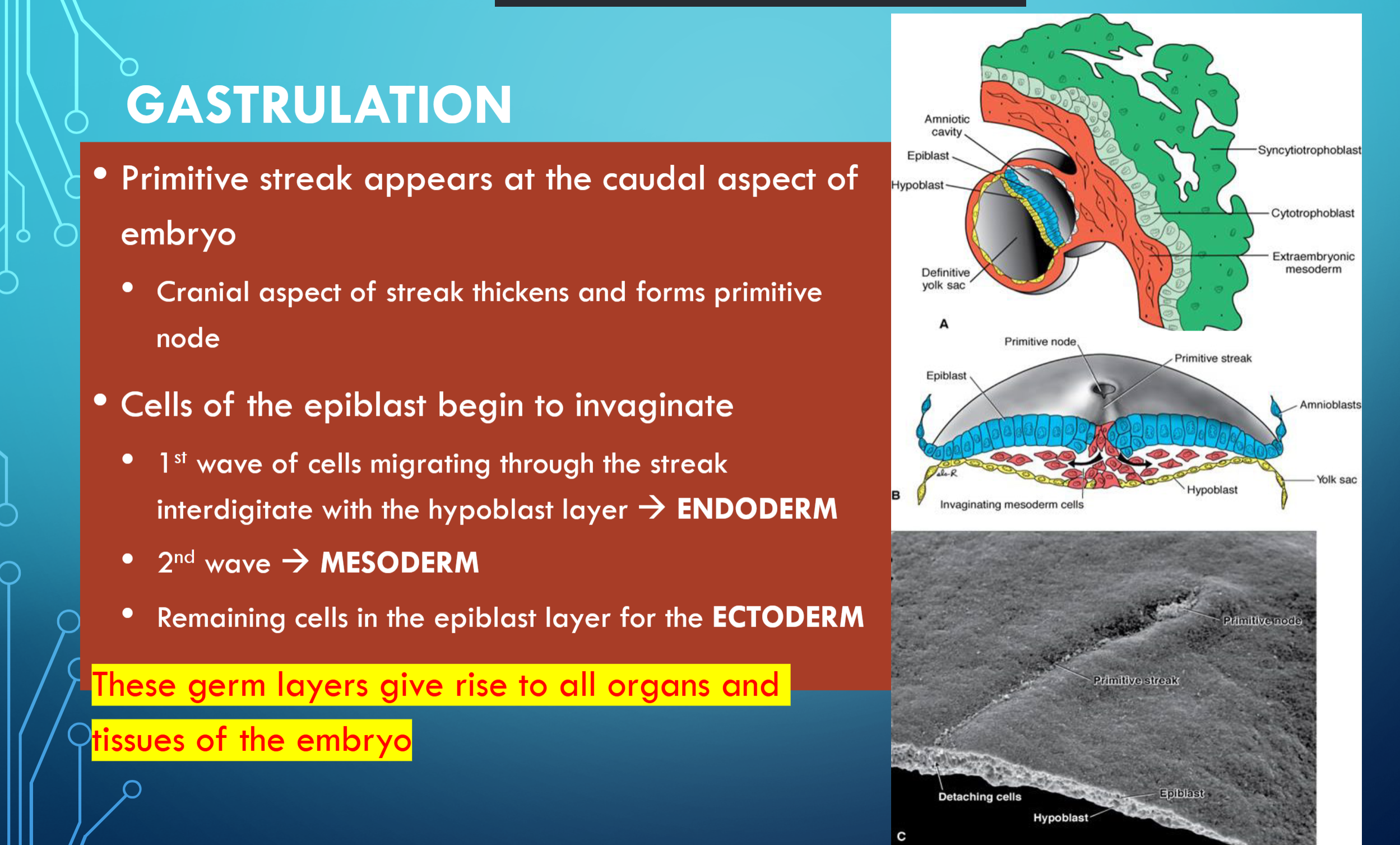
Primitive streak appears at the caudal aspect of
embryo
• Cranial aspect of streak thickens and forms primitive
node
• Cells of the epiblast begin to invaginate
• 1st wave of cells migrating through the streak
interdigitate with the hypoblast layer ENDODERM
• 2nd wave MESODERM
• Remaining cells in the epiblast layer for the ECTODERM
These germ layers give rise to all organs and
tissues of the embryo
FORMATION OF THE NOTOCHORD
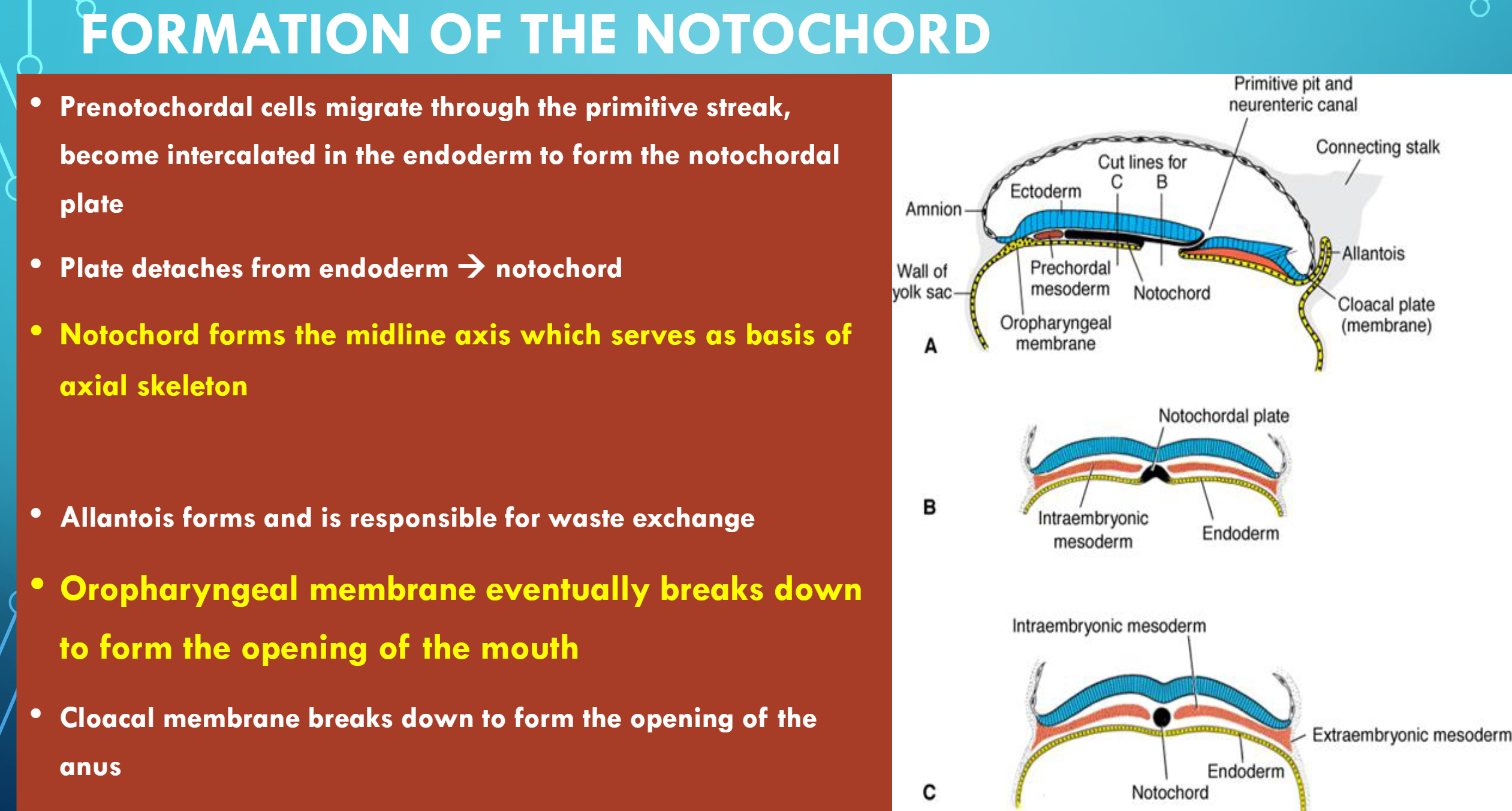
Prenotochordal cells migrate through the primitive streak,
become intercalated in the endoderm to form the notochordal
plate
• Plate detaches from endoderm notochord
• Notochord forms the midline axis which serves as basis of
axial skeleton
• Allantois forms and is responsible for waste exchange
• Oropharyngeal membrane eventually breaks down
to form the opening of the mouth
• Cloacal membrane breaks down to form the opening of the
anus
WEEKS 3-8: THE EMBRYONIC PERIOD
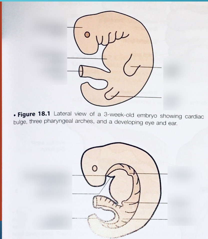
The period of organogenesis!
• By the end of the embryonic period
main organ systems are established
• This is the time at greatest risk for
induction of birth defects
• Sensitive to genetic and environmental
impacts
Three Germ Layers
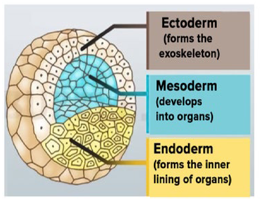
1) Ectoderm
2) Mesoderm
3) Endoderm
Ectoderm (9)
• Central nervous system (Brain & Spinal Cord)
• Peripheral nervous system
• Sensory epithelium of ears, eyes, and nose
• Epidermis
• Hair and Nails
• Subcutaneous glands
• Mammary glands
• Pituitary gland
• Enamel of the teeth
Neurulation
Process by which the Ectoderm [guided by the notochord] forms the neural plate
Which then bends upward and fuses → neural tube
![<ul><li><p>Process by which the Ectoderm [guided by the notochord] forms the neural plate </p></li></ul><p></p><ul><li><p>Which then bends upward and fuses → neural tube</p></li></ul><p></p>](https://assets.knowt.com/user-attachments/4f35178c-b6ad-4338-9e3c-751c5711c385.png)
Neurulation forms (3)
Neurulation results in the segregation of the three ectodermal derivatives:
1) Neural tube,
2) Neural crest
3) Epidermis
Neural Tube (will form)
Neural tube will become the brain [cranial end] and the spinal cord [caudal end]
Epidermis (will form)
the top layer of our skin
Neural Crest Cells Split to 5 Groups
1) Cranial neural crest cartilage, bone, cranial ganglia & connective tissue of the face
2) Cardiac neural crest septum of the outflow tract of the heart & wall of large arteries
3) Vagal neural crest parasympathetic enteric ganglia of the gut
4) Trunk neural crest melanocytes, dorsal root ganglia, sympathetic ganglia & adrenal medulla
5) Sacral neural crest parasympathetic enteric ganglia of the gut
Cranial neural cres
Cardiac neural crest
Vagal neural crest
Trunk neural crest
Sacral neural crest
Mesoderm (8)
All Supporting Tissue
- 1) Muscle
• 2) Cartilage
• 3) Bone
• 4) Dermis of the skin
• 5) Vascular system
• 6) Heart, arteries, veins, lymph vessels, all blood and lymph cells
• 7) Urogenital system: kidneys, gonads, ducts
• 8) Spleen
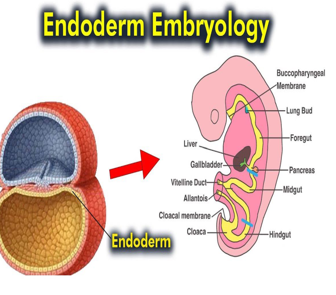
Endoderm
Think of Lining
1) Epithelial lining of: GI tract, respiratory tract, urinary bladder, urethra, tympanic cavity, auditory tube
2) Gastrointestinal tract
3)Thyroid
4) Parathyroid
5) Liver
6) Pancreas
Placenta (two parts)
Fetomaternal organ
1) Fetal component (chorion) derived from the
trophoblast & extraembryonic mesoderm
2) Maternal component derived from the uterine
endometrium
Placenta Morphology & Functions (5)
Flat like a pancake
1) Protection
2) Nutrition
3) Respiration
4) Excretion
5) Hormone production
Amnion
large sac containing amniotic fluid in
which the fetus is suspended by its umbilical cord
• (1) Absorbs jolts
• (2) Allows for fetal movements
• (3) Prevents adherence of the embryo to the
amnion
Amniotic fluid
Derived primarily from maternal blood
• Volume is replaced every 3 hours
• Increases throughout pregnancy from ~30mL at 10 weeks
to 1000 ml near delivery
• Either too much (hydramnios) or too little
(oligohydramnios) is associated with birth
defects
• Fetus drinks about 400ml/day beginning at 5 months
Hydramnios
Too much Amniotic fluid
Oligohydramnios
Too little Amniotic fluid
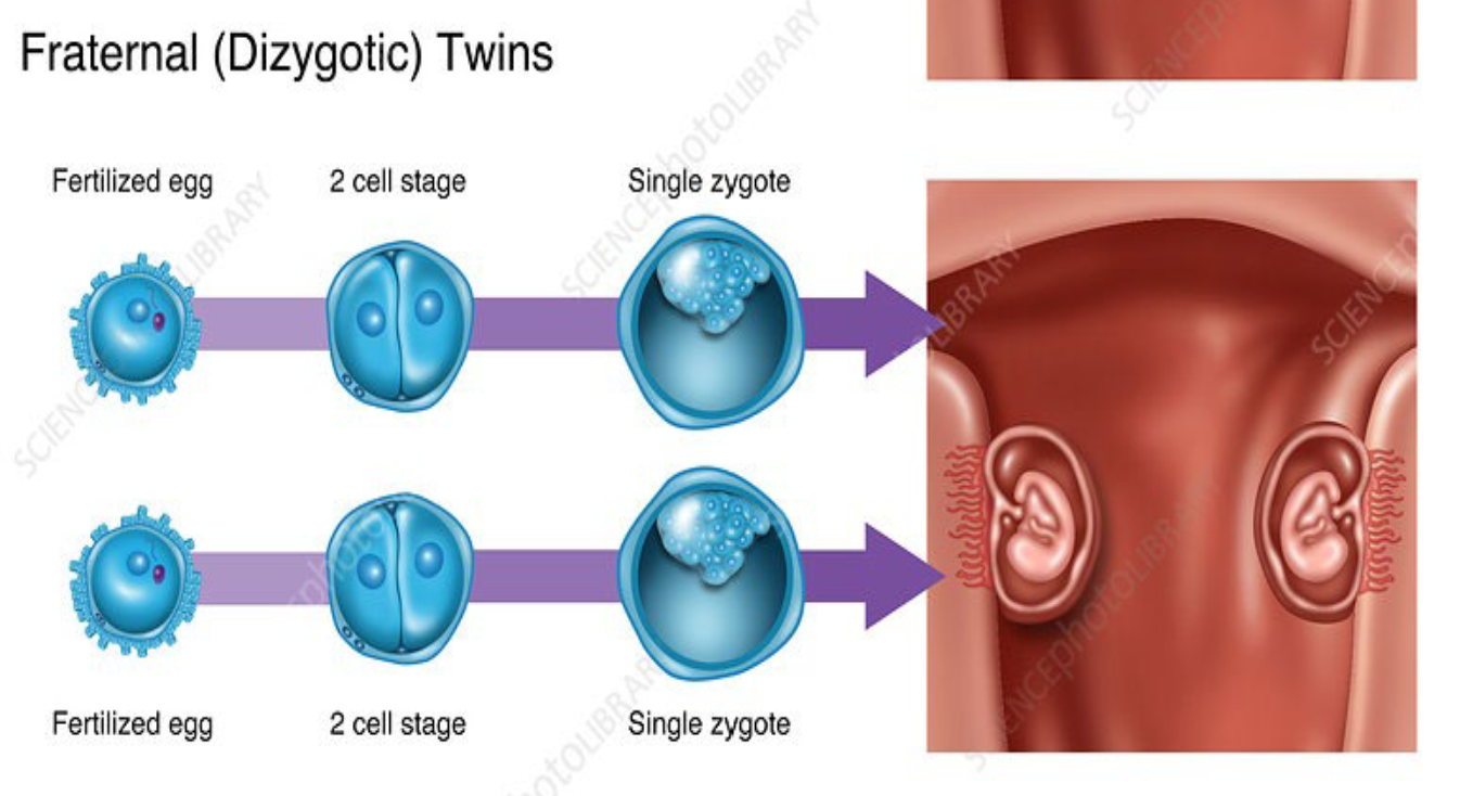
Dizygotic (Twin)
Dizygotic (fraternal) twins = 2 separately
fertilized eggs
• Not genetically identical
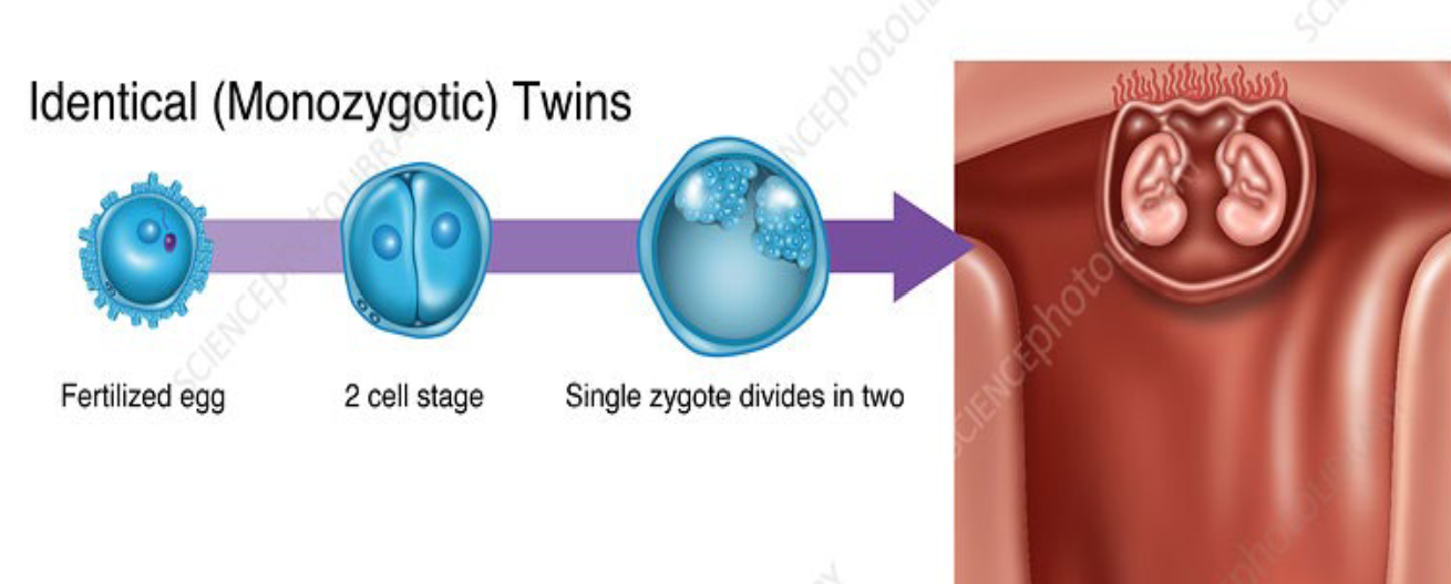
Monozygotic (Twin)
Monozygotic (identical) twins = 1 fertilized egg splits into two embryos
• Genetically identical