Lecture 8: Congenital Disorders of the Abdominal Wall and GI System - Flashcards
 Call Kai
 Learn
Learn Practice Test
 Spaced Repetition
 Match
Match Flashcards
 Knowt Play
1/69

There's no tags or description
Looks like no tags are added yet.
Name | Mastery | Learn | Test | Matching | Spaced | Call with Kai | Chat |
|---|
No analytics yet
Send a link to your students to track their progress
70 Terms
Foregut
• Pharynx
• Lower respiratory system
• Esophagus
• Stomach
• Proximal duodenum
• Liver and the biliary tree
• Pancreas
Midgut
• Distal duodenum
• Jejunum and ileum
• Appendix
• Ascending colon
• Proximal transverse colon
Hindgut
• Distal transverse colon
• Descending colon, sigmoid, and rectum
• Proximal anal canal
Esophageal Atresia (EA)
Caused by a defect in the lateral septation of the foregut into the esophagus and trachea
Tracheoesophageal Fistula (TEF)
thought to derive from a branch of the embryonic lung bud that fails to undergo branching because of defective epithelial-mesenchymal interactions
-congenital heart or genitourinary defects
• VACTERL or CHARGE associations
• Anopthalmia-esophageal genital syndrome
other anomalies that are assoc with EA and TEF
CHARGE
coloboma of the eye, heart anomalies, choanal atresia, retardation, genital and ear anomalies
Polyhydamnios
EA is associated with what during pregnancy
Esophageal Atresia
• Become symptomatic immediately after birth
• Excessive secretions that cause drooling, choking, respiratory distress, and the inability to feed
Tracheoesophageal Fistula (TEF)
-leads to gastric distenstion
-reflux of gastric contents
-aspiration pneumonia and contributes to morbidity
H-Type TEFs
Large defect:
• May present early with coughing and choking associated with feeding as the milk is aspirated through the fistula
H-Type TEFs
Smaller defect:
• May be asymptomatic in the newborn period
• Delay in diagnosis
• prolonged history of mild respiratory distress associated with feeding or recurrent episodes of pneumonia
• Rarely diagnosed in adolescence or adulthood
Esophageal Atresia
-Attempting to pass a catheter into the stomach (NG or OG)
-Catheter cannot be passed further than approximately 10 to 15 cm
confirmed with an AP CXR.
Distal Tracheoesophageal Fistula (TEF)
• Often seen on lateral CXR
• Gas-filled gastrointestinal tract
Esophageal Atresia
Small amount of water-soluble contrast material placed in the esophageal pouch under fluoroscopic guidance will confirm the presence of ____________________
Esophageal Atresia and TEF
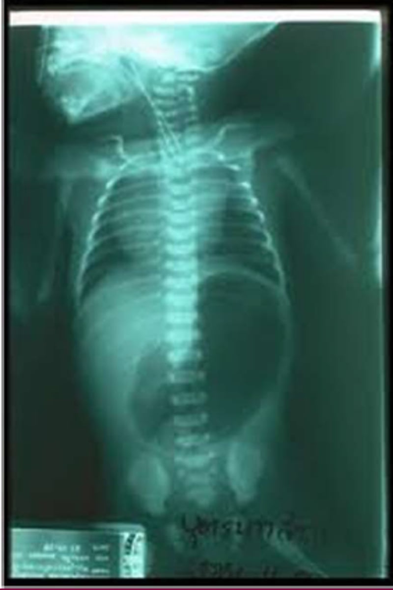
Tracheoesophageal Fistula (TEF)
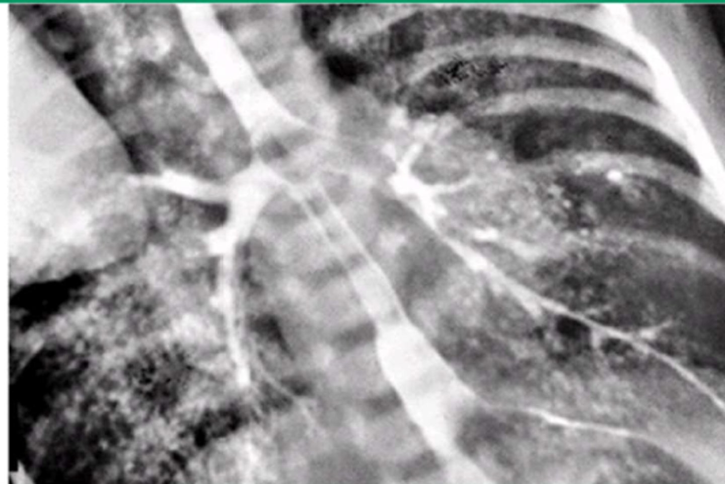
VACTERL
• Vertebral Defects
•Anorectal Atresia
• Cardiac Defects
• Tracheoesophageal Fistula
• Renal Dysplasia
• Limb defects
Prone Position
how to prevent aspiration in EA and TEF?
-Avoid endotracheal intubation
-Surgical correction
-Prone position
management of EA and TEF
Congenital Diaphragmatic Hernia (CDH)
Defined as communication between the abdominal and thoracic cavities with or without abdominal contents in the thorax
5th and 8th weeks
Congenital Diaphragmatic Hernia (CDH) is due to abnormalities in development that occurs during what weeks?
Congenital Diaphragmatic Hernia (CDH)
Causes
• Unknown
• Spontaneous mutation, Rare genetic forms
• Vitamin A deficiency
• Exposure to Thalidomide, anticonvulsants, and quinine
Morgagni Hernia
retrosternial diaphragmatic hernia
Bochdalek
posterolateral diaphragmatic hernia
Congenital Diaphragmatic Hernia (CDH)
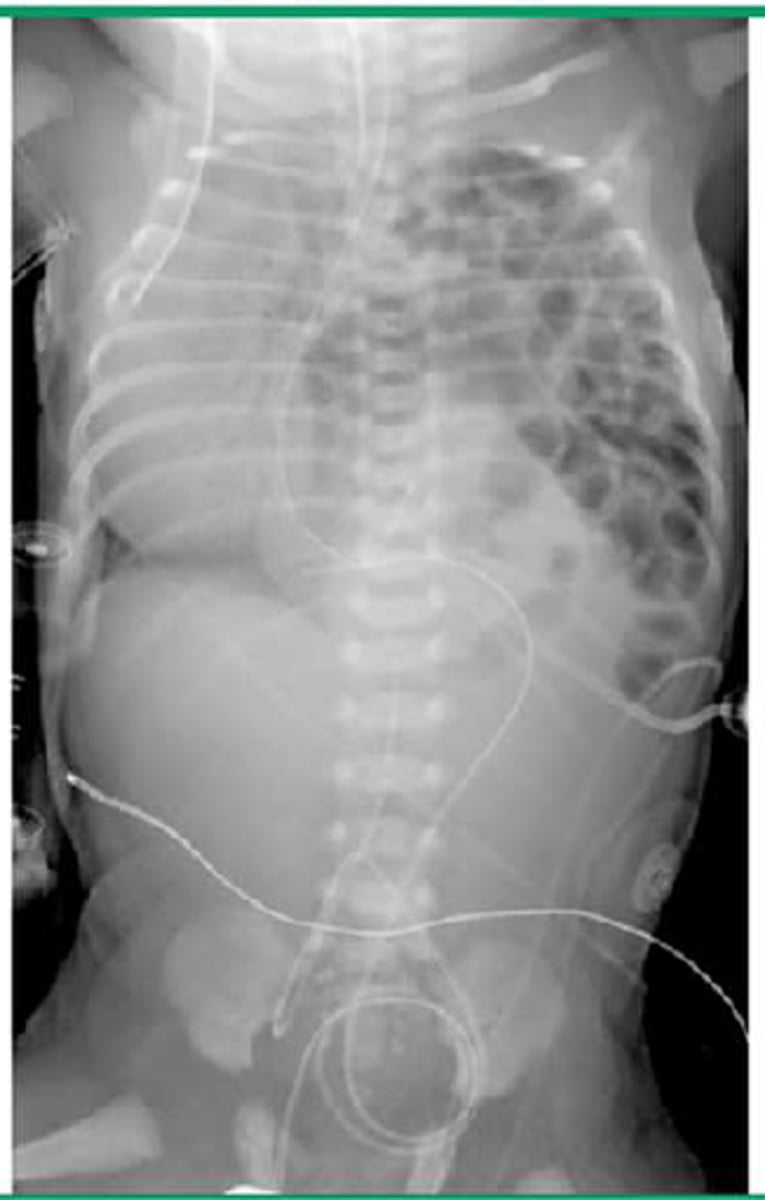
Congenital Diaphragmatic Hernia (CDH)
• Respiratory distress within the first several hours after birth
• Barrel-shaped chest
• Scaphoid abdomen
• Absence of breath sounds on the ipsilateral side
-may hear bowel sounds in chest
-PMI is displaced to right
• Prenatal Ultrasound (polyhydraminos, chest mass, gastric bubble)
• CXR after delivery
how do you diagnose congenital diaphragmatic hernia?
-Immediate Endotracheal Intubation
-Sedation
-NG tube
-Extracorporeal Membrane Oxygenation (ECMO)
management of congenital diaphragmatic hernia
Pulmonary Hypoplasia
the major limiting factor for survival for congenital diaphragmatic hernia
Omphalocele
Herniation of abdominal contents into the base of the umbilical cord
• Sac is covered with peritoneum without overlying skin
Omphalocele
• The abdominal cavity is proportionately small/underdeveloped
-assoc with syndromes like: Beckwith Wiedemann syndrome and Congenital heart defects
4th to 5th week
what weeks of development?
• The flat embryonic disk folds in four directions and/or planes: cephalic, caudal, and right and left lateral
• Each fold converges at the site of the umbilicus, thus obliterating the extraembryonic coelom
4th to 5th week
what weeks of development?
• The lateral folds form the lateral portions of the abdominal wall
• The cephalic and caudal folds make up the epigastrium and hypogastrium
• Rapid growth of the intestines and liver also occurs at this time
6th Week (8 weeks from Last menstrual period)
what weeks of development?
Physiologic midgut herniation: The abdominal cavity temporarily becomes too small to accommodate all of its contents, resulting in protrusion of the intestines into the residual extraembryonic coelom at the base of the umbilical cord
7th-9th weeks
what weeks of development?
Physiologic midgut herniation is sonographically evident
11th week
what weeks of development?
Midgut herniation is no longer physiological
Liver
what organ does not undergo physiologic migration outside the abdominal cavity during development and therefore is never present in midgut herniation?
Lateral folds fail to close
what is the cause of a liver-containing omphalocele?
Omphalocele
associated with a high frequency of fetal aneuploidy when they
contain only small bowel
Week 10
the abdominal contents fo the fetus should return to the abdominal cavity by what week of gestation?
• Maternal serum alpha-fetoprotein concentration (MSAFP) is elevated in 70%
• 2nd trimester Ultrasound
• Sometimes at birth
diagnosis of omphalocele
• Preserve heat and minimize insensible fluid loss
• Orogastric tube to decompress the stomach
• Stabilize the airway
• Tissue must be covered with saline soaked sterile dressings
Management of omphalocele
Mersilene mesh to cover tissue of organs
what do you do if the sac of an omphalocele ruptures
• Primary closure for small defects (<2 cm)
• Staged closure for larger defects (Dacron-reinforced silastic silo)
surgical repairs for omphaloceles
Gastroschisis
• Defect lateral to the median plane of the anterior abdominal wall
• Extrusion of the abdominal viscera without involving the umbilical cord
• There is no peritoneal sac covering
Right
a gastroschisis is usually on what side?
Gastroschisis
• Intestine is usually only herniated organ
• Bowel is often thickened, matted and edematous
• Higher incidence of intestinal atresia, stenosis, perforation, necrosis, or volvulus
Gastroschisis
• Involves defective formation or disruption of the body wall in the embryonic period, with subsequent herniation of bowel
• Typically NOT associated with extra intestinal or chromosomal anomalies
• Elevated maternal serum alpha fetoprotein (MSAFP) level during 2nd trimester in 90%
• Prenatal ultrasound
diagnosis of Gastroschisis
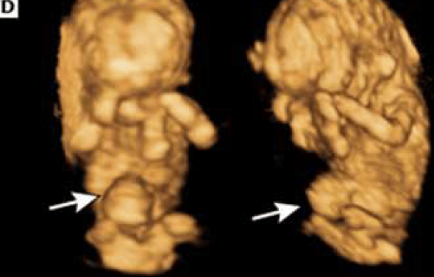
Gastroschisis
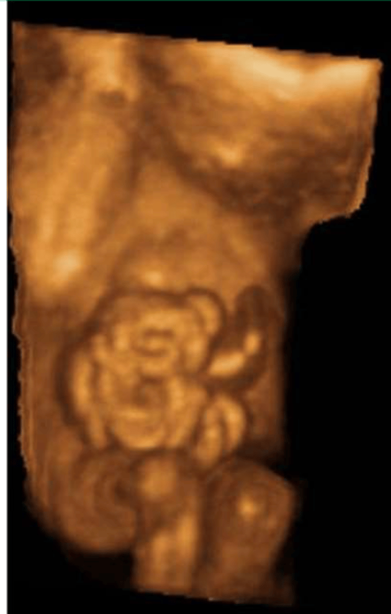
Omphalocele
• Preserve heat and minimize insensible fluid loss
• Orogastric tube to decompress the stomach
• Stabilize the airway
• Safely cover the protruding tissues
Management of Gastroschisis
• Primary closure
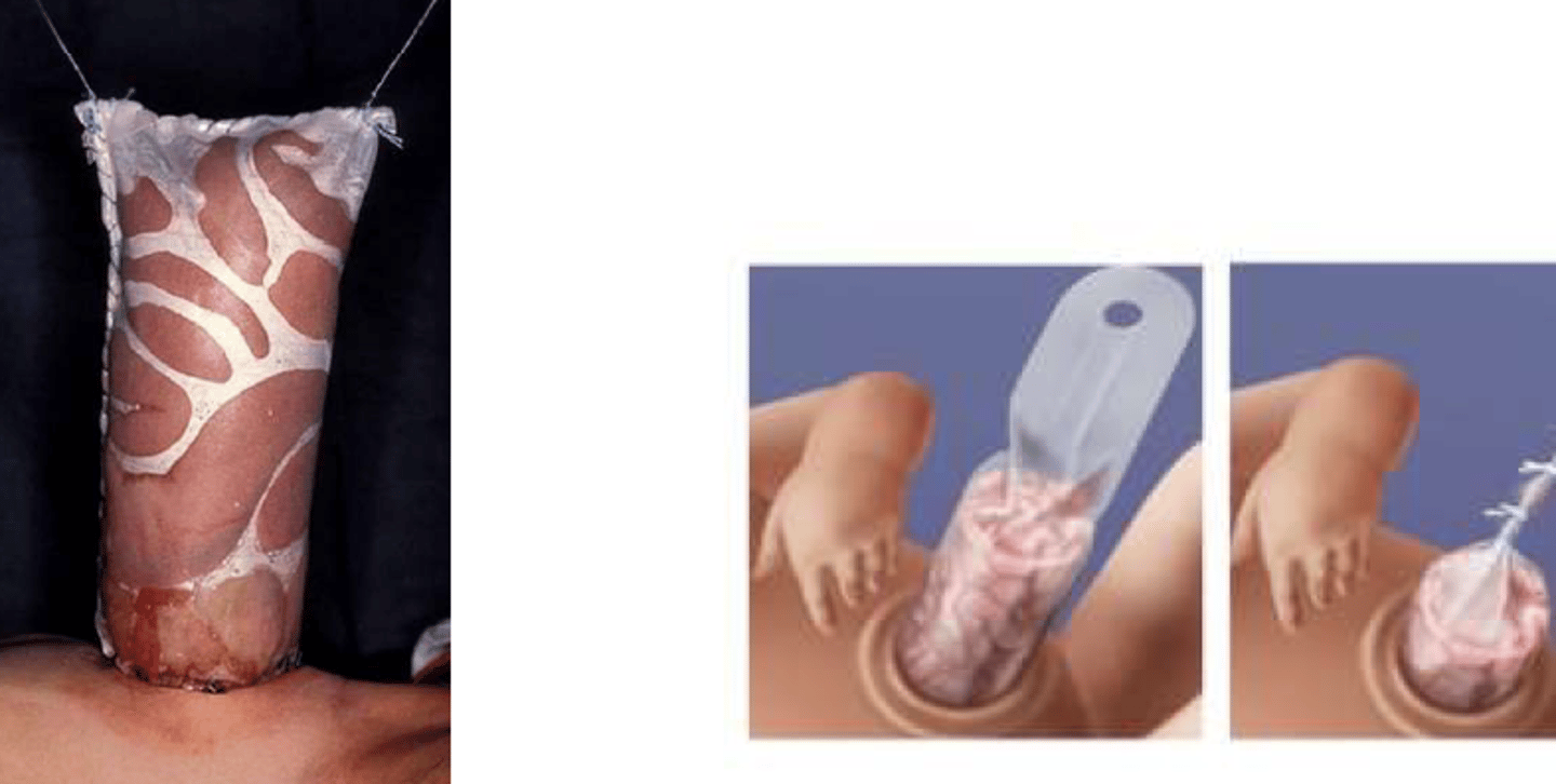
• Staged closure
• Dacron-reinforced silastic silo
• "Gentle touch"
• Slower process allowing gravity to reduce bowel before closure
Surgical repair of gastroschisis
Gastroschisis
is overall survival higher for omphaloceles or Gastroschisis?
Silos
Umbilical Hernia
• Defined as protrusion of intestines through a defect in the abdominal wall muscles
• Also associated with diastasis recti
• Mostly seen in African American children and low birth weight babies
Umbilical Hernia
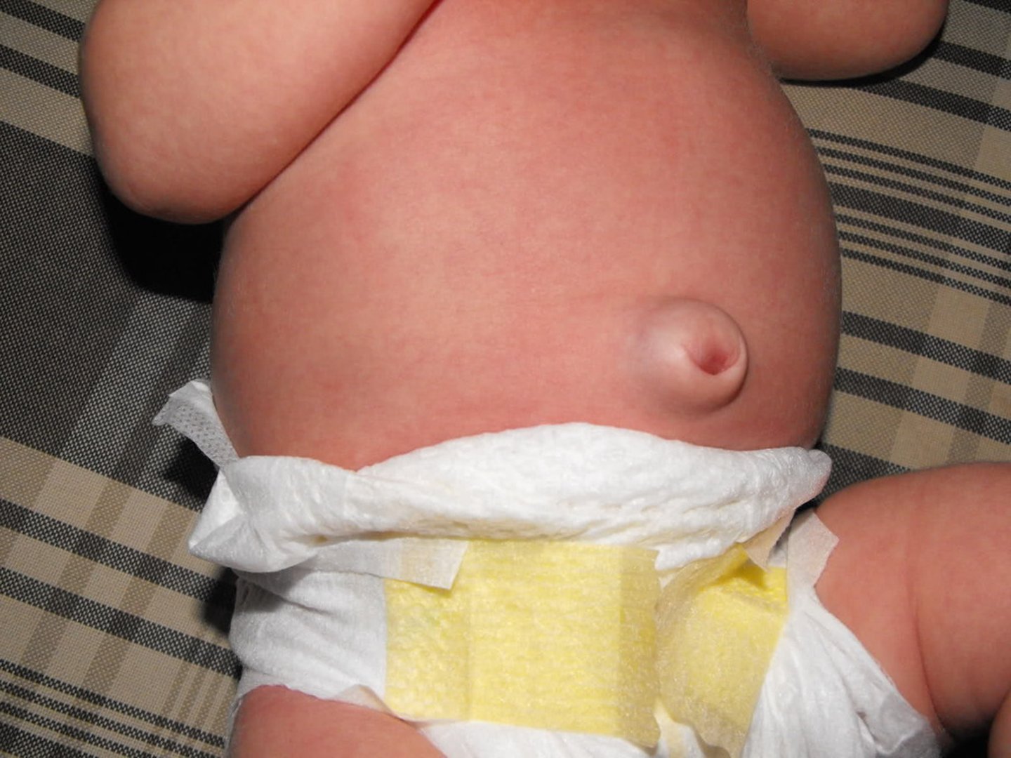
• Typically noted as a soft reducible bulge over the umbilicus which becomes more protuberant during crying, coughing or straining
• Ranges in size from less than 1 cm to as large as 5 cm
• Most resolve with no intervention by 1 year of age
• If still present or worsening, it requires surgical intervention usually around 4-5 years of age
describe treatment for an umbilical hernia
Umbilical Hernia
Prune Belly Syndrome
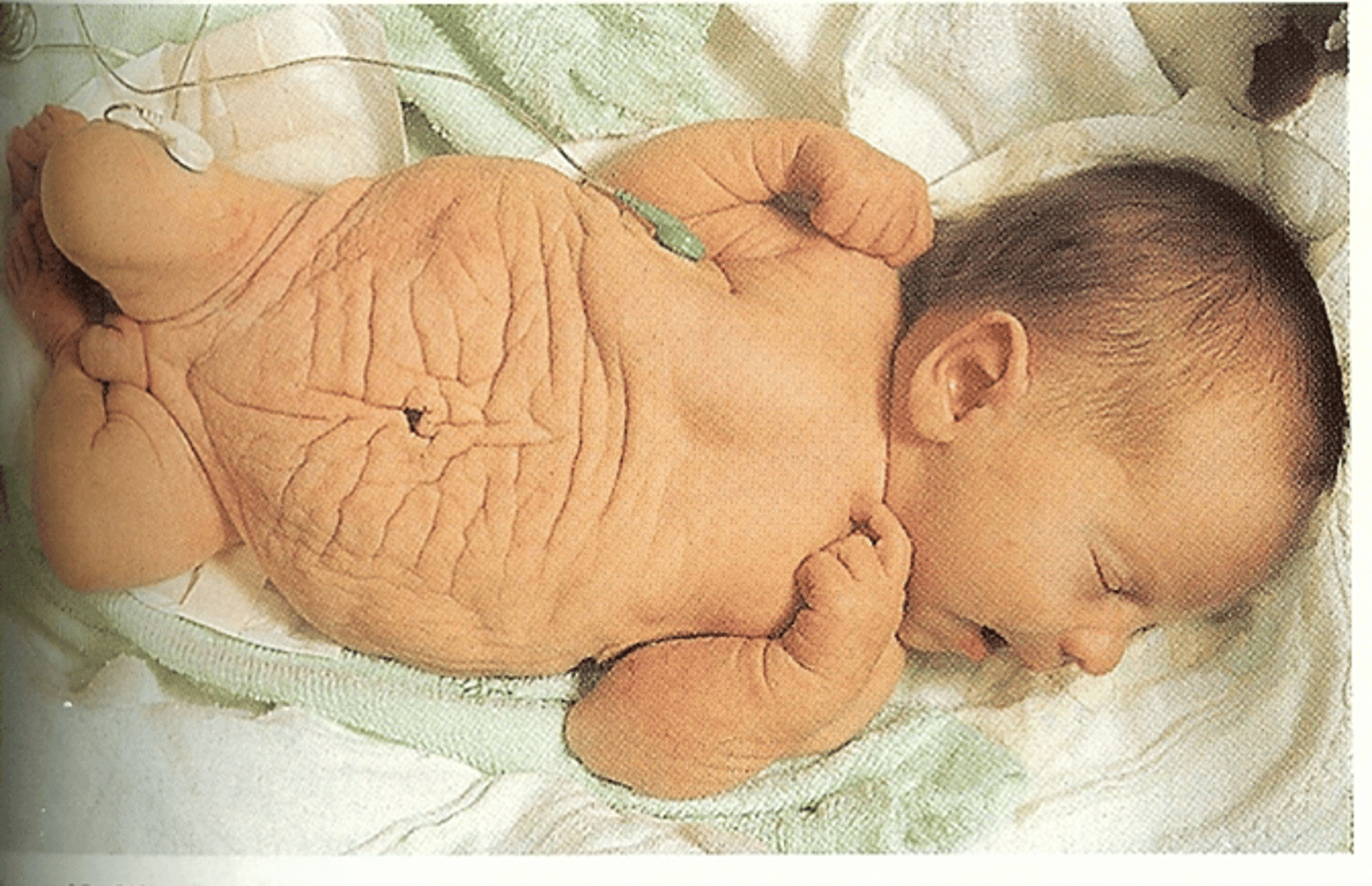
Also called Triad syndrome OR Eagle-Barrett Syndrome
Prune Belly Syndrome
Congenital disorder defined by a characteristic clinical triad:
• Abdominal muscle deficiency
• Severe urinary tract abnormalities
• Bilateral cryptorchidism in males
Prune Belly Syndrome
A primary defect in the intermediate and lateral plate mesoderm affects embryogenesis of the musculature of the abdominal wall, the mesonephric and paramesonephric ducts, and the urinary organs
Prune Belly Syndrome
Associated Anomalies at birth:
• Oligohydraminos and pulmonary hypoplasia are common perinatal complications
• Kidneys show varying degrees of dysplasia
• Malrotation of the bowel is often present
Prune Belly Syndrome
-Antibiotic Prophylaxis (to prevent UTI)
• Orchidopexy is typically done in the first 6 months of life
treatment for prune belly syndrome is there is no urethral obstructions
-Vesicostomy
• Orchidopexy is typically done in the first 6 months of life
treatment for prune belly syndrome is there is a urethral obstructions
Neonatal Intestinal Obstruction
• Meconium Plug (Earlier)
• Intestinal Atresia (Earlier)
• Hirschsprungs Disease (Later)
• Pyloric Stenosis (Later)
• Malrotation with or without volvulus
Intestinal Atresia
most common cause of obstruction in the neonatal period
Bilious Emesis
-Treated as a life-threatening emergency because this is often a symptom of obstruction due to intestinal atresia or mid-gut volvulus