Anatomy
1/38
Earn XP
Description and Tags
random
Name | Mastery | Learn | Test | Matching | Spaced | Call with Kai | Chat |
|---|
No analytics yet
Send a link to your students to track their progress
39 Terms
pinna
aka auricle
located outside the head
acts as a funnel which assists in directing sound further into the ear/auditory canal → tympanic membrane
eustachian tube
middle ear to pharynx
circumvents excess pressure when pressure builds in the ear → equalization of air pressure between middle ear and atmosphere
rapid changes in altitude can result in ear “popping” d/t equalization process
intraocular segment
the axons of the photoreceptors as they exit the eye
shortest segment of the nerve (1mm deep, 1.5mm vertical diameter)
aka optic disc or ONH

intraorbital portion of optic nerve
the segment that extends from globe to optic foramen at the orbital apex
25-30mm in length
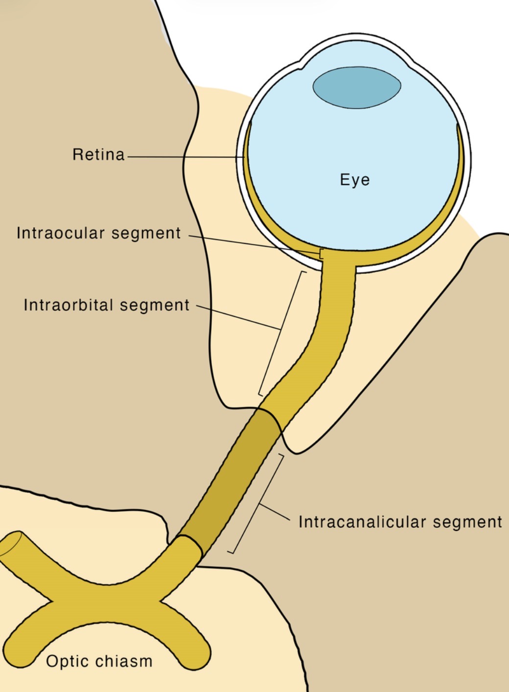
intracanalicular region of the optic nerve
the segment that traverses the optic canal
6mm in length
fixed to the canal due to fusion of the dura material with the periosteum

intracranial segment
the portion that extends to join the optic chiasm
fibers that course posteriorly from optic chiasm are referred to as optic tract
how does a muscle contraction happen?
AP → releases ACh → sarcolemma → uptaken by other side of terminal → AP via T-tubules → release of Ca2+ → tropomyosin and troponin component exposes itself to myosin when Ca2+ is present → now ready to connect to myosin → ATP and bind and release → cycle continues as long as Ca2+ is present
primary and secondary actions for SR
elevate
intort
adduct
SIN RAD
primary and secondary actions for IR
depress
extort
adduct
primary and secondary actions for IO
extort
elevate
abduct
primary and secondary actions for SO
intort
depress
abduct
bones of orbital roof
“Front-less”
Frontal bone
Lesser wing of sphenoid
bones of medial orbit
”SMEL”
sphenoid bone (body; strongest)
maxillary bone
ethmoid bone (thinnest)
lacrimal bone
bones of lateral orbit
“Great Z”
greater wing of sphenoid
zygomatic bone
bones of floor of orbit
“My Pal gets Z’s on the floor”
maxillary bone (weakest)
palatine bone
zygomatic bone
confluence of sinuses is made of
straight sinus
superior sagittal sinus
occipital sinus
transverse sinus
internal carotid artery branches into:
OPAM
ophthalmic a.
posterior communicating a.
anterior cerebral a.
middle cerebral a.
what goes thru superior orbital fissure?
LFT NOA, sov
lacrimal n.
frontal n.
trochlear n.
nasociliary n.
oculomotor n.
abducens n.
superior ophthalmic vein
NOA passes thru SOF and CTR
what goes thru the optic canal?
optic nerve
ophthalmic a.
what goes thru the inferior orbital fissure?
CN V2 (maxillary br. of trigeminal n.)
inferior ophthalmic vein
infraorbital a.
infraorbital v.
infraorbital n.
zygomatic n.
3 layers of tear film
lipid: lubricates, prevent evaporation
meibomian, zeiss, moll
evaporative dry eye, MGD, rosacea, decreased blink rate, poor fitting CL, Accutane
aqueous: nourishes, protects cornea
main lacrimal gland (95%), accessory (Krause, Wolfring)
isotonic solution
increased production due to parasympathetic stimulation
lysozyme, beta lysin, lactoferrin, IgA
lysozyme cleaves peptidoglycan in bacterial cell wall (pcn, cephalosporins, Bacitracin)
mucous: adheres tears to eye
supplied by conj goblet cells
vit A deficiency → decrease in goblet cells → dry eye, bitot spots
vitamins D,E,A,K are fat-soluble
corneal epi cells → glycocalyx
marangoni flow
tears flow from low temperature areas (cornea) to higher temperature areas (lid margins)
compared to typical skeletal muscles, EOMs:
blood supply is denser in EOMs
nere supply is denser and more finely tuned in EOMs
EOM movements are faster and more fatigue resistant
origin of EOMs
common tendinous ring (annulus of zinn) → SR, IR, LR, MR
all recti muscles originate from CTR and insert on the globe to form the Spiral of Tillaux
SR inserts the furthest away, MR inserts the closest to the limbus
maxillary bone → IO
LW of sphenoid → SO
the ___ sheath and ___ sheath combine to form the suspensory ligament of Lockwood
IR and IO
what forms a tight barrier between cells to impede the intercellular movement of particles in the corneal epithelium ?
zonula occludens (tight junctions)
desmosomes
what factors increase the risk of recurrent corneal erosions?
RCE: poor adhesion between epithelium and basement membrane
poor hemidesmosome attachments
EBMD
age-related thickening of BM
characteristics of Bowman’s layer
resists damage
maintains curvature, absorbs UVB
past Bowman’s, nerves lose Schwann covering and pass into epithelium as naked nerves
cannot regenerate, will scar
clinical conditions associated with Bowman’s layer
band keratopathy: calcium deposits (“swiss cheese”)
pterygia
crocodile shagreen (bilateral)
Reis-Buckler’s dystrophy
keratoconus: begins in Bowman’s
advanced → hydrops in Descemet’s
refractive surgery
flap created in LASIK = epithelium and Bowman’s
PRK = laser thru Bowman’s → post-op haze
limbal girdle of Vogt: Bowman’s degeneration (d/t Ca2+ deposits)
anterior 1/3 of collagen fibrils in corneal stroma vs. posterior 2/3
anterior 1/3
thinner lamellae
more covalent bonding (cross-linking)
more structure — branching, interweaving
posterior 2/3
larger lamellae
more regular arrangement
components of the corneal stroma
collagen fibrils (type 1 collagen)
keratocytes → fibroblasts
make collagen, ECM
ground substance
proteoglycans (protein + GAGs)
attracts water
blood cells (WBC, lymphocytes, macrophages, leukocytes)
stromal stem cells
which corneal layers can potentially scar? which layers can regenerate?
will scar — Bowman’s, stroma
can regenerate — epithelium, stroma, Descemet’s
what properties of drugs allow it to penetrate the cornea?
small in size
uncharged
lipid soluble
weak base
what layer(s) of the cornea are hydrophobic? hydrophilic? lipophilic?
epithelium: hydrophobic, lipophilic
stroma: hydrophilic
endothelium: lipophilic
where is type 1 collagen seen in the body?
Bowman’s layer of cornea
bones
stromal layer of cornea
sclera
associated with Ehlers-Danlos, osteogenesis imperfecta
ERG waves
A wave: photoreceptors
B wave: bipolar cells, Mueller cells
oscillations are due to amacrine cells
C wave: RPE
D wave: off bipolar cells
what structures are in the forebrain? midbrain? hindbrain?
forebrain:
cerebrum
olfactory lobes
thalamus
hypothalamus
limbic system
pituitary gland
pineal gland
midbrain:
tectum
substantia nigra
red nucleus
hindbrain:
cerebellum
pons
medulla oblongata