Circulatory System: Heart - Notes
 Call Kai
 Learn
Learn Practice Test
 Spaced Repetition
 Match
Match Flashcards
 Knowt Play
1/66

There's no tags or description
Looks like no tags are added yet.
Name | Mastery | Learn | Test | Matching | Spaced | Call with Kai | Chat |
|---|
No analytics yet
Send a link to your students to track their progress
67 Terms
Cardiovascular System: Function
Transporting blood throughout the body
Delivering oxygen and nutrients
Removal of carbon dioxide and other waste products
Goal: provide adequate perfusion to all body tissues
Cardiovascular System: Components
Heart: Major organ that pumps blood
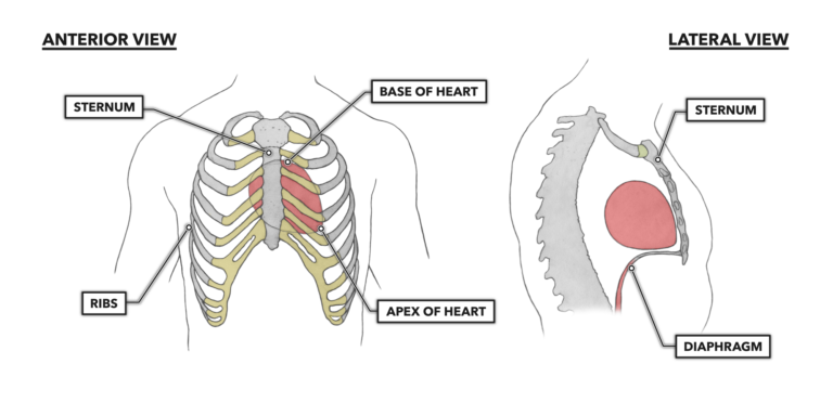
Location: Directly behind the sternum.
Orientation: Base is superior, apex is inferior.
Mediastinum:
Region located between the lungs.
Blood Vessels: Include arteries, veins, and capillaries
Blood
Artery
Transport away from the heart
Thick-walled, elastic vessels that transport high-pressure blood away from the heart
They branch into smaller arterioles
Vein
Transport blood toward the heart
Thinner-walled vessels with larger lumens and often containing valves, transporting low-pressure blood toward the heart.
They originate from venules
Capillaries
Responsible for the exchange of gases and nutrients between blood and systemic cells, as well as alveoli in lungs
Microscopic, thin-walled vessels forming extensive networks within tissues
The Heart by the numbers
Chambers of the Human Heart: 4 chambers
Average Heartbeats per Day: Approximately 100,000 times
Blood Pumped per Beat: About 70 mL
Average Weight of Human Heart: 300g
Ventricles
Receives the Blood
Atria
Pump blood to the lungs and body tissue
Function of the Heart
Acts as a powerful, rhythmic, and continuous pump, generating pressure to propel blood through the pulmonary and systemic circuits, ensuring unidirectional flow
Function of Blood Vessels
Vessels provide a closed, dynamic pathway for blood circulation, distributing blood to tissues and collecting it for return to the heart. They also regulate blood flow and pressure through vasoconstriction and vasodilation
Structure of Blood Vessels
Most vessels have three layers (tunics):
Tunica Intima (innermost): Endothelium (simple squamous epithelium) with underlying connective tissue
Tunica Media (middle): Smooth muscle and elastic fibers; controls vessel diameter
Tunica Externa (outermost): Connective tissue sheath; anchors vessel
Pulmonary Circuit
Carries blood from the heart → lungs → heart
Blood flows from the right side of the heart to the lungs and back to the left side of the heart
Systemic Circuit
Carries blood from the heart → body → heart
Blood flows from the left side of the heart to the rest of the body and returns to the right side of the heart.
Blood Flow Through Pulmonary Circulation
Deoxygenated blood enters the right atrium from the venae cavae (superior and inferior vena cavae).
Blood passes through the right atrioventricular (AV) valve (tricuspid valve).
Blood enters the right ventricle.
Blood passes through the pulmonary semilunar valve into the pulmonary trunk.
Blood travels through right and left pulmonary arteries to lungs.
Gas exchange occurs in pulmonary capillaries, oxygenating the blood.
Oxygenated blood enters right and left pulmonary veins.
Blood returns to the left atrium of the heart.
Blood Flow Through Systemic Circulation
Oxygenated blood enters the left atrium from the pulmonary veins.
Blood passes through the left AV valve (bicuspid or mitral valve).
Blood enters the left ventricle.
Blood passes through the aortic semilunar valve into the aorta.
Blood is distributed throughout the body via systemic arteries.
Blood enters systemic capillaries for nutrient and gas exchange.
Deoxygenated blood drains into systemic veins, returning to the heart
Blood enters the right atrium, completing the circuit.
Anatomy of the Heart: Size and Position
Average size: Approximately the size of a fist
Location:
Lies in the mediastinum, posterior to the sternum and anterior to the vertebral column
Extends from the level of the 2nd rib to the superior surface of the diaphragm
Majority located to the left of the body's midline
Orientation of the Heart
The heart has two parts:
Base: Superior, broader portion where vessels arise, points toward the right shoulder.
Apex: Inferior, narrower portion resting on the diaphragm, points toward the left hip.
Orientation: Right side is more anterior; left side is more posterior.
Chambers of the Heart
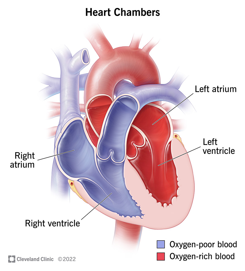
The human heart has four chambers:
Two atria: Receiving chambers for blood.
Two ventricles: Pumping chambers for blood.
Heart Coverings and Wall Structure
Pericardium: Surrounds the heart.
Fibrous Pericardium: Composed of dense irregular connective tissue
Serous Pericardium: Contains two layers:
Parietal Layer: Lines the fibrous pericardium.
Visceral Layer (Epicardium): Covers the heart.
Pericardial Cavity: Contains pericardial fluid, composed of simple squamous epithelium and underlying areolar connective tissue.

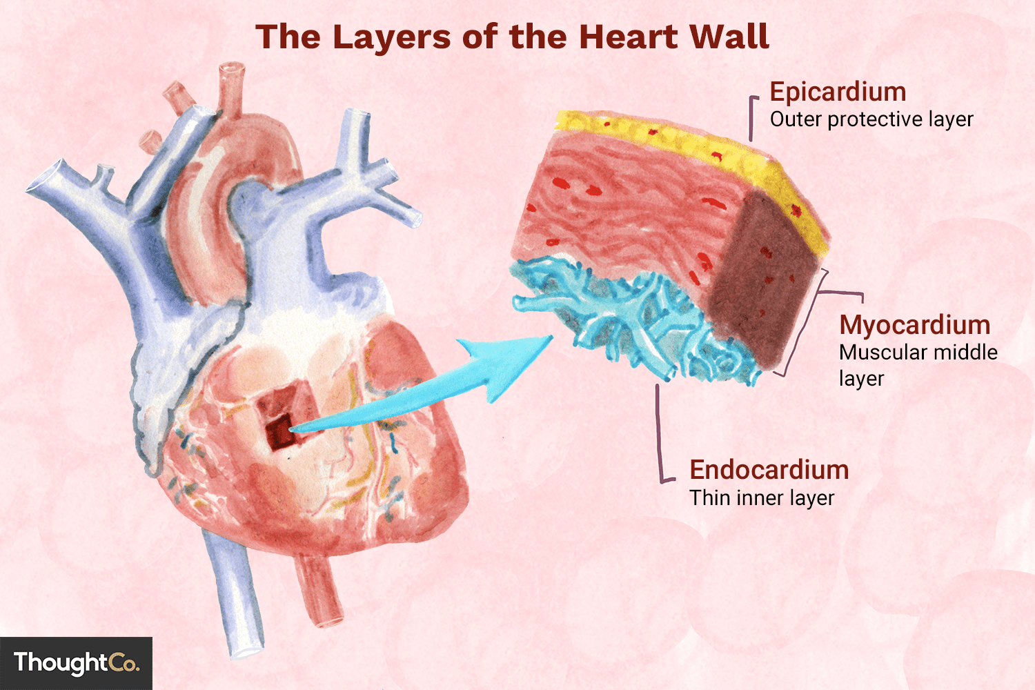
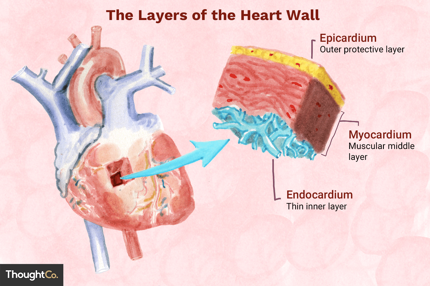
Layers of the Heart Wall: Epicardium
The outermost layer that is also called the visceral layer of the serous pericardium
Layers of the Heart Wall: Myocardium
Middle layer, composed predominantly of cardiac muscle.
Thickest layer, responsible for contracting and pumping blood.
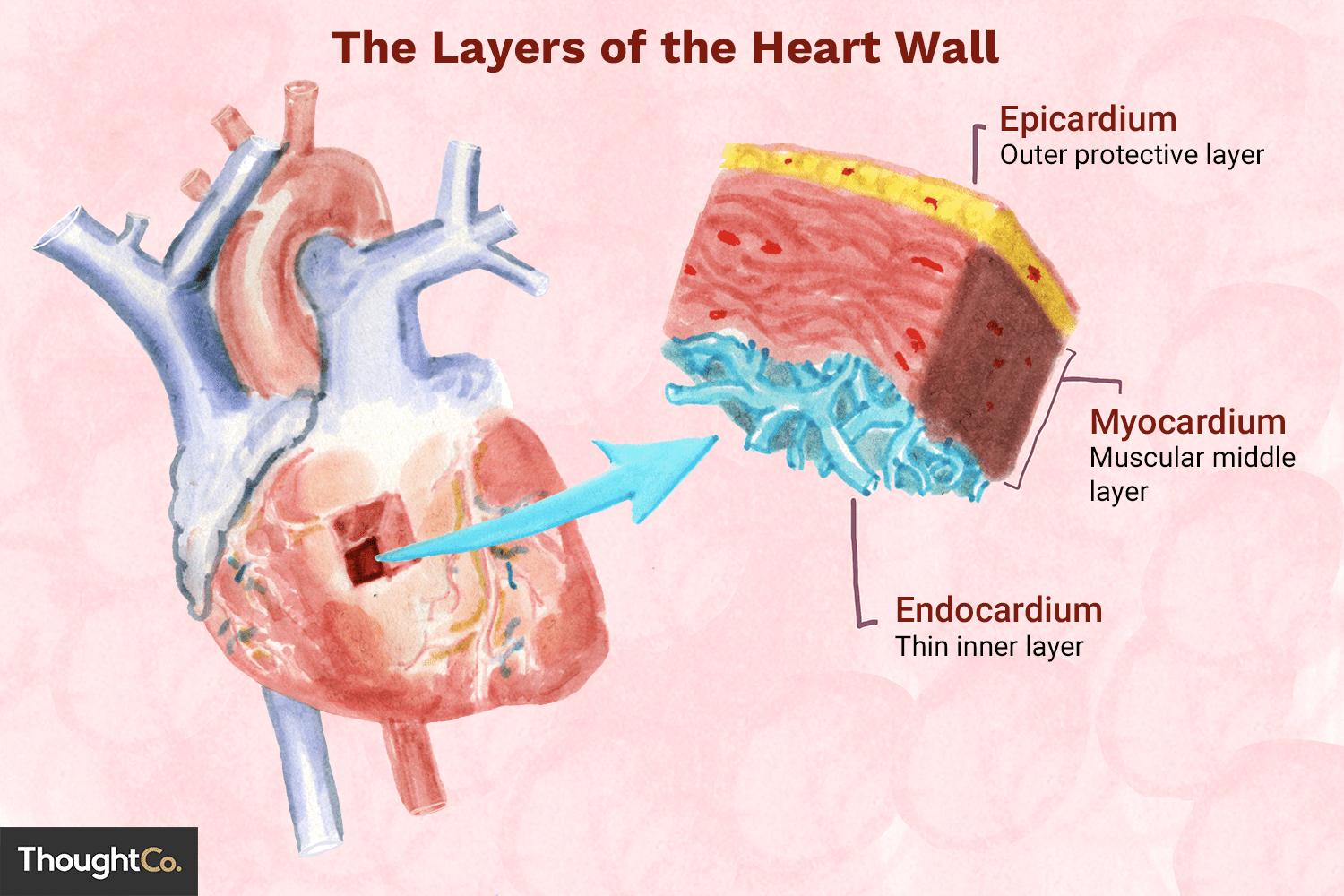
Layers of the Heart Wall: Endocardium
Covers the internal surface of the heart and the external surfaces of the heart valves
It is continuous with the inner lining of blood vessels
Fibrous Skeleton of the Heart
Composed of dense irregular connective tissue.
Provides structural support, anchors cardiac muscle cells and valves, acts as an electrical insulator between heart chambers
Between atria and ventricles
Heart functions as a double pump
Left Side: Handles oxygen-rich blood, pumping it to the body
Right Side: Handles oxygen-poor blood, pumping it to the lungs
Blood Moving one-way
Blood moves in one direction through the heart due to valves preventing backflow.
The heart is regulated by pressure differences caused by contraction and relaxation of chambers.
Atria: Receive blood and push it into ventricles.
Ventricles: Pump blood out of the heart.
Path of the Blood Flow
Arteries: Transport blood away from the heart, branch into arterioles.
Capillaries: Sites of nutrient and gas exchange.
Veins: Transport blood back toward the heart.
Anatomy of the Heart: Valves
Valves: Ensure one-way blood flow and prevent backflow.
Atrioventricular (AV) Valves: Between atria and ventricles (tricuspid and bicuspid/mitral valves).
Semilunar Valves: Between ventricles and arteries (pulmonary and aortic valves).
Tendinous Cords: Anchor the AV valves to papillary muscles in ventricles, preventing inversion
Valves Mechanism
AV Valves: Open during atrial contraction; close during ventricular contraction.
Semilunar Valves: Open during ventricular contraction; close during ventricular relaxation
Coronary Circulation
Coronary Arteries: Blood vessels that supply blood to the heart wall.
Right coronary artery and left coronary artery arise from the ascending aorta.
Coronary Veins: Great cardiac vein, middle cardiac vein, and small cardiac vein drain into the coronary sinus
Cardiac Muscle Structure
Striated muscle, shorter than skeletal muscle with one or two centrally located nuclei
Cardiac Muscle: Features for Energy Demand
Extensive blood supply, myoglobin, high levels of mitochondria.
Ability to utilize multiple energy sources (fatty acids, glucose).
Intercalated Disks
Connections between cardiac muscle cells, providing strength (desmosomes) and allowing electrical continuity (gap junctions).
Autorhythmicity: Some cardiac cells can depolarize spontaneously and generate action potentials.
Intercalated Disks: Desmosomes
Act as mechanical junctions to prevent cardiac muscle cells from pulling apart
Intercalated Disks: Gap Junctions
Provide a low-resistance pathway for the flow of ions between cardiac cells
Allow an action potential to move continuously along the sarcolemma of cardiac muscle cells, resulting in synchronous contraction of that chamber.
Cardic Conduction System
Special cells that generate and propagate electrical impulses controlling contractions:
Components: SA node (pacemaker), AV node, AV bundle, bundle branches, and Purkinje fibers.
Innervation of the Heart
Myogenic initiation of contraction but modulated by the autonomic nervous system:
Parasympathetic: Decreases heart rate via vagus nerves.
Sympathetic: Increases heart rate and contractility.
Physiological Events of the Heartbeat
Sinus Rhythm: Normal heart rate (70-80 bpm).
Electrical Events:
Depolarization, plateau, and repolarization actions for muscle contraction cycle
Electrocardiography (ECG/EKG)
Recording of electric currents in the heart:
Main waves: P wave (atrial depolarization), QRS complex (ventricular depolarization), T wave (ventricular repolarization).
The Cardiac Cycle
Sequence of events from one heartbeat to the next, involving contraction (systole) and relaxation (diastole)
Heart sounds (S1, S2) associated with valve closure.
Ventricular Volume: End-diastolic volume (EDV) ~ 130 mL, End-systolic volume (ESV) ~ 60 mL, Stroke volume ~ 70 mL.
Heart Sounds
S₁ (lub) - AV valves close (tricuspid & mitral valve)
S₂ (dub) - semilunar valves close (pulmonary semilunar & aortic semilunar valves)
Heart Rate (HR)
Average 70-80 bpm influenced by multiple factors
Stroke Volume (SV)
Influenced by venous return, inotropic agents, and afterload
Blood Pressure Dynamics
Preload: Volume of blood in ventricles before contraction, determined by venous return.
Frank-Starling Principle: Greater preload increases contractile strength.
Afterload: Resistance in arteries affects how much blood is ejected
Heart Attack
Also known as myocardial infarction (MI)
Resulting from prolonged ischemia (lack of blood flow) to a part of the heart muscle, usually due to a blockage in one or more coronary arteries. This leads to the death of cardiac muscle tissue
Symptoms: severe chest pain (angina pectoris), often radiating to the left arm, jaw, or back; shortness of breath (dyspnea); cold sweat; nausea; and lightheadedness.
Right Side of the Heart
Receives deoxygenated blood from the body
Pumps it to the lungs
Blue shading
Left Side of the Heart
Receives deoxygenated blood from the body
Pumps it to the body
Red shading
Blood Pressure
The force exerted by blood against the walls of blood vessels.
Measured in millimeters of mercury (mmHg).
Systolic Pressure
Peak arterial pressure during ventricular systole (contraction)
Diastolic Pressure
Minimum arterial pressure during ventricular diastole (relaxation)
Regulation of Blood Pressure: Neural Mechanism
Neural Mechanisms: Baroreceptors (detect changes in pressure) and chemoreceptors (detect changes in blood gas/pH) in the carotid sinuses and aortic arch.
Vasomotor Center in the medulla oblongata controls vasoconstriction and vasodilation.
Hypertension (High Blood Pressure)
Persistently high arterial pressure (e.g., above 140/90 mmHg)
Increases risk of heart attack, stroke, kidney disease, heart failure
Often asymptomatic initially
Hypotension (Low Blood Pressure)
Abnormally low arterial pressure (e.g., below 90/60 mmHg)
Great Vessels
Large arteries and veins that directly connect the specific chambers of the heart
Semilunar Valves: Cusps
Each valve has three pocket-like flaps, or cusps, that look like half-moons
Describe the specific areas of the heart supplied b their major branches
Three major coronary arteries off the right side of the heart (RMP): the right coronary artery splits into the right marginal artery and posterior interventricular artery
Three major coronary arteries off the left side of the heart (LAC): the left coronary artery, the anterior interventricular artery, and the circumflex artery
Atrioventricular valves
The right AV valve covers the right atrioventricular opening, and it has three cusps; the left AV valve covers the left atrioventricular opening, but it only has two cusps.
When open, the cusps of the valve extend into the ventricles, allowing blood to move from an atrium into the opening of a ventricle. This causes the AV valves to close.
The papillary muscles secure the thin chordae tendineae that attach to the lower surface of each AV valve cusp, preventing blood flow back into the atrium.
Semilunar valves
The pulmonary semilunar valve is located between the right ventricle and the pulmonary trunk, and the aortic semilunar valve is located between the left ventricle and the aorta.
Each valve is composed of three cusps
Neither papillary muscles nor chord tendineae are associated with these valves.
The semilunar valves open when the ventricles contract and the force of the blood pushes the AV valves open and blood enters the arterial trunks
The valves close when the ventricles relax and the pressure in the ventricle becomes less than the pressure in a great arterial trunk.
Blood in the arteries begins to fall backward toward the ventricle and is caught in the cusps of the semilunar valves, and they close, preventing the back flow of blood into the ventricle.
Trabeculae Carnae
Irregular, muscular ridges and beams lining the inner walls of both the right and left ventricles. They help the heart pump efficiently and prevent suction
Papillary Muscles
Cone-shaped, nipple-like muscular projections shooting up from the ventricular walls. They anchor the heart and contract to tense the
Chordae tendineae
Strong, fibrous strings (often called "heart strings") attached from the papillary muscles to the cusps of the atrioventricular (tricuspid and mitral) valves. They stop the valves from flipping backward into the atria during a heartbeat
Interventricular Sucli
Grooves on the outside surface of the heart that mark the division line between the right and left ventricles.
Mitral (Bicuspid) Valve
Left atrioventricular valve
Tricuspid Valve
Right atrioventricular valve
Structure of the Conduction System
SA node: located in posterior wall of right atrium, adjacent entrance of the superior vena cava.
AV node: located on the floor of the right atrium between right AV valve and the opening for coronary sinus
AV bundle: extends from AV node into and through the interventricular septum (left and right bundle branches)
Subendocardial Branches (Purkinje Fibers): extend from left to right bundle branches. Beginning at the apex heart and extend throough the walls of the ventricles
SA Node
Initiates heartbeat