sleep - p2 Arousal (alterness)
1/23
Earn XP
Description and Tags
(alertness) - what aspects of our brain keep us alert/ awake
Name | Mastery | Learn | Test | Matching | Spaced | Call with Kai |
|---|
No analytics yet
Send a link to your students to track their progress
24 Terms
Major Brain Subdivisions involved in keeping the brain aroused/ attentional state
are located in the :
• The Brainstem Reticular Formation (BRF) : a group of dozens of nuclei running through medulla, pons and tegmentum (reticular = looks like a net)
• Part of this BRF makes up/ is the Reticular Activating System (RAS) – activating = i.e getting you alert
list Neurotransmitters involved in Arousal (i.e. keeping us awake/ alert and influencing our EEG patterns) within the Reticular Activating System:
• Acetylcholinergic
• Noradrenergic
• Serotonergic
• Histaminergic
• Hypocretinergic
explain the Mechanism of Arousal: Acetylcholine
Regarding sleep/ wake states there are two groups of acetylcholinergic neurons which are important :
– One group is part of the Reticular Activating System and based in the Pons (Metencephalon brain region)
– One group is based in in the Basal Forebrain (Telencephalon)
They have very strong neuro modularity influences: they strongly influence the state of lots of the cortex – when these groups of neurons are active and releasing acetylcholine around cortical neurons, its when the EEG looks wakefulness/ REM stage
Both have long axons that can release Acetylcholene all over the brain, and infleucne most of the cortical neurons in the brain –
refernce evidence which supports that acetylcholine neurotransmitters are responsible for wake like states
study -
studies suggest Acetylcholine is responsible for wake like neural activity during REM sleep stage
when people are given acetycholinergic agonists (drugs which mimimc or enhance acetylcholine activity) participants are more likely to enter a REM sleep like state
conversely, giving participants acetylcholinergic antagonists (which block ACh receptors) cn supress REM sleeo, preventing the brain from attain that same highly active, desynchronised level which characterises REM sleep
we cna infer ACh is mechanistically causally involved for at least that aspect of REM sleep, and possibly Explains why ACH is low during slow wave sleep, how
agonist
a substance that enhances or mimics the action of a neurotransmitter.
antagonist
a substance that inhibits or blocks the action of a neurotransmitter.
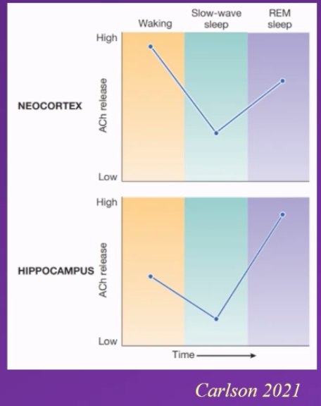
diagram depicting amount of acetylcholine (ACH) being released into the neocortex and hippocampus
describe results
neocortex – high when in wakeful stage, significant drop during slow wave sleep and high again during REM sleep
hippocampus: moderate / medium levels during wakefulness, drops significantly during slow wave sleep, highest significantly increase during REM sleep
describe how Noradrenaline (Norepinephrin) is a Mechanisms of Arousal
Noradrenaline comes From Locus Coeruleus (located in RAS in Pons) – Locus Coeruleus – translates to the blue place because it looks blue –
Has long axons that make synapses In large brain regions. – influencing a large brain region via noradrenaline
• Related to Vigilance, induced by external stimuli: i.e it is active when we engage in vigilance e.g. hear a loud bag and get startled turn around activates the locus coeruleus
Carlson 2021- quite active when we are being vigilant
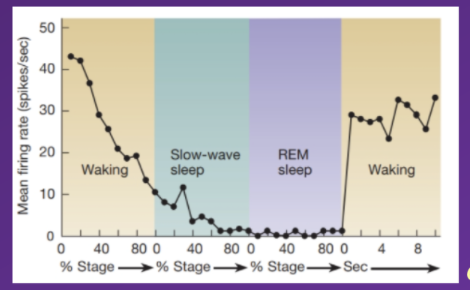
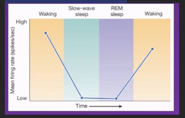
Diagram depicting firing rate of neurons (and subsequently release of noradrenaline via these action potentials) in the locus coeruleus.
describe Typical acitivity in the locus coeruleus is
how does this evidence noradrenalines involvement in arousal ?
Typically – acitivity in the locus coeruleus is
High when awake
Continuously declines until the individual enters slow wave sleep
Continues to decline during slow wave sleep (not reaching 0 though)
During rem sleep firing rate is reduced to 0, and locus coeruleus becomes inactive during REM sleep
When individual wakes up activity significantly increases, spikes as locus coeruleus activates again
As no activity in locus coeruleus during REM sleep, no noradrenaline released during this time, indicating that no noradrenaline being released is a crucilal condition for REM sleep to occur
might contribute to the switch between slow wave and REM sleep, as its only when low enough threshold is attained can we transition from slow wave to REM sleep
evidences Noradrenaline levels ned to be very low during REM sleep for it to occur
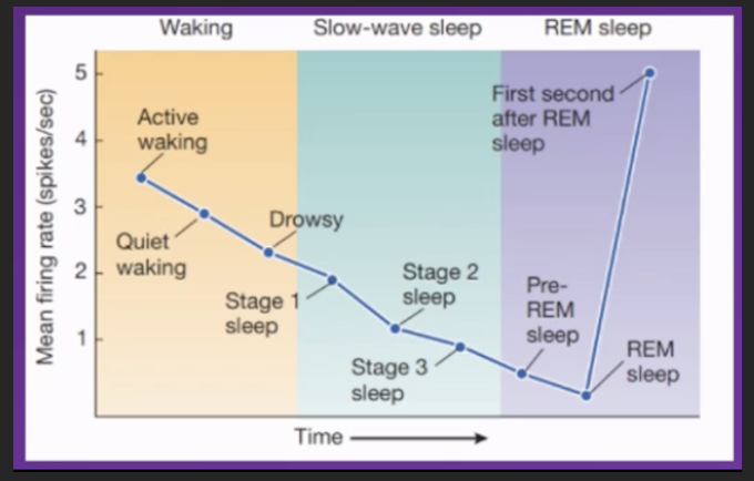
how Serotonin a Mechanism of Arousal:
where does serotonin come from
what evidence supports its involvement
◦ Where does sertononin come from?
Serotonin comes From Raphe Nuclei (which axons release serotonin into the brain) (is part of the RAS in Pons and Medulla)
Influences locomotion and cortical arousal, but not sensitive to external stimuli
Is highly active when awake and continually decrease in activity as the individual transitions from wakeful to slow wave sleep
during
Slow wave sleep: Continues to decrease in activity – mean firing rate of neurons releasing serotonin reduces.
REM sleep: Slowly after entering REM sleep stage the lowest activity rate occurs
First second after individual enters REM sleep stage activity significantly spikes and increases
Conclusion
Can infer there's an incompatibility between serotonin levels being high and REM sleep happening.
Serotonin levels need to be low during slow wave sleep and very low during REM sleep for these stages to occur
Where is serotnonin used in clinical settings artificially?
It is the same serotonin, the same source of serotonin that we believe to be involved in depression and in antidepressant drug treatment
How does serotonin differ from noreadrenaline/ what does noradrenaline do differently
Both contribute to keeping us awake (if they are high we will be awake/ not sleep, and need to be low for us to fall asleep) and influence out alertness and vigilance but under different conditions/ circumstances – what they share in common
Where do they differ?
While noradrenaline seems to be involved in vigilance that is driven by external stimuli triggered by external stimuli
Serotonin tends to be involved in vigilance and alertness that's internally driven e..g consciously choosing to pay close attention to something driven by personal motivation, crashing noise will not cause raphae nuclei to fire and subsequently release increased serotonin, it will only cause locus coeruleus neurons to fire.
how is Histamine neurotransmitter a Mechanism of Arousal:
where does serotonin come from
what evidence supports its involvement
◦ Where are histamine releasing neurons located?
By a group of neuons in the In the Tuberomammilary Nucleus (in the Hypothalamus)
◦ High during waking, low during sleep – i.e involved in keeping us awake
Anti-histamines put you to sleep. - evidenced via medicine administration, infer histamines do the opposite
Why would people take anti-histamines?
Allergies – because histamine is involved in allergic reactions: it is a signalling molecule all through your body and involved in the inflammatory response and allergic reactions
Wakefulness – it is a neurotransmitter in the brain involved in keeping us awake
Can be prescribes as a sleep inducing agent
How do antihistamines influence wakefulness?
If antihistamine crosses the blood brain barrier (some do, some don't) then it will not just have an effect on the allergy allergic reaction but will also influence our wakefulness, and cause drowsiness
how is Hypocretin (also called Orexin) neurotransmitter a Mechanism of Arousal:
Where are hypocretin producing neurons located?
Located and produced in the lateral hypothalamus
Function relating to wakefulness – contributes to keeping us alert and awake via its
◦ excitatory (hypocretinergic) connections to (i.e increases activity in) the:
◦ Locus coeruleus
◦ Raphé nuclei
◦ Tuberomammillary nucleus
◦ Dorsal Pons
◦ Basal Forebrain
◦ Cerebral cortex
◦ … which contribute to keeping us awake
• is Active during active waking and exploration, So keeps us alert
Hypcretin must be high when awake and really low during slow wave sleep and REM sleep , gradualy increase during REM sleepunitl threshold activity has been surpassed and wakefullness can be attained
explain evidence which supports that hypocretin is involved and is a mechanism of arousal
Seems to be master controller
( why does Hypocretin (also called Orexin) have two names?
Because different people discovered the same neurotransmitter and named it independently of each other (i.e unaware it had already been named)
Extra – anecdotal info. I think
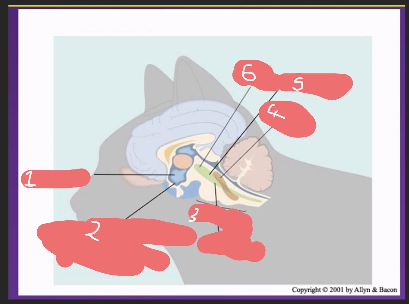
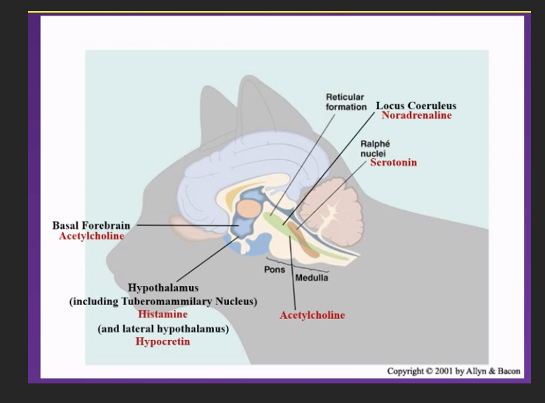
fill in the blanks to identify thevbrain areas in volved in arousal and their locations
p.s
He says prioritise understnaidng the mechanisms over the names of each neurotransmitter and its location – but you should know both! Hes justh idintg at what will be in exam potentially
Good explanation of how it works could be potential SAQ
Norepinephrine / noradrenalin are he same thing
new topic transitions into sleep = SWS
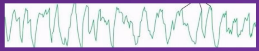
The person from whom this EEG trace was recorded is...
A)..asleep, their neurons are firing independently
B)awake, their neurons are firing independently
C)asleep, their neurons are firing synchronously
D)awake, their neurons are firing synchronously
Answer- c
Which is the correct combination of brain area and neurotransmitter?
A) Locus coeruleus – norepinephrine / noradrenaline
B) Raphe nuclei – histamine
C) Basal forebrain – noradrenaline
D) Lateral hypothalamus – serotonin
E) Tuberomammilary nucleus – hypocretin
Answer – A
Because correct answer for all answers would be
B - Raphe nuclei – serotonin
c- Basal forebrain – acetylcholine
d - Lateral hypothalamus – hypocretin
e- Tuberomammilary nucleus – histamine
Mechanisms of Transition to Sleep:
evidence
Ventrolateral Preoptic Area (vlPOA / VLPA) primary system
located in hypothalamus
animal studies found that this region is very quiet when awake and active when asleep – presumably the same occurs in humans
In animals if u stimulate this brain region, they fall asleep, if this brain region is removed/ lesioned they will not fall asleep and eventually die
Showing sleep is a crucial brain state for survival , and h ighlightingthat this is a region tht is integral to enabling sleep
how does Ventrolateral Preoptic Area (vlPOA / VLPA) control sleep