Neural Systems Plasticity and Potentiation
1/32
There's no tags or description
Looks like no tags are added yet.
Name | Mastery | Learn | Test | Matching | Spaced |
|---|
No study sessions yet.
33 Terms
Neural Plasticity
change to neuronal circuitry that leads to changes in neural processing
Why is plasticity required?
store information (memory) which requires change (learning), circuit development (axons and dendrites growing), recover after injury, adaptation
Gill-withdrawl reflex in Aphysia
changes in synapses result in behavioral changes
Synaptic Plasticity
change in strength of synapse; pre-(vesicle release) or postsynaptic (receptors) or both (structural)
Intrinsic Plasticity
physiological response to EPSP/IPSPs; membrane resistance and density of ion channels
Structural Plasticity
anatomical change to alter signalling; synapse, spine, dendrite, axon morphology
Long-Term Potentiation
increased synaptic strength; more receptors
Long-Term Depression
decreased synaptic strength; less receptors
Hebbian Plasticity
neurons that fire together, wire together; cell A synapsing to cell B and helping drive firing, something is changing so cell A is more likely to drive firing of cell B (less input to get B going)
Dentate Gyrus
consists of many granule cells; main input to hippocampus
Trisynaptic Circuit of the Hippocampus
Dentate gyrus receives input from entorhinal cortex, CA3 receives input from dentate gyrus, CA1 receives input from CA3
Field Recordings
recording electrode near dendrites stem electrode forces neurons to fire action potentials, axons release glutamate which binds to AMPAR and NMDAR causing them to open and allow Na+ in away from electrode
Field EPSP
opposite of whole cell EPSP
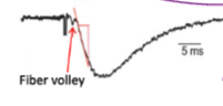
Fiber Volley
electrical signal of presynaptic action potentials; positive charges flowing in
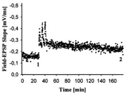
LTP Experiment
fEPSPs recorded before and after bursts of strong stimuli; fiber volley does not change; large jump in slope then stabilizes but still higher than baseline
High Frequency Stimulation (HFS)
100Hz for 1 second, repeated; effective but not physiological
Theta Bursts
patterned input with timing based on natural brain rhythms; burst if 4 pulses at 100 Hz repeated in 200 ms intervals; more physiological and efficient
LTP Synaptic Strength
presynaptic stimulation + postsynaptic depolarization repeatedly = increased strength of synapses, more likely to drive firing of postsynaptic cell
LTP Experiment Steps
collect baseline fEPSPs once every 30 seconds, average baseline, 100 Hz presynaptic stimulation (theta burst or HFS), collect fEPSPs
Input-Specificity
only synapses that are stimulated are potentiated
NMDARs in LTP
required; prolong response; coincidence detectors (need glutamate and depolarization)
Ca2+ in LTP
cannot occur without
LTP Pathway
NMDARs open, calcium entry, calmodulin (CaM) activated, CaMKII activated, AMPAR phosphorylated (greater conductance, more receptors, increased synaptic strength)
CaMKII
Ca/CaM-dependent protein kinase II; 12 subunits that open after binding CaM; long-lasting because of autophosphorylation
CaMKII subunit domains
association (heteromer formation), regulatory (activation, folds to block catalytic part), catalytic (acting on substrates)
CaMKII in LTP
direct phosphorylation of AMPAR and AMPAR auxiliary subunits
AMPAR Phosphorylation
increases current flowing into the synapse; bigger synaptic response
TARPs
transmembrane AMPAR regulatory proteins; anchors AMPARS to posy synaptic membrane
Silent Synapses
contain only NMDARs; unsilenced by translocation of AMPARs to synapse through LTP
Gene Expression in LTP
Ca2+ influx and CaMKII activates cAMP, activates CREB TF, induces gene expression, effector proteins maintain LTP
ACT-D
blocks transcription; causes loss of long lasting LTP
Animycin
blocks translation; causes loss of long lasting LTP
LTP Mainentance
formation of new and larger dendritic spines