Bontrager Chapter 1- Part 2
1/153
There's no tags or description
Looks like no tags are added yet.
Name | Mastery | Learn | Test | Matching | Spaced | Call with Kai |
|---|
No analytics yet
Send a link to your students to track their progress
154 Terms
Analog Imaging
-analog images provide a two dimensional image of anatomic structure
- the image device is a film screen system that consists of a pair intensifying screens with a film in between them.
hard copy image
also referred to as the film image
- is composed of a deposit of metallic silver on a polyester base and is permanent; it cannot be altered.
analog image receptors are best described as
self regulating systems with a limited dynamic range.
-they are also described using the term exposure latitude.
what are exposure factors also known as?
technique factors
What are the exposure factors?
Kilovoltage (kV)
Milliamperage (mA)
Exposure time (ms)
What is kilovoltage?
Controls the energy ( penetrating power) of the X-ray beam.
-can also be referred to as kilovoltage peak (kVp)
What is the kilovoltage peak? kVp
-The maximum electrical potential used to create the X-ray photons within the tube.
-(the penetrator)
-kilovoltage peak
What is milliamperage?
Controls the number of X-rays produced
What is exposure time?
Controls the duration of the exposure usually exposed in milliseconds
What are the four image quality factors?
Density/brightness
Contrast/adjacent shades
Resolution/detail
Distortion
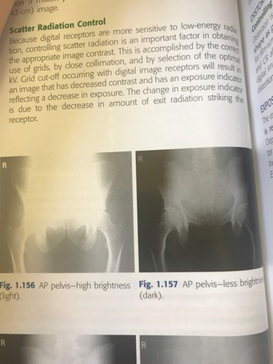
What is density?
The amount of blackness on the processed radiograph.
-when an image with high density is viewed, less light is transmitted through the image.
If your image is too dark?
1/2 the mass or decrease the KV by 15%
if your image is too light?
double the mass or increase the KV by 15 %
15 % change rule in KV example:
80 KV x .15= 12 kv
12+80=90KV
What is the primary controlling factor of film density?
mAs
It controls density by controlling the quantity of X-rays emitted from the tube and the duration of the exposure.
What is SID?
Source to image receptor distance
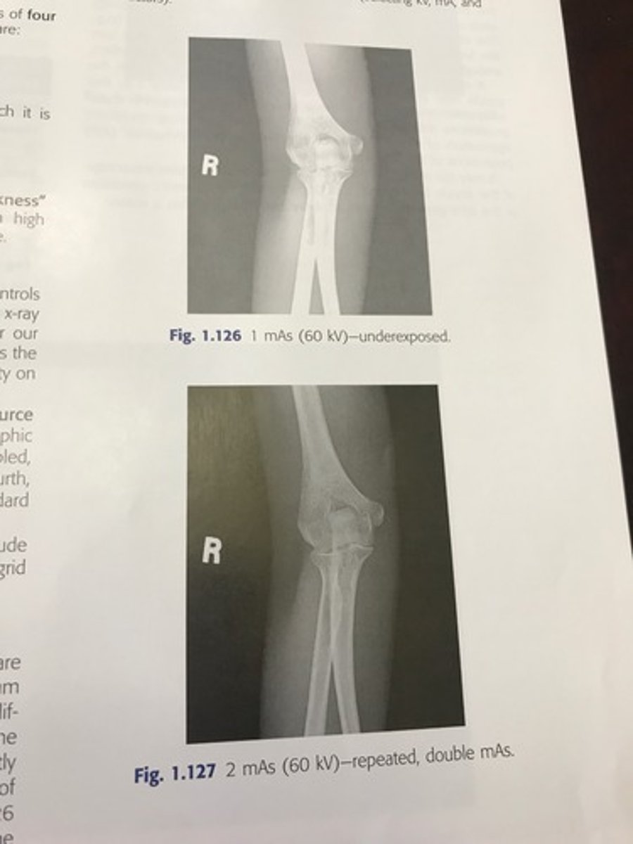
What is the general rule for under or over exposed repeat images?
Minimum of 25-30% change in mAs is required to make a visible difference.
* sometimes even doubling mAs is necessary
Examples of over/under exposed
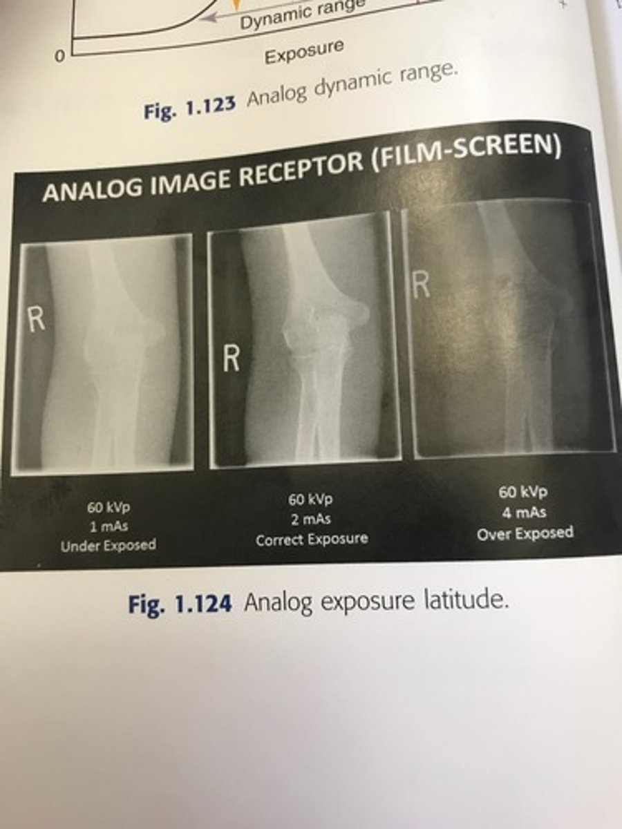
Example of over/under exposed
What is the anode heel effect?
The intensity of radiation emitted from the cathode end of the tube is greater than that emitted at the anode end.
-greater attenuation or absorption of xrays occurs at the anode because of the angle of the anode.
Application: the patient is positioned so that the thicker parts are under the cathode of the tube and the thinner parts are under the anode.
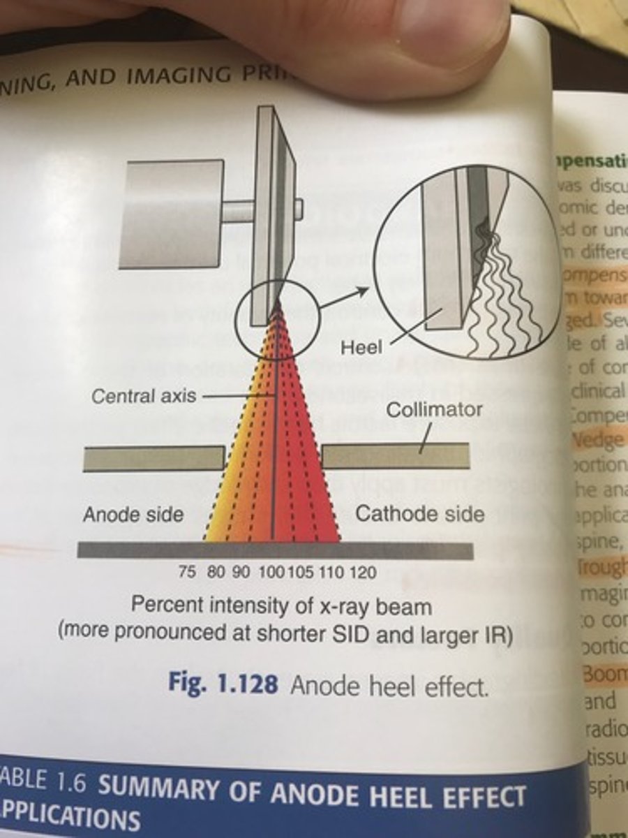
Angle of anode
xrays are emitted from deeper within the anode and must travel through more anode material before exiting.
How should you position the patient using the anode heel effect?
The thicker portion of the part should be positioned at the cathode, thinner part under anode.
anode heel effect image
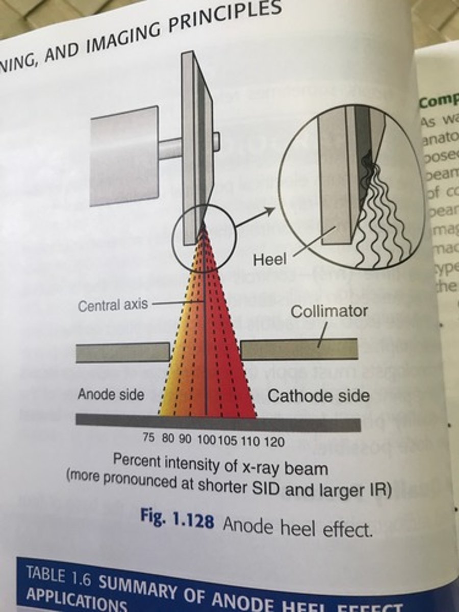
summary of anode heel effect application
look at chart on page 40
What is a compensating filter?
It filters out a portion of the primary beam toward the thin or less dense part of the body that is being imaged.
wedge filter
-mounts on the collimator; thicker portion of the wedge is placed toward the least dense part of the anatomy to even out the densities.
-thicker part under the toes for a foot
-numeric applications: most common include the AP foot, AP thoracic spine, and axiolateral hip
trough filter
-mounts on the collimator and is used for chest imaging
-has a filter on both sides
-the thicker portions of the filter are placed to correspond to anatomically less dense lungs
-the thinner portions of the filter correspond to the mediastinum
Boomerang filter
placed behind the patient and is used primarily for a shoulder and upper thoracic spine
-it improves visualization of soft tissues
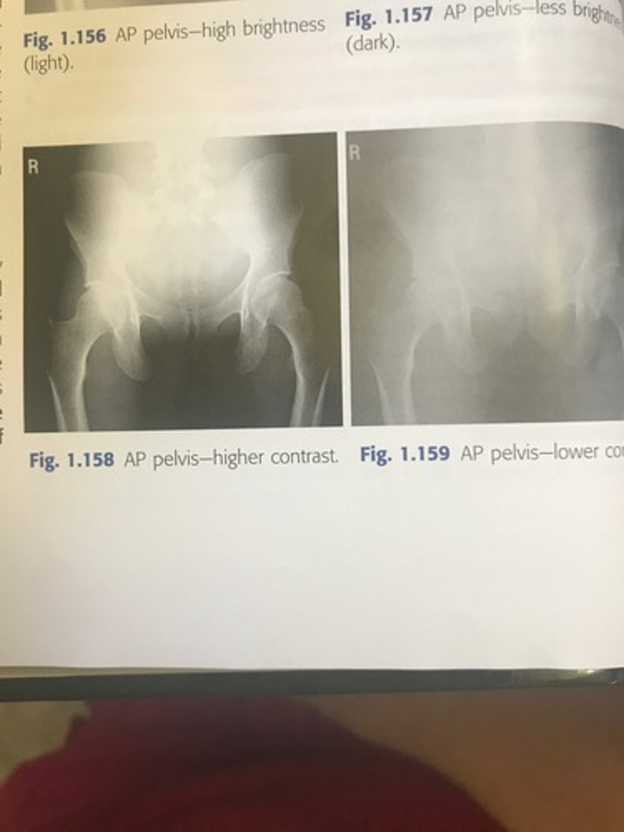
What is radiographic contrast?
Differences in density level between adjacent structures of a radiographic image.
Controlling factor is kV
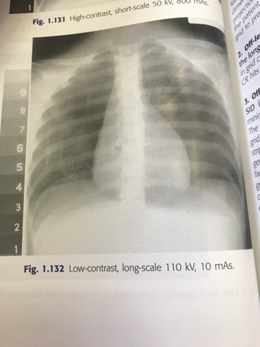
-can be referred to as long scale or short scale contrast referring to the total range of optical densities from the lightest to the darkest part of the radiographic image
When the density difference is large the contrast is what?
High contrast - short scale
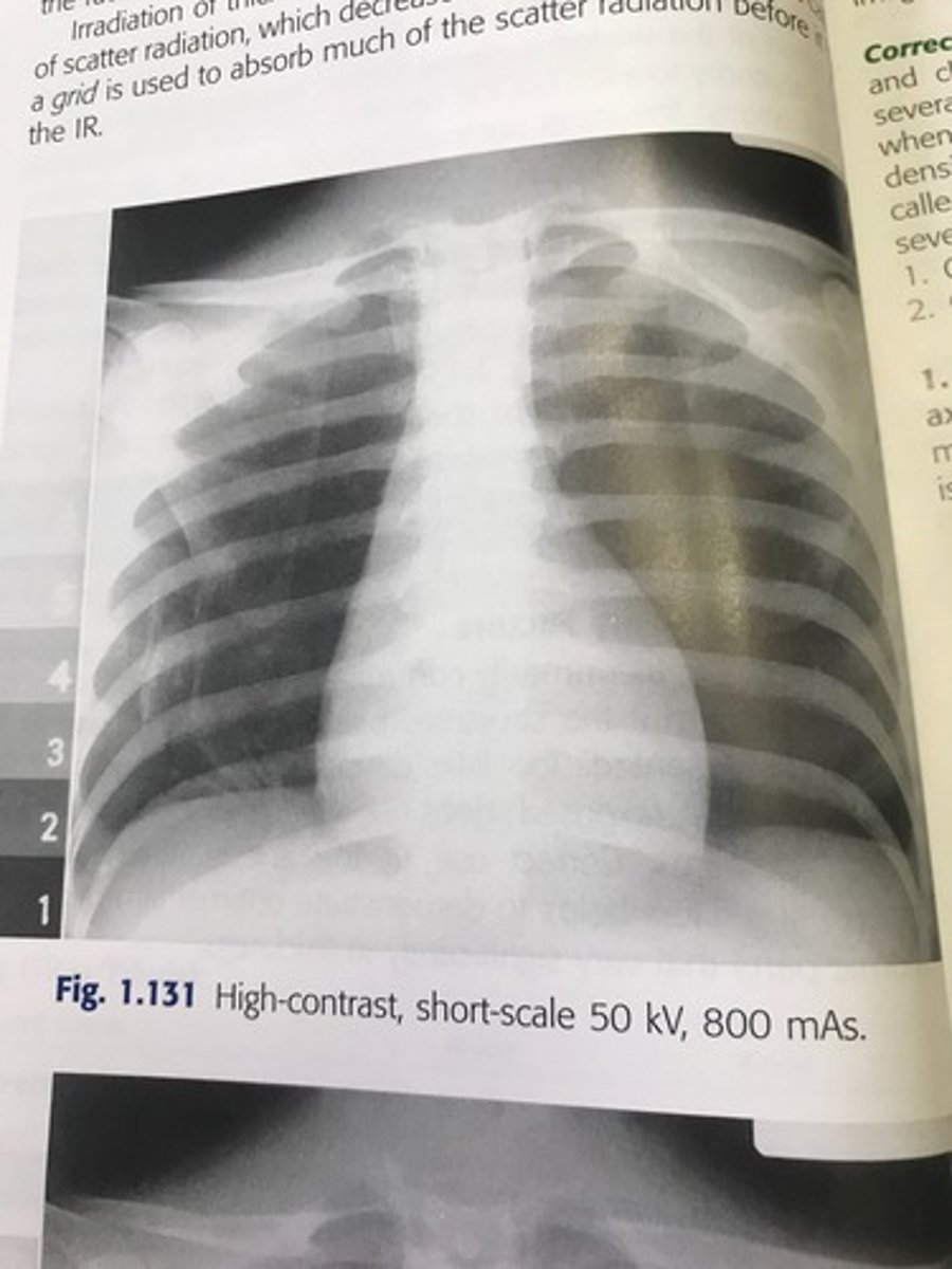
When the density difference is small the contrast is what?
Low contrast -long scale
Why is radiographic contrast important?
It allows you to see anatomic detail. It allows you to visualize a quality image.
primary controlling factor for contrast in film based imaging is:
kV
-kV controls the energy or penetrating power of the xray beam
high kV produces less attenuation resulting in ______?
lower contrast
When is it recommend that you use a grid?
On any body part that is thicker than 10 cm. Grid absorbs scatter radiation
-it is positioned between the patient and the IR and absorbs much of the scatter radiation before it hits the IR.
Compton scattering
Interaction with matter in which a higher energy photon strikes a loosely bound outer electron, removing it from its shell, and the remaining energy is released as a scattered photon
-the scattering we as techs protect ourselves from
grid cutoff
incorrect use of grids resulting in loss of density across all or part of the radiographic image
4 causes of grid cut off
1. off-center grid
2. off-level grid
3. off-focus grid
4. upside-down grid
off center grid
-The CR must be centered along the center axis of the grid.
-if it is not lateral decentering is said to occur
-the more the CR is off center the greater the cutoff
results in overall decrease in image density
off-level grid
-with angling, the CR must be angled along the long axis of the lead strips.
-transverse tilted grid, results in overall decrease of image density
off focus grid
-a focused grid must be used at a specific SID if grid cutoff is to be prevented
-grids usually have a min and max SID called the focal range
-the focal range is determined by the grid frequency and grid ratio
upside down focused grid
-each grid is labeled to indicate the side that must be positioned to face the x ray tube
-the lead strips are tilted or focused to allow the xray beam to pass unimpeded
-if the grid is positioned upside down the image will show severe cutoff (grid not correctly placed; the right side is not up)
What is spatial resolution?
-Recorded sharpness of structures on the image.
-resolution is known as detail, image sharpness, or definition (being able to see the smallest of things)
-lack of visible sharpness or resolution is known as blur or unsharpness
Spatial resolution is controlled by what?
Geometric factors
Film screen system
Motion
What is the greatest deterrent to image sharpness?
Motion.
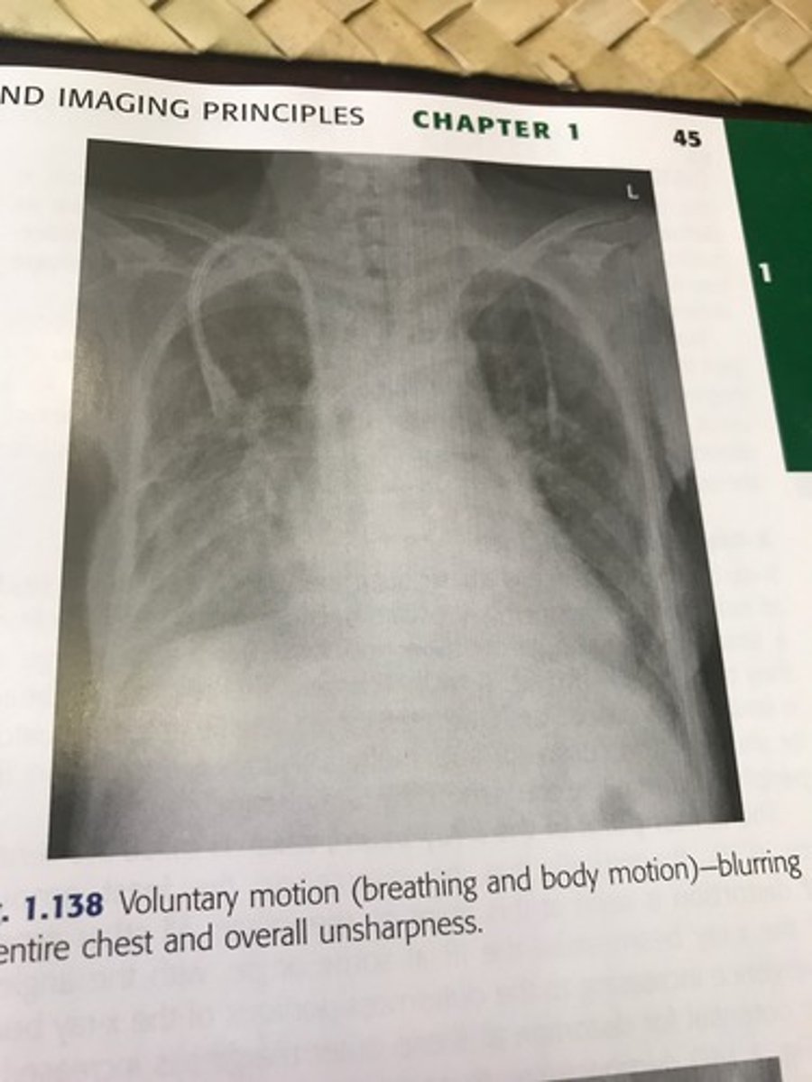
Voluntary and involuntary
Voluntary motion vs. involuntary
-Voluntary-motion in which the patient can control; motion from breathing or movement of body parts during exposure can be prevented or minimized by controlled breathing and patient immobilization such as tape, sponges, sandbags.
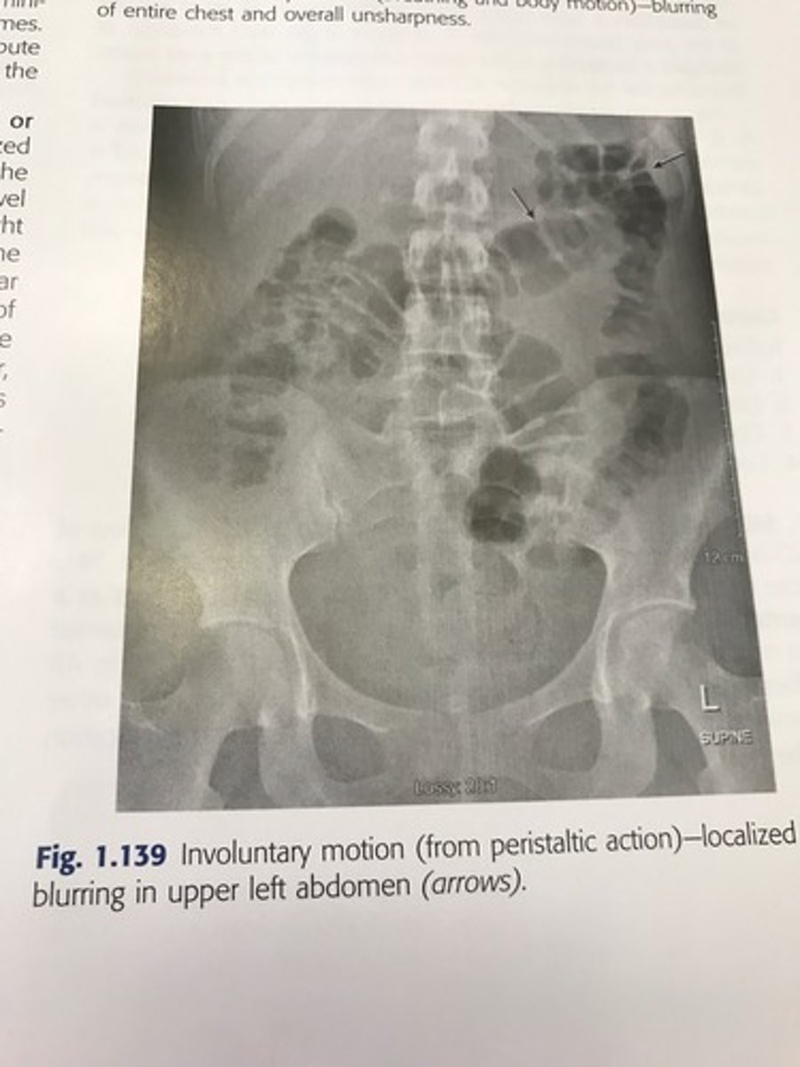
-Involuntary motion: includes peristaltic action, tremors, or the chills and is almost impossible to control.
what is the best way to reduce voluntary motion on a radiograph?
give the patient a thorough explanation of the procedure with well explained instructions.
what is the best way to control voluntary motion on a radiograph?
short exposure time
Voluntary motion image
Involuntary motion image
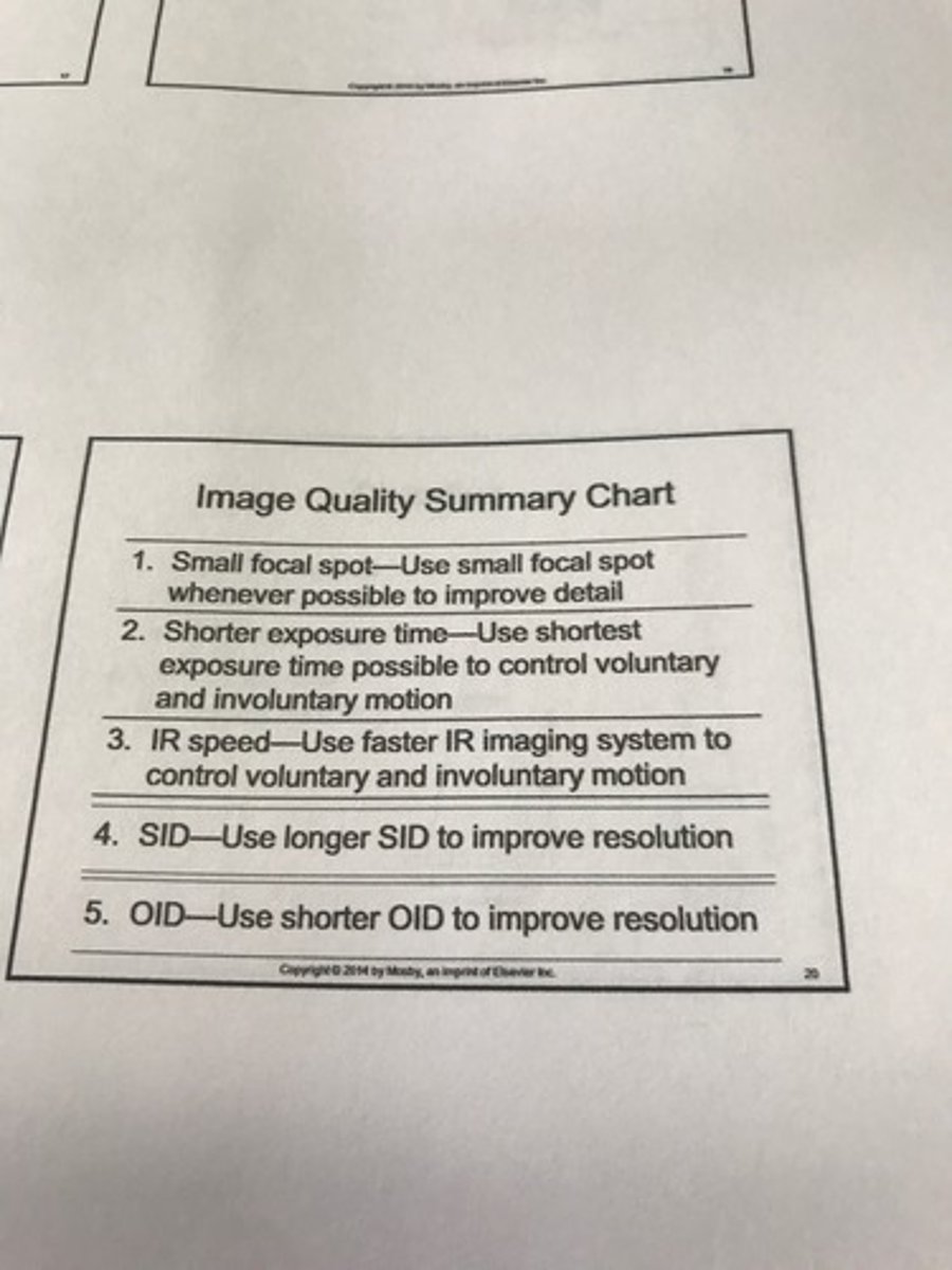
4 ways to control image quality
1) small focal spot- use whenever possible to improve detail
2) short exposure time- used to control voluntary and involuntary motion
3) SID- use longer SID to improve resolution
4) OID- use shorter OID to improve resolution
What do you need to get the best image?
1- small focal spot
2- long SID (increase SID)
3- short OID (decrease OID)
small focal spot
results in less geometric unsharpness or (penumbra)
- makes a clear image; with greater detail
large focal spot
results in more geometric unsharpness or (penumbra)
-makes a fuzzy or blurry image
penumbra
unsharp edges in the projected image
What is distortion?
a misrepresentation of object size and shape
two types:
1. size distortion (magnification)
2. shape distortion
central ray
the center point of the x ray beam
What are the 4 primary controlling factors of distortion?
1) source image receptor distance (SID)
2) object image receptor distance (OID)
3) object image receptor alignment
4) central ray alignment
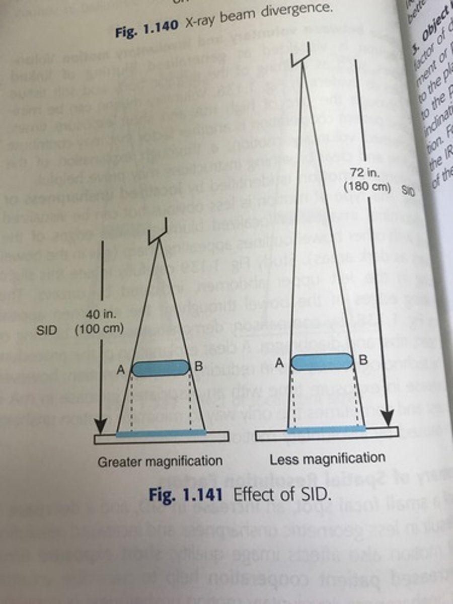
1.) SID
-less magnification occurs at a greater SID than at a shorter SID
-this is the reason chest x rays are performed at 72 inches instead of 40 inches because of less magnification
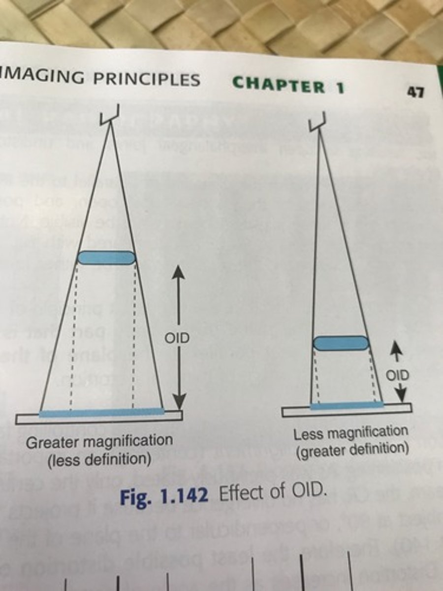
2. OID
the closer the object being radiographed is to the IR, the less are the magnification and shape distortion and better is the resolution.
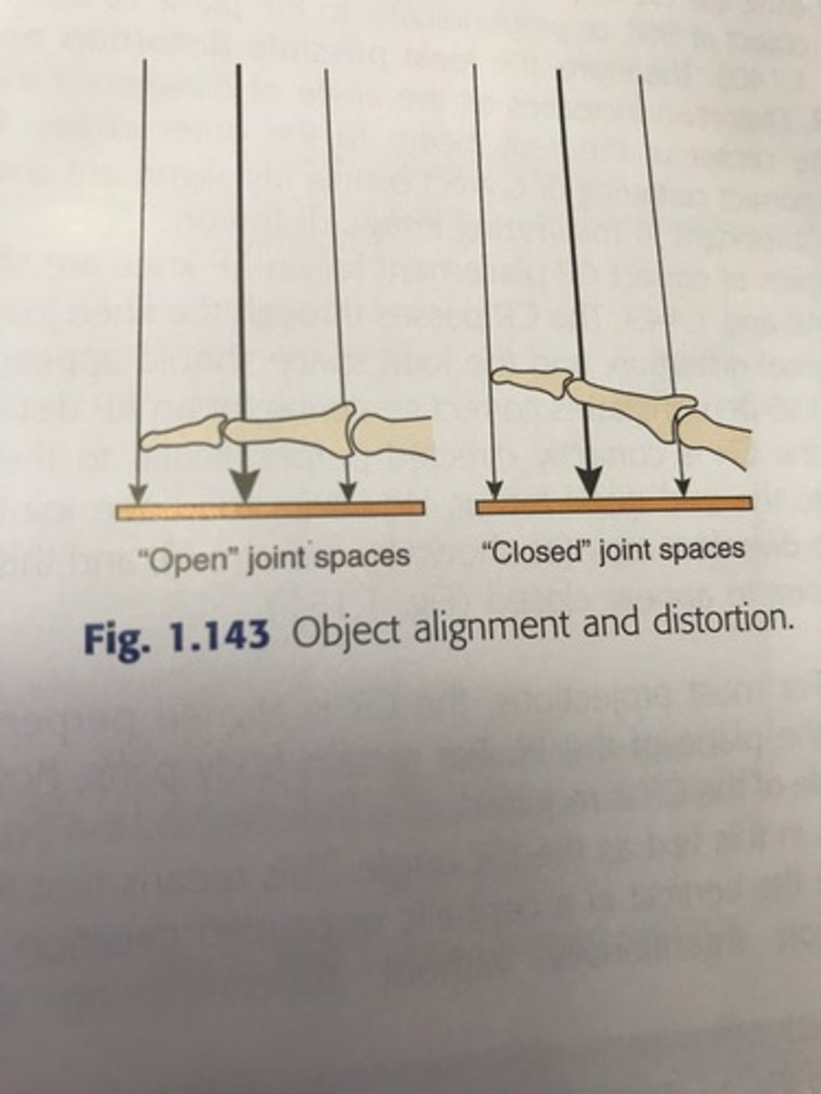
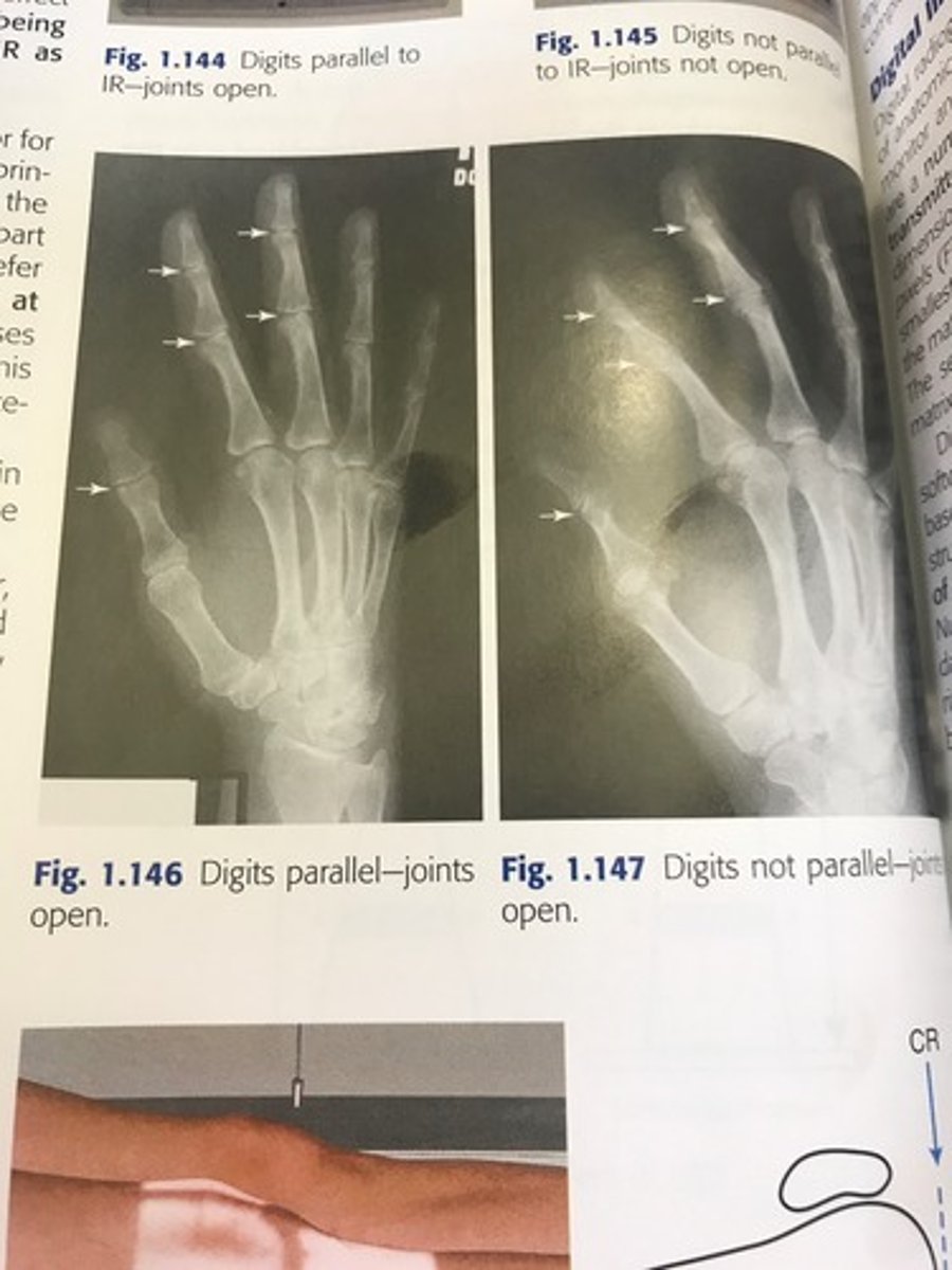
3. Object image receptor alignment
the alignment or plane of the object that is being x rayed in relation to the plane of the IR.
-if the object is not parallel to the plane of the IR it causes distortion.
4. central ray alignment
the least possible distortion occurs at the Central ray.
It is important to have correct positioning
How is a digital image formed?
By a matrix of pixels.
Pixel is the smallest unit, they make up rows and colums which forms the matrix.
The higher number of pixels you have makes what better?
Resolution!
mA
controls the number of x rays produced
mAs (mAx time)
refers to the number of xrays and the duration of exposure.
kV
controls the penetrating power of x-rays
What are the image quality factors in digital radiography?
1- brightness
2- contrast resolution
3- spatial resolution
4- distortion
5- exposure indicator
6- noise
What is brightness?
Intensity of light representing individual pixels in an image
What are the controlling factors for brightness?
in contrast to the linear relationship between mAs and density in film-screen imaging changes in mAs do not have a controlling effect on digital image brightness
pixels and bit depth
The greater the bit depth of a system, the greater is the contrast resolution (ie, the greater is the number of possible shades of gray that a pixel can have)
What is contrast resolution?
Differences in brightness between light and dark areas of an image
spatial resolution
in digital imaging is defined as the recorded sharpness or detail of structures on the image.
-controlling factors: pixel size (inherent to the IR. the smaller the pixel size the greater the spatial resolution)
-resolution is controlled by the display matrix (depends on the capabilities of the monitor)
What is distortion in digital radiography?
Misrepresentation of object shape or size.
What are the controlling factors for distortion?
SID
OID
CR alignment
*same as analog
What is an exposure indicator?
A numeric value that is representative of the exposure the image receptor received.
-may be inversely or directly proportional to radiation striking the IR
- key in verifying optimal digital image is obtained with least dose to patient
How do you find out if your exposure indicator number is valid?
Each institution will have a chart that your number must fall between to be an acceptable image. Varies on who makes the equipment
What is noise?
Random disturbance that obscures or reduces image clarity.
-A high SNR is desirable in imaging, in which the signal is greater than the noise so that low contrast soft tissue images can be demonstrated.
- a low SNR is undesirable in imaging, a low signal with accompanying high noise obscures soft tissue detail and produces a grainy mottled image.
What is post processing?
Changing or enhancing the electronic image to improve diagnostic quality.
-Algorithms applied to improve diagnostic quality of image.
-post-processing cannot improve a low SNR image
What are post processing options?
1- windowing
2- smoothing
3- magnification
4- edge enhancement
5- equalization
6- subtraction
7- image reversal
8- annotation
windowing
two types of adjustment are possible:
1. window width: controls the contrast of the image
2. window level: controls the brightness of the image
smoothing
specific image processing is applied to reduce the display of noise in an image.
magnification
all or part of an image is magnified
subtraction
background anatomy can be removed to allow visualization of contrast media-filled vessels (used in angiography)
image reversal
The dark and light pixel values of an image are reversed-the x-ray image reverses from a negative to a positive
annotation
text may be added to images
What was one of the first applications of computers in radiography?
Computed tomography
PSP photostimulable storage phosphor
-was the first widely implemented digital imaging system for general radiography; commonly called computed radiography
-may be cassette based or cassette less
technologist work station
-includes a bar code reader, a monitor to display the image, a keyboard with a mouse
Application for using Computed Radiography or Digital systems
1. collimation- close and accurate collimation helps reduce the amount of radiation exposure to the patient
2. Accurate centering of part and IR- the body part should be centered to the IR and properly displayed.
3. use of lead masks- use of lead masks or a blocker is recommended when a cassette based CR is used
4. use of grids- use a grid for body parts thicker than 10 cm
direct radiography (DR) (Flat-Panel detector with thin-film transistor)
1. direct radiography is the second type of digital imaging system
2. commonly referred to as direct radiography or direct digital radiography
What does PACS stand for?
What is it?
Picture- digital medical images
Archiving- electronic storage of images
Communication- routing and displaying of images
System- specialized computer network that manages the complete system.
What does PACS do?
Sends images to different areas of the hospital
-an electronic storage system for patient information and pictures
what are the advantages of PACS?
1. elimination of less efficient traditional film libraries and their inherent problem of physical space requirements for hard copy images
2. convenient search and retrieval of images
3. rapid transfer of images with hospital
4. ease in consulting outside specialists
5. viewing images at multiple locations
6. elimination of misplaced or damaged films
what does PACS stand for?
Picture Archiving Communication System
What is DICOM?
The current standard that ensures all manufactures and types of equipment are able to communicate and transmit images effectively.
what does HIS stand for?
Hospital Information System
what does HL7 stand for?
Health Level 7
What does RIS stand for?
Radiology Information System
What does IP stand for?
image plate