Lecture 2.1: Ligand Binding & Intro to Soluble Transport Proteins
 Call Kai
 Learn
Learn Practice Test
 Spaced Repetition
 Match
Match Flashcards
 Knowt Play
1/95

There's no tags or description
Looks like no tags are added yet.
Name | Mastery | Learn | Test | Matching | Spaced | Call with Kai | Chat |
|---|
No analytics yet
Send a link to your students to track their progress
96 Terms
What is a ligand?
A ligand is a molecule or ion that binds specifically to a protein.
What is a ligand-binding site?
A ligand-binding site is the specific region or pocket on a protein where a ligand binds.
Why does protein function often require binding partners?
Many proteins only work when they interact with specific molecules, such as ligands, substrates, cofactors, or other proteins.
What are the five major classes of protein function shown in Lecture 2.1?
Structural proteins, transport proteins, metabolic enzymes, cell signaling proteins, and genomic caretaker proteins.
What do structural proteins do?
Structural proteins provide support, shape, and organization, such as in the cytoskeleton or extracellular matrix.
What do transport proteins do?
Transport proteins move molecules from one place to another; examples include membrane transporters and soluble transport proteins like oxygen-binding proteins.
What do metabolic enzymes do?
Metabolic enzymes catalyze chemical reactions in the cell, including reactions involving energy transfer.
What do cell signaling proteins do?
Cell signaling proteins detect and relay signals, such as membrane receptors and kinases that modify target proteins.
What do genomic caretaker proteins do?
Genomic caretaker proteins help maintain and use genetic information, including DNA replication, chromatin organization, and RNA synthesis.
What does binding specificity mean?
Binding specificity means that a protein binds certain ligands much better than others.
What determines whether a ligand fits a protein-binding site?
The ligand must complement the binding site in size, shape, charge, and hydrophobic or hydrophilic character.
What is the lock-and-key model?
The lock-and-key model says that the binding site is already rigidly shaped to fit the ligand, like a key fitting into a lock.
What is the major assumption of the lock-and-key model?
It assumes that the complementary surfaces of the protein and ligand are preformed before binding occurs.
What is the induced-fit model?
The induced-fit model says that ligand binding can cause the protein to change shape so the ligand fits more tightly.
How does induced fit improve binding?
Induced fit allows the protein to adjust around the ligand, creating many weak interactions that strengthen binding.
Why is induced fit important in multisubunit proteins?
A shape change in one subunit can affect the conformation and binding behavior of neighboring subunits.
Which binding model emphasizes protein flexibility?
The induced-fit model emphasizes structural flexibility and adjustment during binding.
What is the general reversible ligand-binding equation?
P + L ⇌ PL, where P is protein, L is ligand, and PL is the protein-ligand complex.
What does Ka measure?
Ka is the association constant; it measures how strongly protein and ligand associate to form PL.
What is the formula for Ka?
Ka = [PL]/([P][L]).
What does a high Ka mean?
A high Ka means strong association and high affinity between the protein and ligand.
What does Kd measure?
Kd is the dissociation constant; it measures how easily the protein-ligand complex separates into free protein and ligand.
What is the formula for Kd?
Kd = ([P][L])/[PL].
What does a low Kd mean?
A low Kd means tight binding and high affinity.
What does a high Kd mean?
A high Kd means weaker binding and lower affinity.
How are Ka and Kd related for simple 1:1 binding?
They are inversely related: Ka = 1/Kd and Kd = 1/Ka.
What is theta (θ) in ligand binding?
Theta is fractional saturation, meaning the fraction of total binding sites occupied by ligand.
What is the formula for theta using protein-ligand complex concentration?
θ = [PL]/([PL] + [P]).
What is the formula for theta using ligand concentration and Kd?
θ = [L]/([L] + Kd).
What does θ = 0 mean?
No binding sites are occupied.
What does θ = 0.5 mean?
Half of the binding sites are occupied.
What does θ = 1 mean?
All or nearly all binding sites are occupied.
What happens to ligand binding as ligand concentration increases?
Fractional saturation increases until the binding sites become saturated.
What happens when [L] equals Kd?
θ = 0.5, so the protein is 50% saturated with ligand.
If Kd = 0.2 mM, what is θ when [L] = 0.2 mM?
θ = 0.5, because [L] equals Kd.
What happens when [L] is much greater than Kd?
The protein approaches full saturation, so θ approaches 1.
What happens when [L] is much less than Kd?
Very few binding sites are occupied, so θ is low.
Why is Kd useful?
Kd lets you compare how tightly different proteins bind their ligands.
If Protein A has Kd = 10 nM and Protein B has Kd = 60 nM, which has higher affinity?
Protein A has higher affinity because it has the lower Kd.
On a Kd scale, which side represents stronger binding?
The lower-Kd side represents stronger binding and higher affinity.
Which biological interaction shown has extremely high affinity? Why?
Biotin-avidin has extremely high affinity and a very low Kd. This is due to the strong non-covalent interactions that occur between the two molecules. Avidin has four binding sites for biotin, and the structure of avidin creates a highly specific fit for biotin, allowing for stable binding.
What does a larger Kd value generally indicate on the biological Kd scale?
A larger Kd value indicates lower affinity and weaker binding.
What is the difference between specificity and affinity?
Specificity describes which ligand a protein prefers; affinity describes how tightly the protein binds that ligand.
What are myoglobin and hemoglobin?
Myoglobin and hemoglobin are oxygen-binding proteins that use heme to bind O2.
Where is myoglobin mainly found?
Muscle
Where is hemoglobin mainly found?
Erythrocytes, or red blood cells
What is the main role of myoglobin?
Myoglobin mainly stores oxygen in muscle and acts as an oxygen reserve.
What is the main role of hemoglobin?
Hemoglobin mainly transports oxygen in the blood.
How many subunits does hemoglobin have and what are they?
Hemoglobin has four subunits: two alpha subunits and two beta subunits.
How many heme groups are shown in hemoglobin?
Hemoglobin has four heme groups, one in each subunit.
How many heme groups does myoglobin have?
Myoglobin has one heme group.
What is a globin fold?
A globin fold is a compact globular structure made from eight alpha helices.
What do myoglobin and hemoglobin subunits have in common structurally?
They share a similar globin fold, even though their amino acid sequences are not very similar.
What does it mean that myoglobin and hemoglobin have little sequence similarity but similar structure?
Their exact amino acid sequences differ, but they still fold into a similar 3D shape.
Why is oxygen chemically difficult for cells to handle?
Oxygen is nonpolar, poorly soluble in water, and does not diffuse through tissues easily.
Why do organisms need proteins to transport and store oxygen?
O2 is poorly soluble and no amino acid side chain can reversibly bind O2 well on its own.
Can amino acid side chains alone reversibly bind O2 effectively?
No. O2 binding requires a helper group, especially heme.
What is a cofactor?
A cofactor is a non-protein helper that allows a protein to perform a function it could not perform as well by itself.
What are the two types of cofactors?
Organic cofactors and inorganic cofactors.
What are examples of inorganic cofactors?
Metal ions such as Cu2+, Zn2+, K+, and Mg2+.
What are examples of organic cofactors?
Coenzymes such as NAD+ and prosthetic groups such as heme.
What is a prosthetic group?
A prosthetic group is a cofactor that is tightly associated with a protein, often permanently or covalently.
What is a cosubstrate?
A cosubstrate is a cofactor that binds transiently and then leaves during the reaction cycle.
What type of cofactor is heme?
Heme is an organic prosthetic group.
What does heme do in myoglobin and hemoglobin?
Heme enables reversible O2 binding.
What is heme made of?
Heme consists of a porphyrin ring with a coordinated Fe2+ ion in the center.
Which part of heme directly interacts with oxygen?
The Fe2+ ion directly binds oxygen.
Why is Fe2+ important in heme?
Fe2+ is the iron form that allows reversible oxygen binding in heme proteins.
Where does heme bind in myoglobin?
Heme binds in a hydrophobic pocket inside myoglobin.
Why is the heme pocket hydrophobic?
The hydrophobic pocket helps position heme properly and creates an environment suitable for O2 binding.
Which amino acid is required for stabilizing heme and facilitating O2 binding in myoglobin?
Histidine; specifically, the proximal histidine and distal histidine are key.
What is the proximal histidine?
The proximal histidine is His F8, also called His93 in myoglobin; it directly coordinates the Fe2+ of heme.
What is the distal histidine?
The distal histidine is His E7; it sits near the O2-binding site and helps stabilize bound oxygen.
Which histidine directly binds the Fe2+ in heme? Why?
The proximal histidine, His F8/His93, directly binds Fe2+.
Which histidine helps stabilize O2 through hydrogen bonding?
The distal histidine, His E7, helps stabilize the bound O2 ligand.
What do the labels His E7 and His F8 mean?
They identify histidines located on the E and F helices of the globin fold.
What is pO2?
pO2 is the partial pressure of oxygen, a measure of oxygen availability.
Why is pO2 used for oxygen-binding proteins?
Because oxygen is a gas, its availability is often described by partial pressure instead of ordinary concentration.
What is P50?
P50 is the pO2 at which an oxygen-binding protein is 50% saturated.
How is P50 related to Kd?
P50 is the oxygen-binding version of Kd; lower P50 means higher oxygen affinity.
What is the oxygen-binding equation using pO2 and P50?
θ = pO2/(pO2 + P50).
What does it mean when pO2 = P50?
The protein is 50% saturated with O2, so θ = 0.5.
What does a low P50 mean?
A low P50 means the protein has high affinity for oxygen.
Why does myoglobin have high oxygen affinity?
Myoglobin has a very low Kd/P50 for O2 binding.
What shape is the myoglobin O2-binding curve?
The myoglobin O2-binding curve is hyperbolic.
Why does the myoglobin binding curve plateau?
The curve plateaus because most myoglobin molecules become saturated with O2 at higher pO2.
What happens to myoglobin saturation as muscle pO2 decreases during activity?
Myoglobin releases some O2 as pO2 decreases, but it remains fairly highly saturated because it binds O2 tightly.
Why is myoglobin better for oxygen storage than long-distance oxygen transport?
Myoglobin binds O2 very tightly and releases only a limited amount unless pO2 becomes very low.
Could myoglobin efficiently transport O2 from lungs to resting tissues by itself?
No. Its affinity is too high, so it would hold onto O2 and not unload enough at resting tissue pO2.
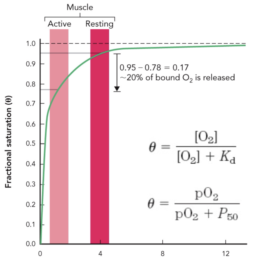
What do the active and resting muscle bars on the myoglobin graph show?
They show that lower pO2 in active muscle causes more O2 release than resting muscle, but myoglobin still remains substantially saturated.
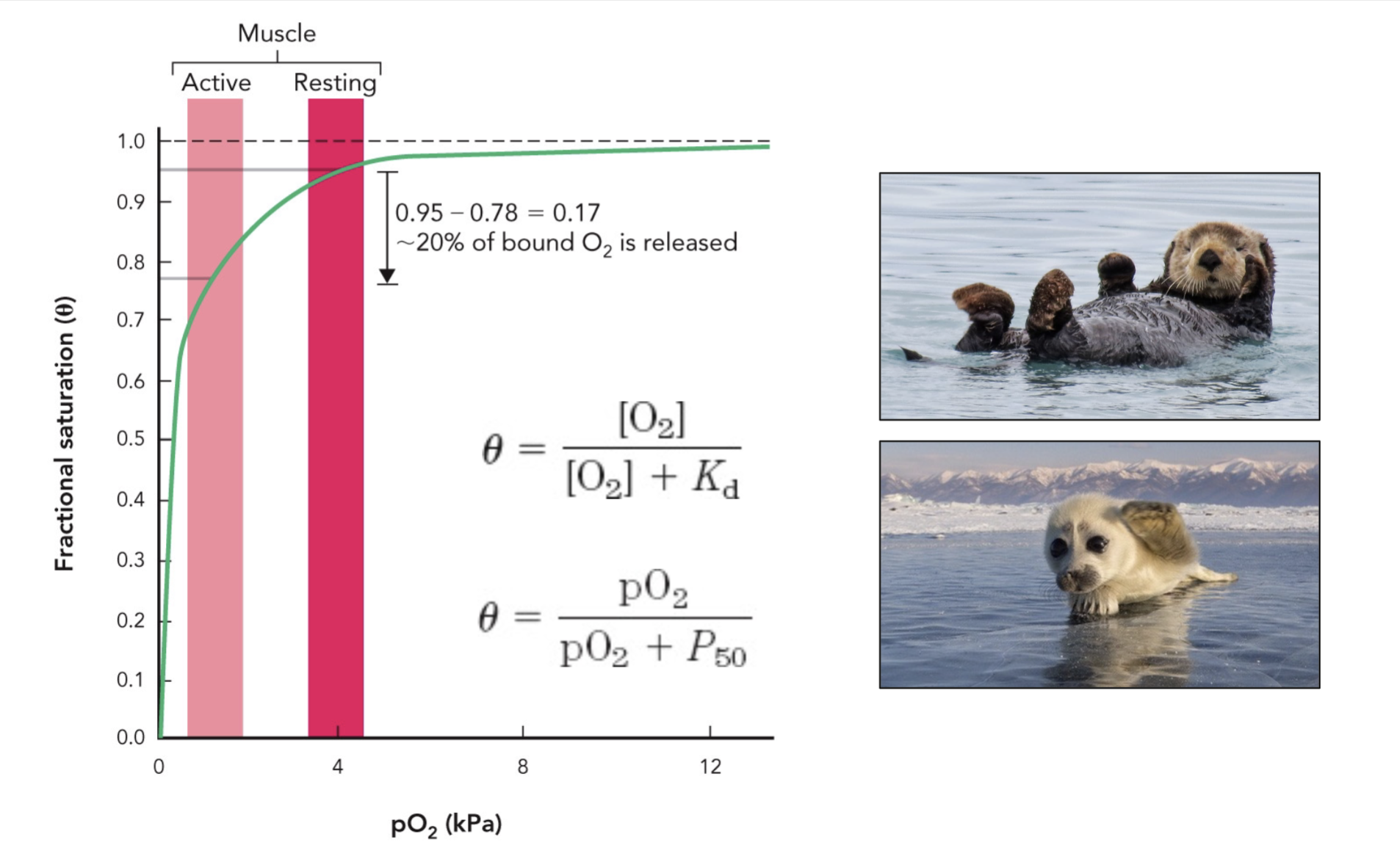
What do the diving animal images on the myoglobin slide emphasize?
They emphasize myoglobin's role as an oxygen-storage protein in muscle when oxygen availability is limited.
For reversible binding between a protein and a ligand, the larger the Ka, the ______ (higher/lower) the affinity between the protein and ligand.
For reversible binding between a protein and a ligand, the larger the Kd, the _______ (higher/lower) the affinity between the protein and ligand.
higher, lower
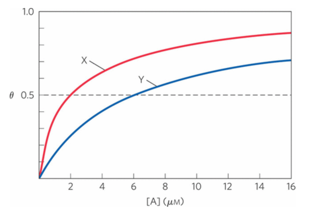
Two proteins, X and Y, bind to the same ligand, A, with the binding curves shown below.
Which protein has a higher affinity for ligand A?
A) Protein X
B) Protein Y
A) Protein X
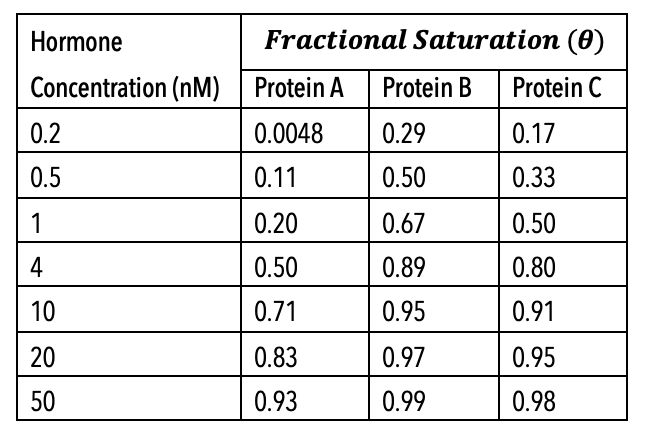
Three membrane receptor proteins bind to the same hormone. Use the data in the table below to answer the following 4 questions regarding these binding interactions.
Based on the data presented in the table, which protein (A, B, or C) binds least tightly to the hormone being studied?
Protein A
A particular genomic caretaker protein, Protein X, can bind to both Ligand Y and Ligand Z.
When 0.23 μM of Protein X is mixed with 0.11 μM of Ligand Y, the solution reaches equilibrium with 0.14 μM free Protein X, 0.02 μM free Ligand Y, and 0.09 μM protein–ligand complex.
However, when 0.23 μM of Protein X is mixed with 0.11 μM of Ligand Z, the solution reaches equilibrium with 0.20 μM free Protein X, 0.09 μM free Ligand Z, and 0.02 μM protein–ligand complex.
Based on the information provided, which of the following is true and why?
a. Protein X has a higher affinity for Ligand Z than Ligand Y.
b. Protein X has a higher affinity for Ligand Y than Ligand Z.
c. Protein X has an equal affinity for both Ligands Y and Z.
d. It is not possible to determine with the given information.
B
Protein X forms more complex with Ligand Y than with Ligand Z, meaning Ligand Y binds more tightly and has the lower Kd.
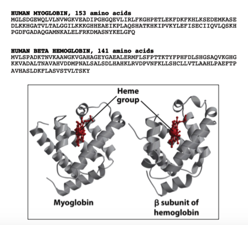
The figure shows the amino acid sequences and folded structures of human myoglobin and the β subunit of human hemoglobin. Although the two proteins have different sequence lengths and many different amino acids, both contain a similarly positioned heme group within a mostly α-helical fold. Which conclusion is best supported by the figure?
a. Myoglobin and β hemoglobin must have identical oxygen-binding affinities because both contain heme.
b. Myoglobin and β hemoglobin likely evolved from a common ancestral globin protein, because they preserve a similar tertiary fold and heme-binding architecture despite substantial differences in primary sequence.
c. Myoglobin and β hemoglobin have unrelated functions because their amino acid sequences are too different to produce similar protein folds.
d. The β subunit of hemoglobin binds oxygen independently in the same way as myoglobin, so hemoglobin cannot display cooperative oxygen binding.
e. Myoglobin and β hemoglobin have similar primary structures because both sequences contain the same number of amino acids and identical residues around the heme group.
B
The diagram shows that the primary structures are different, meaning the exact amino acid sequences are not very similar. However, the folded shapes are clearly similar: both proteins are mostly α-helical and hold a heme group in a comparable position. This supports the idea that proteins can maintain a similar tertiary structure and functionally important binding site even when their sequences have changed over evolution. The strongest interpretation is that myoglobin and β hemoglobin are related globin proteins that preserve the same overall fold and heme-binding role.