Session 1: Cell Injury
1/106
There's no tags or description
Looks like no tags are added yet.
Name | Mastery | Learn | Test | Matching | Spaced | Call with Kai | Chat |
|---|
No analytics yet
Send a link to your students to track their progress
107 Terms
Three ways we can visualise cell injury
1) Naked eye = gross appearance
2) Light microscopy = microscopic features
3) Electron microscopy = ultrastructural features
List some causes of cell injury
1) Hypoxia
2) Chemical agents/drugs
3) Infection
4) Immune-mediated processes
5) Nutritional imbalance
6) Genetic derangement
7) Physical agents e.g., trauma
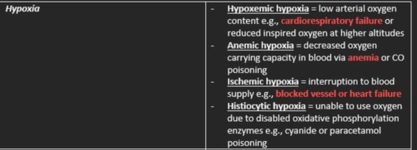
The four types of hypoxic cell injury
1) Hypoxemic hypoxia = low arterial O2 concentration e.g., cardiorespiratory failure
2) Anemic hypoxia = decreased oxygen carrying capacity e.g., anaemia
3) Ischemic hypoxia = interruption to blood supply e.g., blocked vessel
4) Histiocytic hypoxia = unable to use oxygen due to disabled oxidative phosphorylation enzymes (e.g., cyanide poisoning)
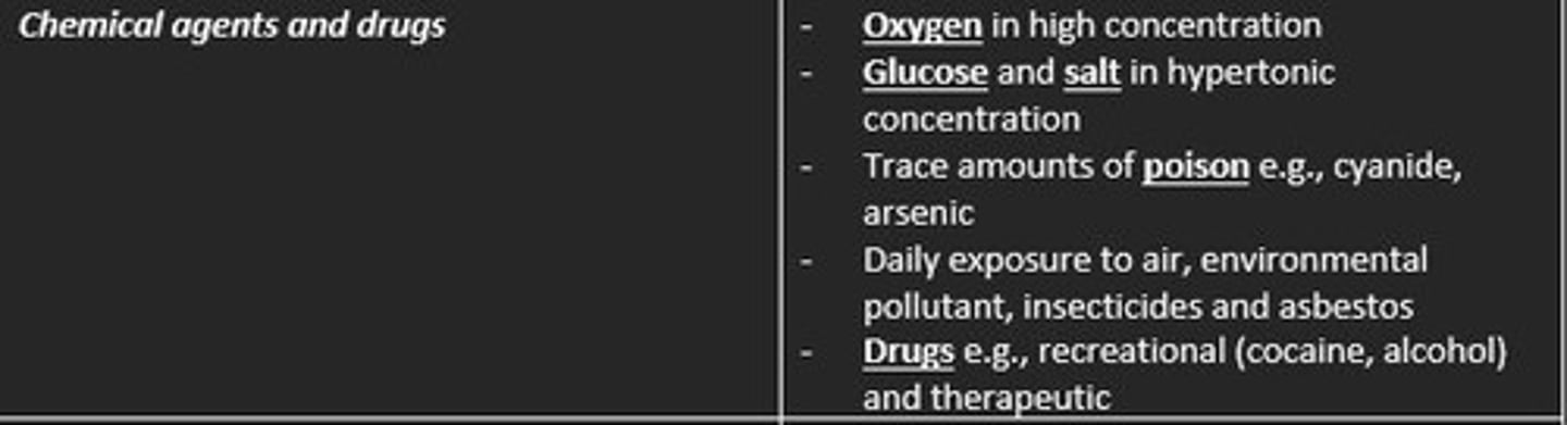
Examples of chemical agents or drugs that can cause cell injury
1) Oxygen in high concentration
2) Glucose & salt in hypertonic concentrations
3) Trace amount of poison (arsenic, cyanide)
4) Daily exposure to air/pollutant/insecticide/asbestos
5) Drugs (recreational e.g., cocaine, therapeutic)
Examples of immune-mediated processes that can cause cell injury
1) Autoimmune disease = reacting to endogenous self-antigens
2) Hypersensitivity reactions = allergies are a result of a strong immune reaction in host tissue damage e.g., urticaria and hives
Examples of nutritional imbalance and how this can cause cell injury
1) Dietary insufficiency = malnourished states in deprived population e.g., kwashiorkor, marasmus. Self-imposed insufficiency e.g., anorexia
2) Dietary excess = obesity, diabetes, atherosclerosis, cancer
Examples of physical agents e.g., trauma that can cause cell injury
- Mechanical trauma
- Extreme temperatures
- Sudden change in atmospheric pressure
- Radiation
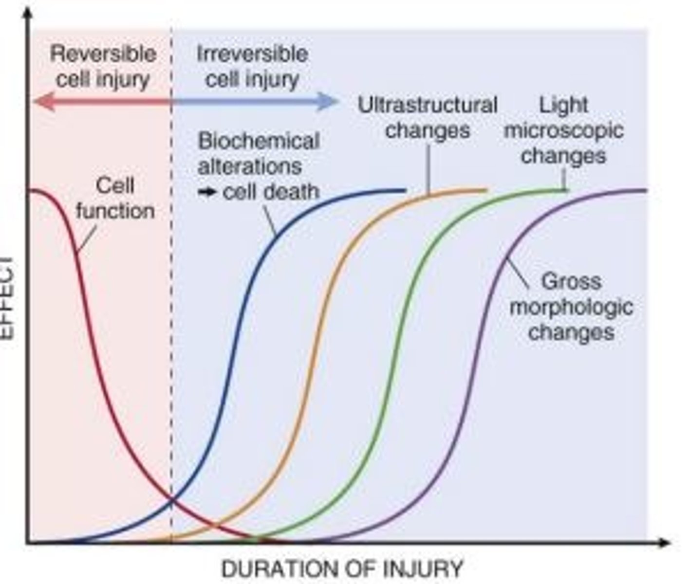
Irreversible cell injury is usually encompassed by major ___ changes
Morphological
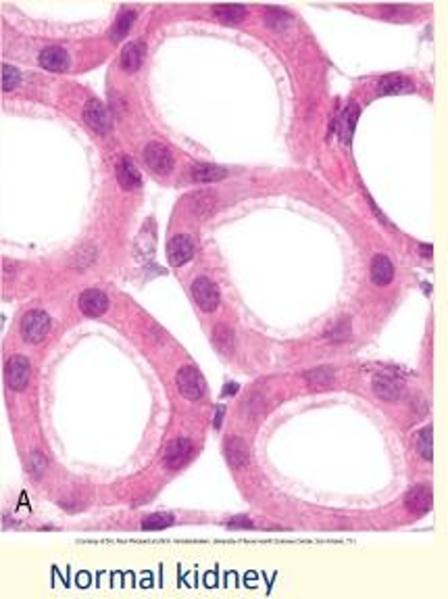
Normal kidney cells
Reversible kidney cell damage
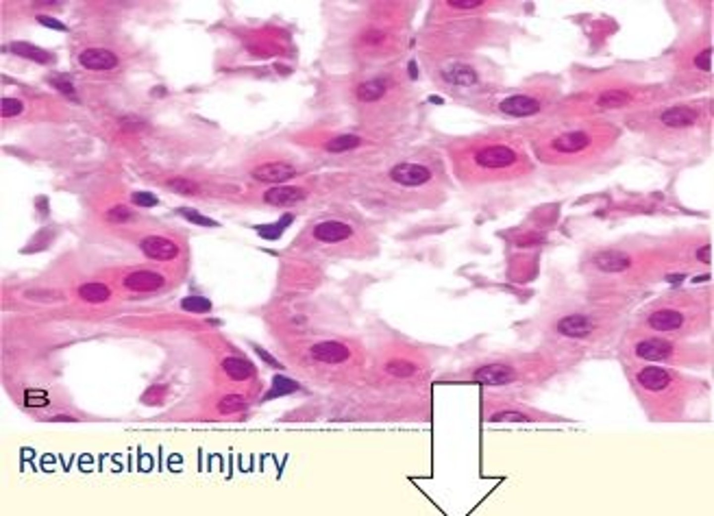
Irreversible kidney cell damage
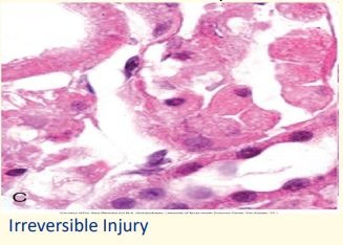
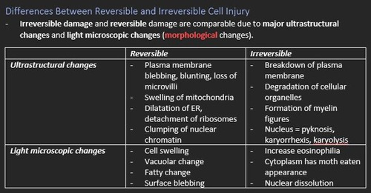
Ultrastructural changes are responsible for morphological changes in irreversible cell damage.
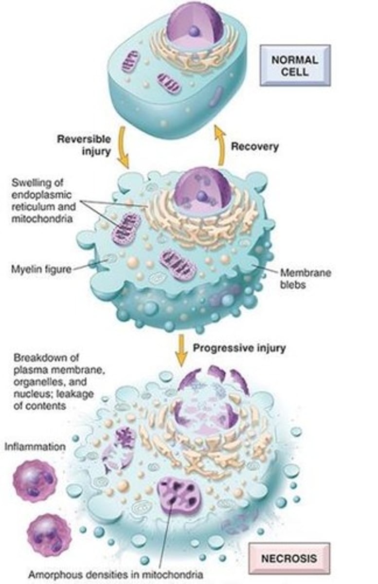
What components of the cell are damaged in irreversible cell injury?
1) Cell membranes = plasma membrane & organelle membrane
2) Nucleus = DNA
3) Proteins e.g., enzymes
4) Mitochondria = oxidative phosphorylation
In the irreversible cell injury example - there is breakdown of the plasma membrane, organelles and nucleus as well as leakage of contents. This is known as necrosis.
Necrosis
Tissue death
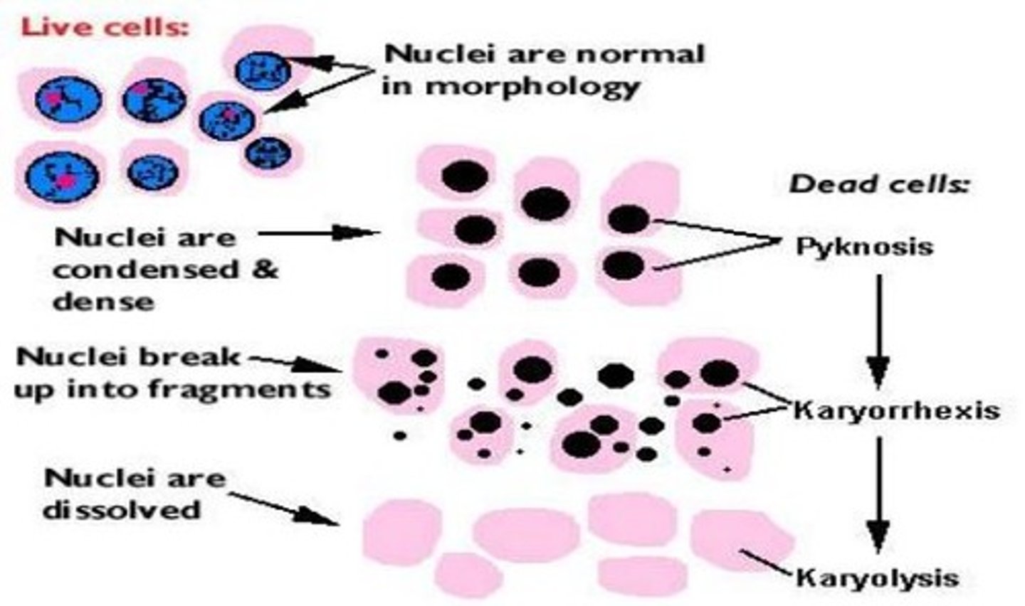
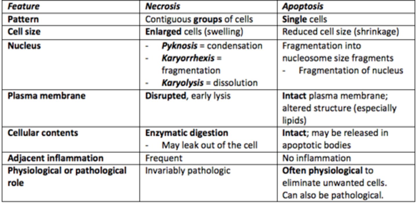
What are the three characteristic nuclear changes that occur in irreversible cell injury?
1) Pyknosis = nuclei condensed
2) Karyorrhexis = nuclei fragmentation
3) Karyolysis = nuclei dissolved
Differences between irreversible and reversible cell injury
ATP production sources
1) Mitochondria = oxidative phosphorylation (aerobic)
2) Glycolytic pathway (anaerobic)
3) Glycogenolysis
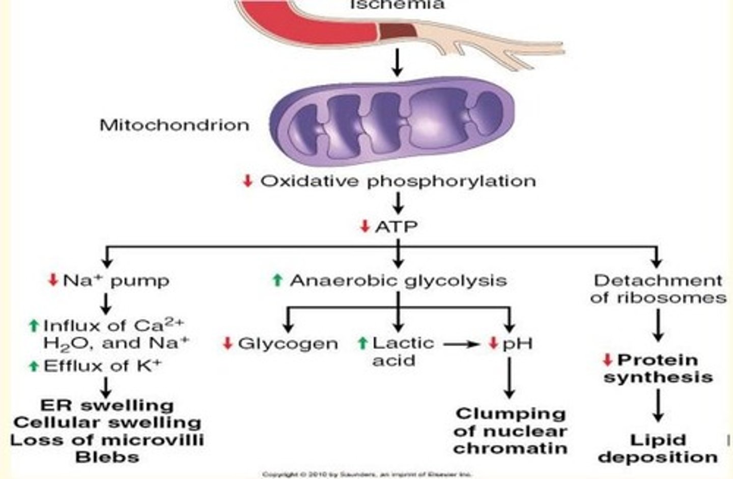
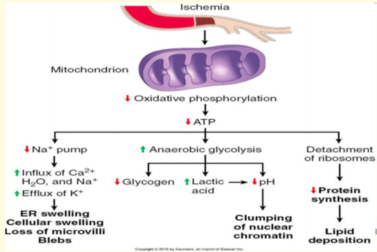
Reversible damage to the mitochondria
Decreased oxidative phosphorylation leading to decreased ATP production
Decreased ATP production leads to...
- Decreased functioning of Na+ pump = swelling/blebbing
- Detachment of ribosomes = lipid deposition
- Increased anaerobic glycolysis = clumping of nuclear chromatin
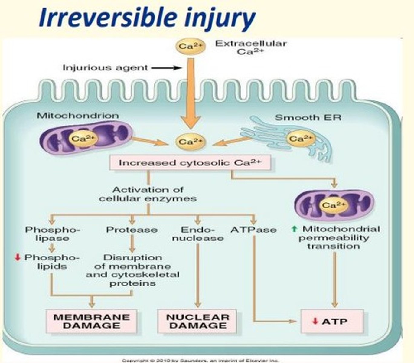
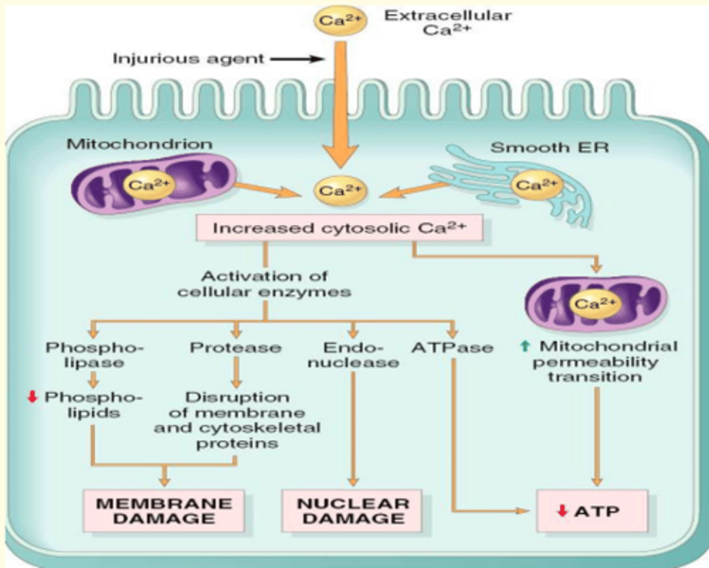
Irreversible damage to the mitochondria
Irreversible injury leads to calcium ion influx. This leads to increased cytosolic calcium concentration.
Activation of cellular enzymes
- Phospholipase & protease = membrane damage
- Endonuclease = nuclear damage
- ATPase = decreased ATP
Free radicals
Highly reactive molecules with unpaired electron
Examples of some free radicals
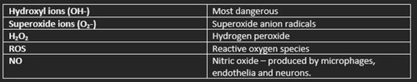
Free radicals are present at low concentrations in normal healthy cells. They are important for...
- Killing bacteria/pathogens
- Cell signalling
At high concentrations, free radicals are damaging.
How do they produce cell damage?
1) Attack lipids = lipid peroxidation
2) Damage protein/carbohydrates
3) Damage nucleic acids = mutagenic
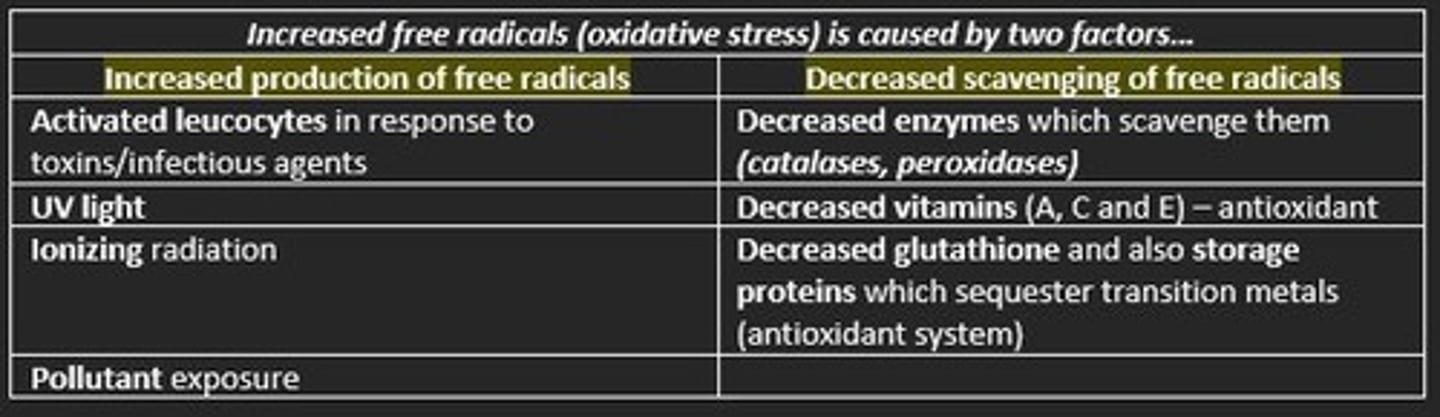
Increased free radicals (oxidative stress) is caused by two factors.
Explain how they accumulate.
1) Increased production of free radicals
Sourced from activated leukocytes in response to toxins, UV light, ionizing radiation, pollutant exposure
2) Decreased scavenging of free radicals
Decreased levels of scavenging enzyme (catalase, peroxidase)
Decreased vitamins A, C, E (antioxidants)
Decreased glutathione
Heat shock proteins (HSPs) are ___ (class of molecular chaperones)
Chaperonins
What three functions do HSPs fulfill?
1) Provide optimal condition for denatured protein folding
2) Prevent protein aggregation
3) Label misfolded proteins for degradation at proteasome
Give an example of a HSP
Ubiquitin
What does the heat shock response aim to do?
The heat shock response aims to mend misfolded proteins using HSPs and maintain the viability of cells
Two main cellular processes seen in necrosis?
1) Denaturation of intracellular proteins
2) Enzymatic digestion by lysosomes
The earliest microscopic evidence of necrosis may not become apparent until ___ hours
4-12 hours
List the five different types of necrosis
1) Coagulative necrosis
2) Liquefactive necrosis
3) Caseous necrosis
4) Fat necrosis
5) Fibrinoid necrosis
Most common form of necrosis
Coagulative necrosis
- Occurs in most organs
- Results from protein denaturation

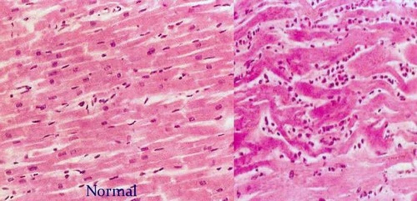
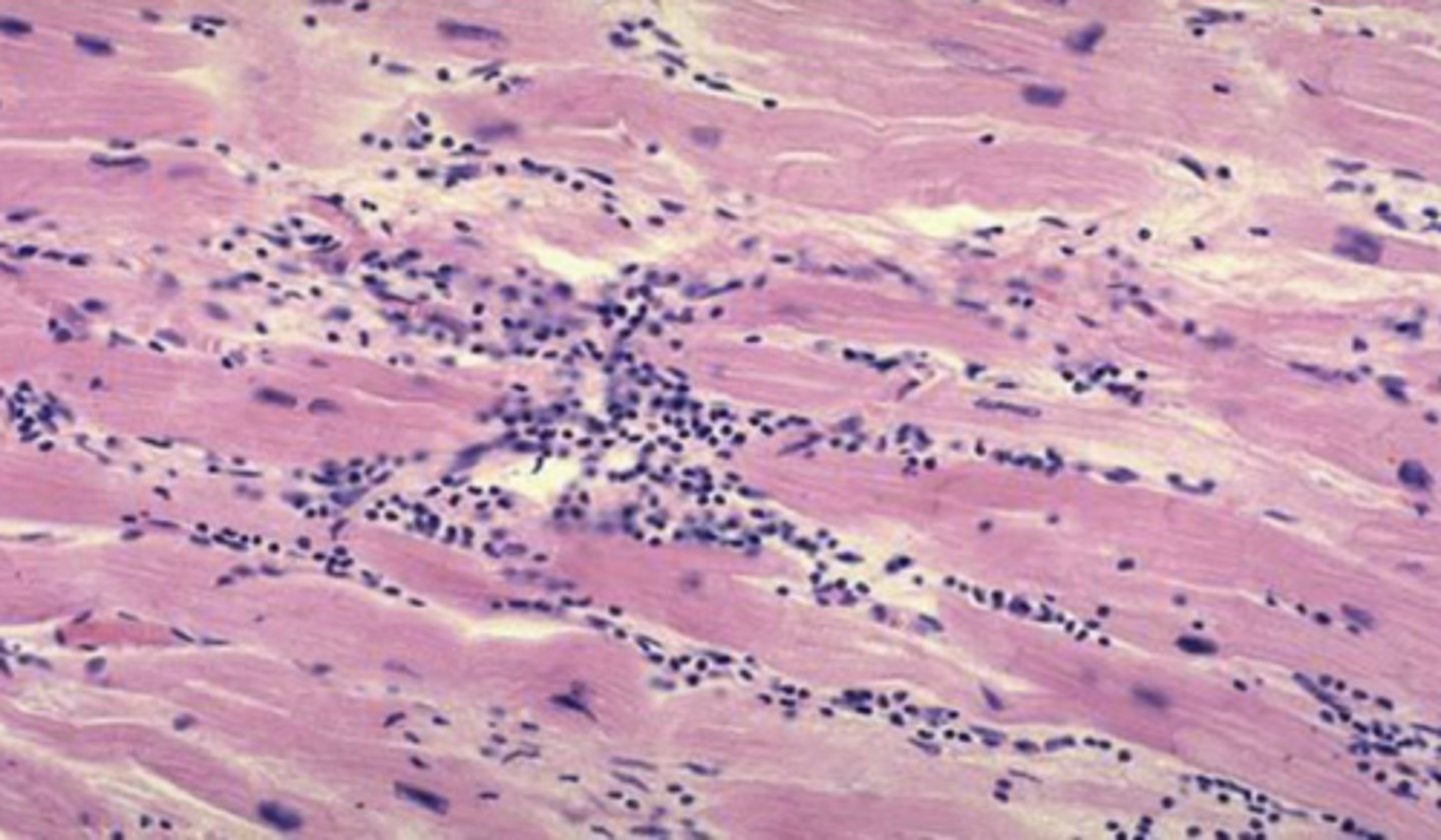
What type of necrosis can be seen here (heart)?
Coagulative necrosis of the heart
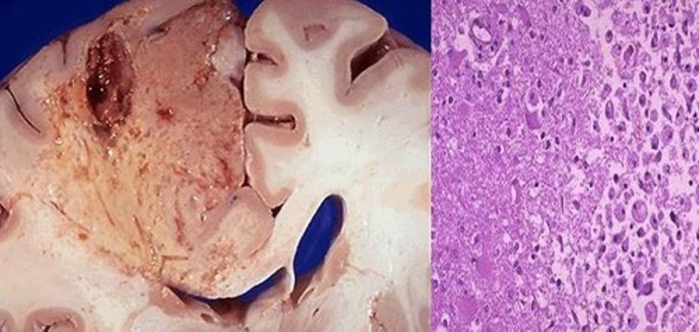
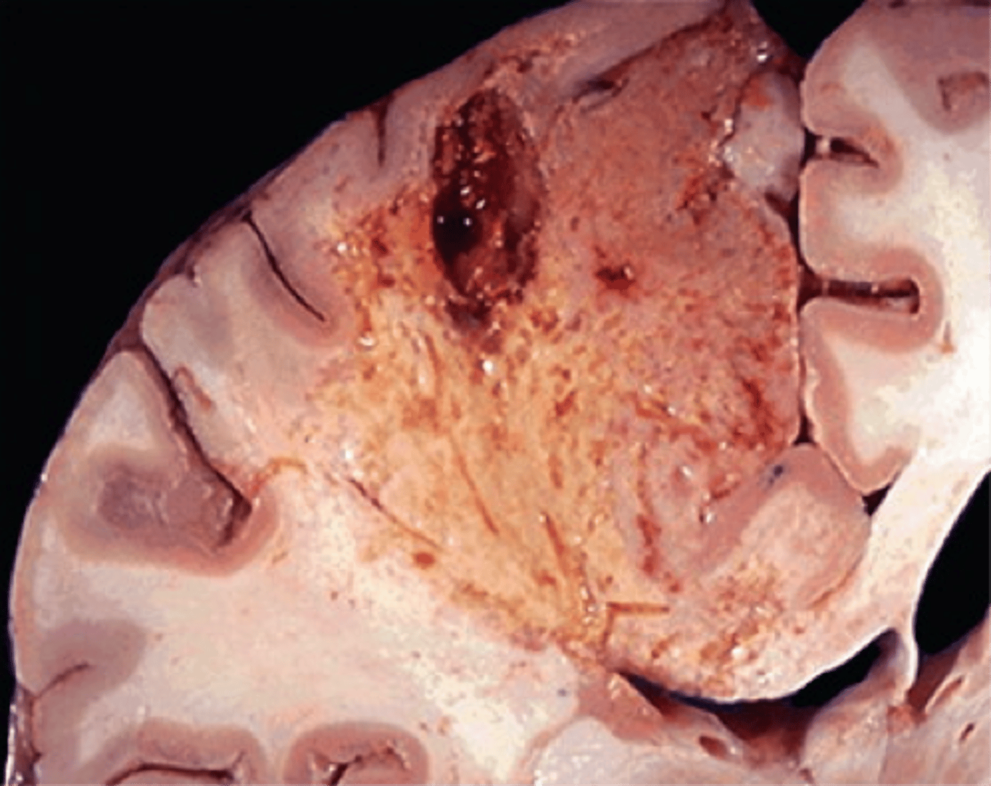
What type of necrosis can be seen here (brain)?
Liquefactive necrosis of the brain
What type of necrosis...
- Usually seen in the brain
- Seen in infections leading to abscess formation
- Tissue degradation by enzymes
- Creamy yellow necrotic tissue due to presence of dead leukocytes (pus)
- Presence of neutrophils
Liquefactive necrosis
What type of necrosis...
- Commonest form
- Occurs in most organs
- Due to protein denaturation
- Firm, pale wedge of tissue
Coagulative necrosis
What type of necrosis...
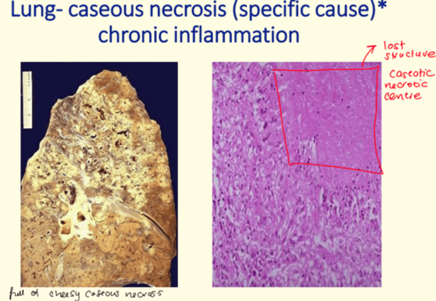
- Cheese-like gross appearance
- Amorphous debris surrounded by histiocytes
- Granulomatous inflammation
- Associated with tuberculosis
Caseous necrosis
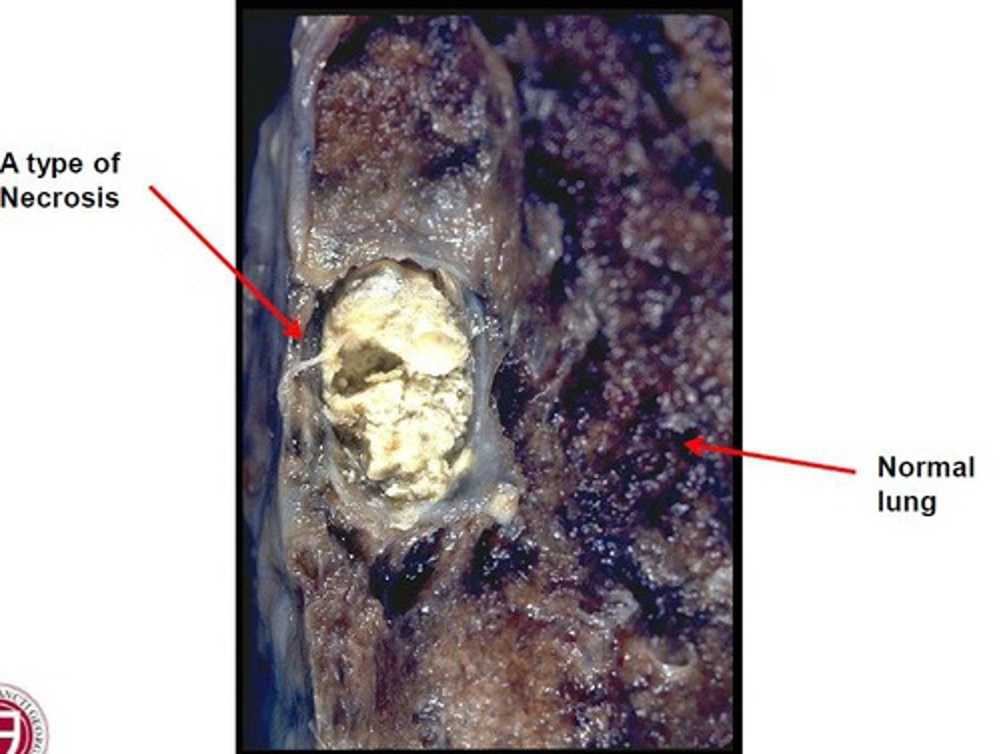
What type of necrosis can be seen here (lung)?
Caseous necrosis of the lung
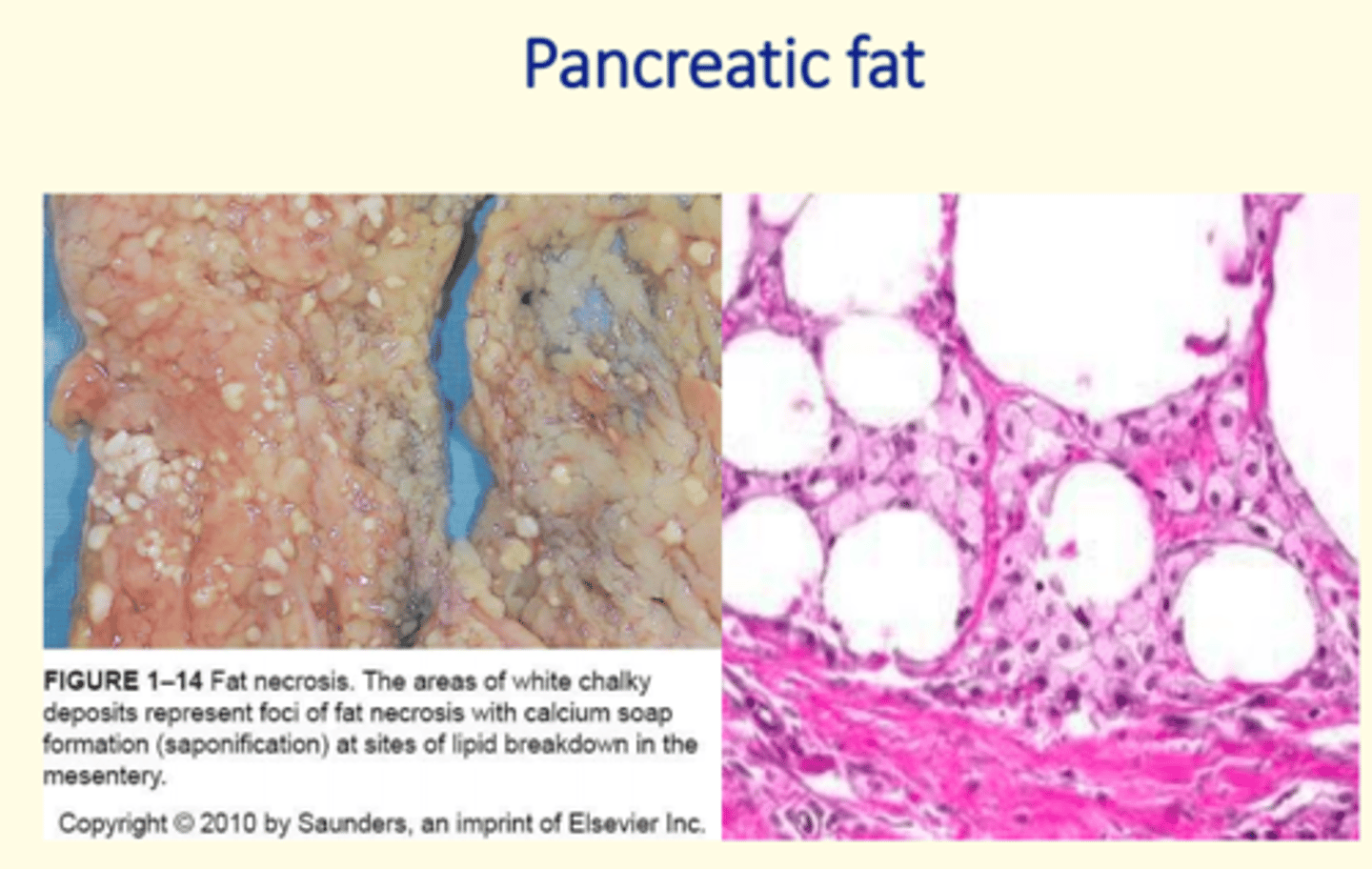
Fat necrosis primary cause
Consequence of trauma to adipocytes
Fat necrosis secondary cause
Release of lipases from damaged pancreatic tissue
Fat necrosis produces ___ ___ which react with ___ to form white deposits in fatty tissue. These deposits can sometimes mimic breast tumours when found in breast tissue on radiology. These must be biopsied to exclude cancer.
Fat necrosis produces fatty acids which react with calcium to form white deposits in fatty tissue. These deposits can sometimes mimic breast tumours when found in breast tissue on radiology. These must be biopsied to exclude cancer.
What type of necrosis can be seen here (pancreas)?
Fat necrosis of the pancreas
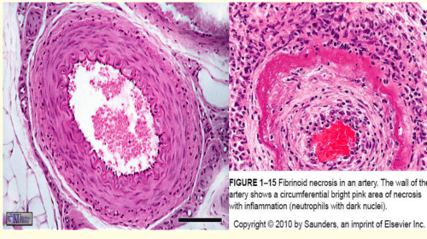
What type of necrosis...
- Usually seen in immune reactions
- Usually involves blood vessels
- Associated with vasculitis
- Bright pink appearance on H&E called "fibrinoid"
Fibrinoid necrosis
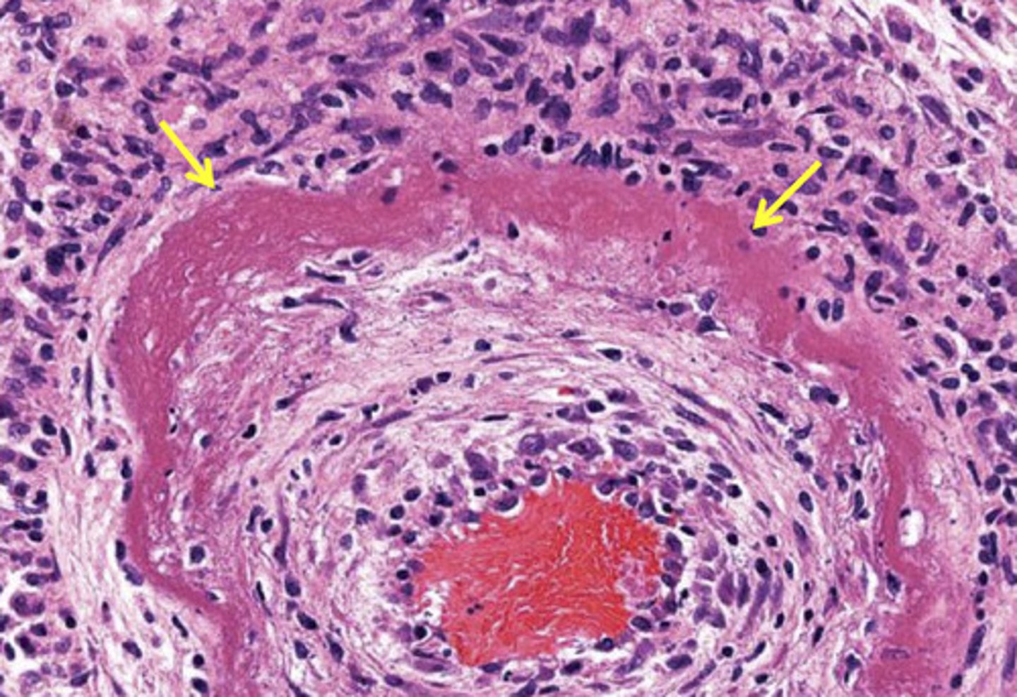
What type of necrosis can be seen here (blood vessel)?
Fibrinoid necrosis of a blood vessel
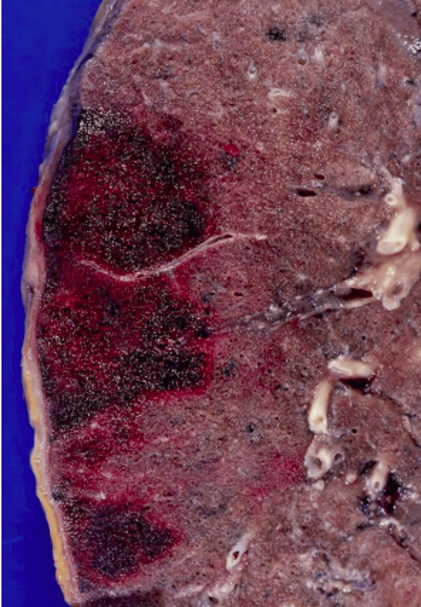
Infarction is a cause of necrosis (coagulative and liquefactive necrosis).
What are the two main types of infarction?
White infarct
Red (haemorrhagic) infarct
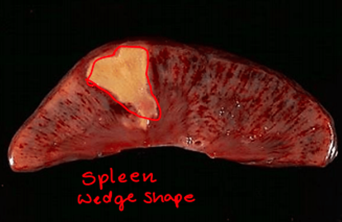
White infarct
- Occurs in tissue with a single blood supply
- Pale, little bleeding into organ affected
- Solid organs like kidney, spleen, heart
- Leads to arterial occlusion
Red (haemorrhagic) infarct
- Occurs in organs with dual blood supply
- Occurs in organs with numerous anastomoses
- Characteristic of lung and GIT
- Can be caused by venous occlusion

Gangrene
Visible tissue necrosis caused by loss of blood supply
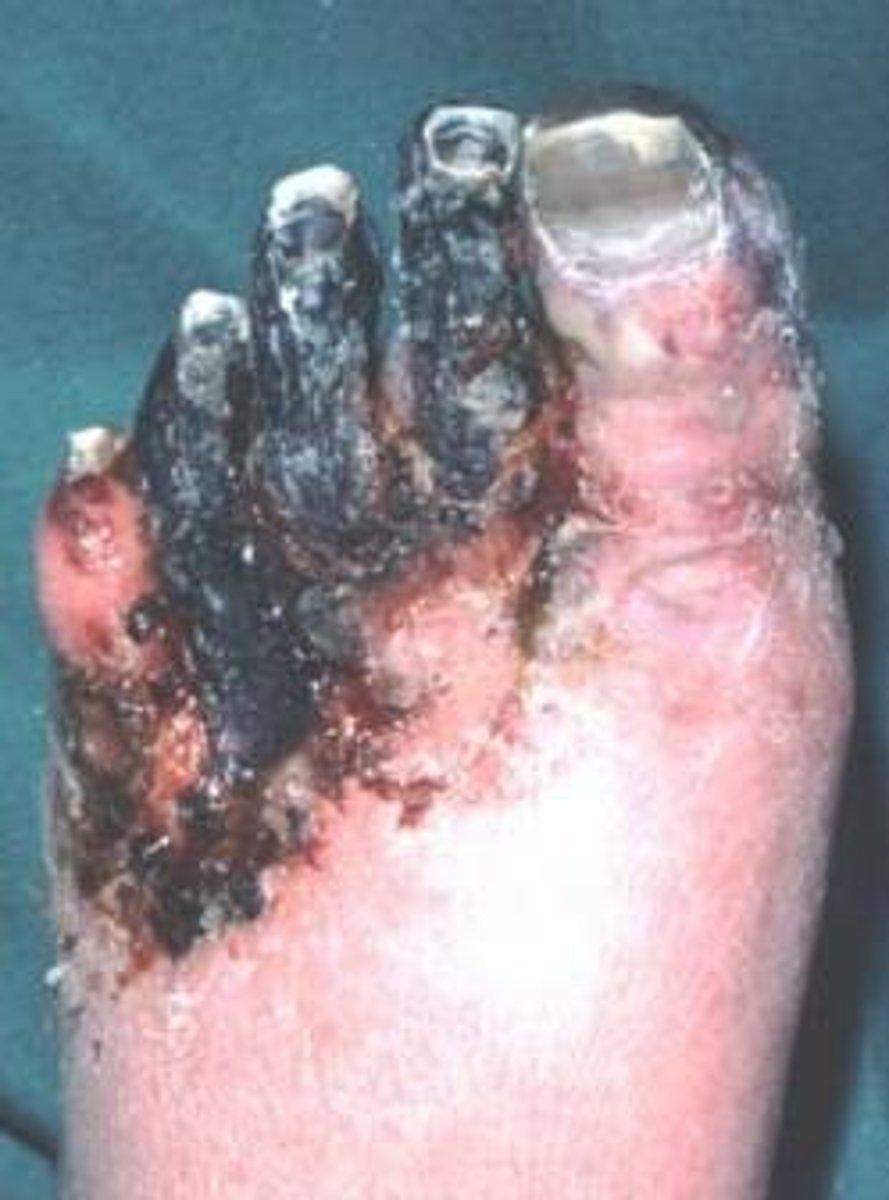
Types of gangrene
Wet, dry, gas
Dry gangrene
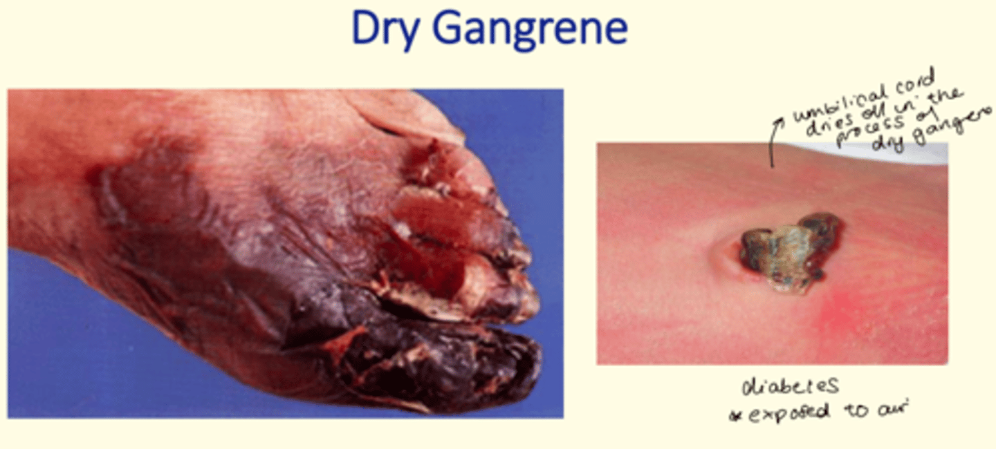
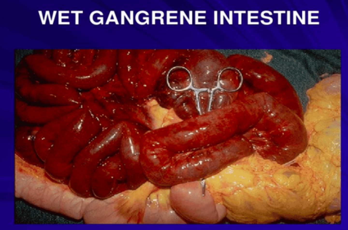
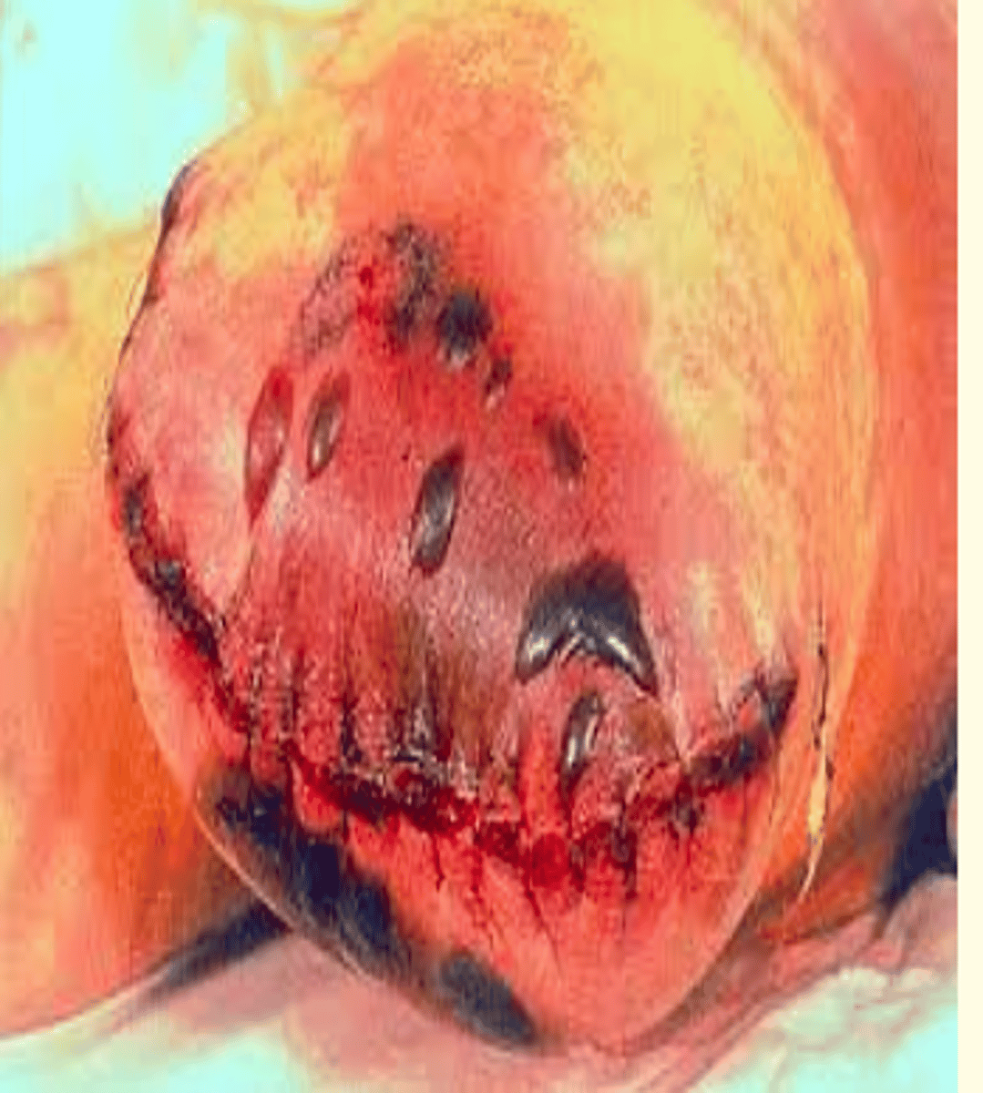
Wet gangrene
Area of gangrene with secondary bacterial infection and pus
Gas gangrene caused by what bacteria
Clostridium perfringens
What is apoptosis
Programmed cell death (cell suicide) which uses ATP
Does apoptosis result in an inflammatory response?
No
Only necrosis results in an inflammatory response
Two types of apoptosis
Physiological and pathological
Examples of physiological apoptosis
1) Embryogenesis/fetal development = loss of hand webbing
2) Shedding of endometrium (menstruation)
3) Death of cells that have fulfilled purpose e.g., neutrophils/lymphocytes
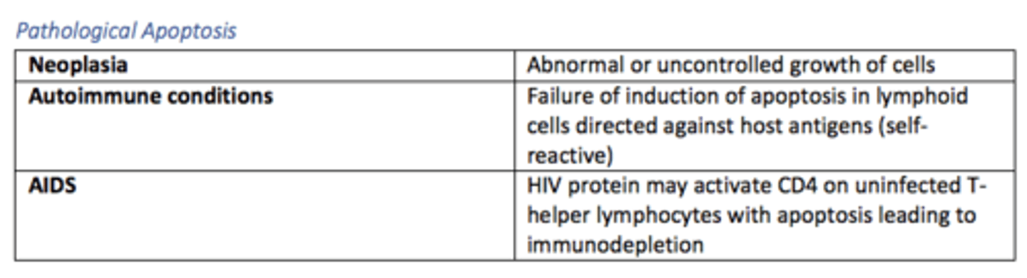
Examples of pathological apoptosis

Regulation of apoptosis - name three modes of regulation
- Genes
- Inhibitors e.g., growth factors
- Inducers e.g., growth factor withdrawal
Extrinsic apoptosis pathway
Death receptor dependent
Receptor-ligand interactions = Fas and TNF-receptor
Intrinsic apoptosis pathway
Non-receptor mediated
Withdrawal of growth factors/hormones cause molecules to be released from mitochondria = Bcl2, Bax, p53
Necrosis vs Apoptosis
What types of molecules are charcteristically released after cell injury/death (bloods)
- Electrolytes = potassium, calcium
- Enzymes e.g., troponin-I in MI
- Myoglobin
What enzymes are heightened during myocardial infarction (MI) and are a marker for cardiac damage?
Troponin I
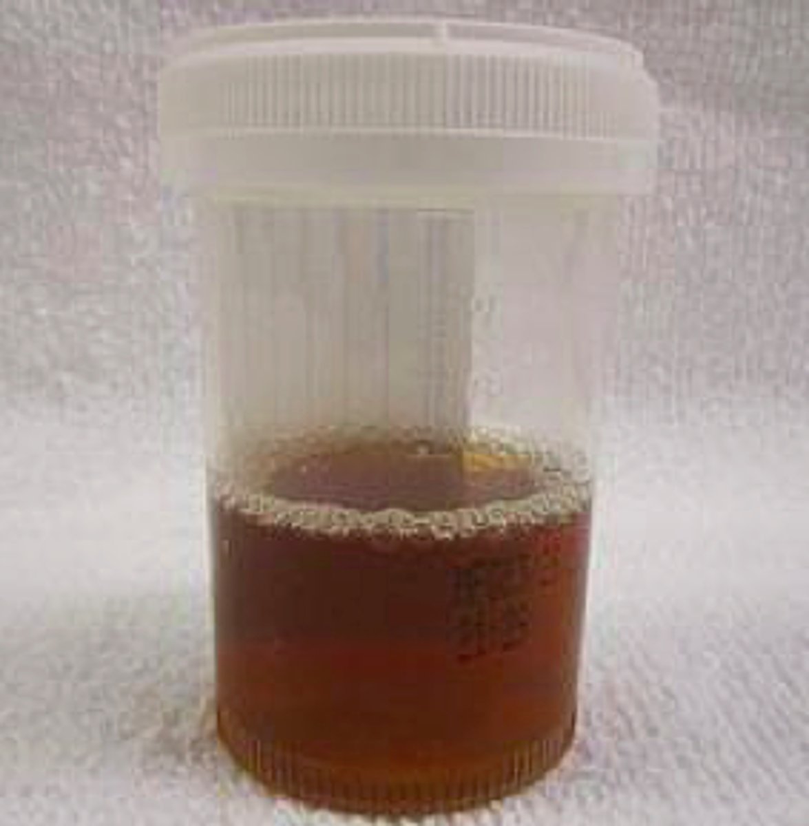
Rhabdomyolysis
Destruction of muscle to produce myoglobin
Symptoms of rhabdomyolysis
Classic triad
- Muscle pain
- Weakness
- Dark coca cola urine (myoglobinuria)
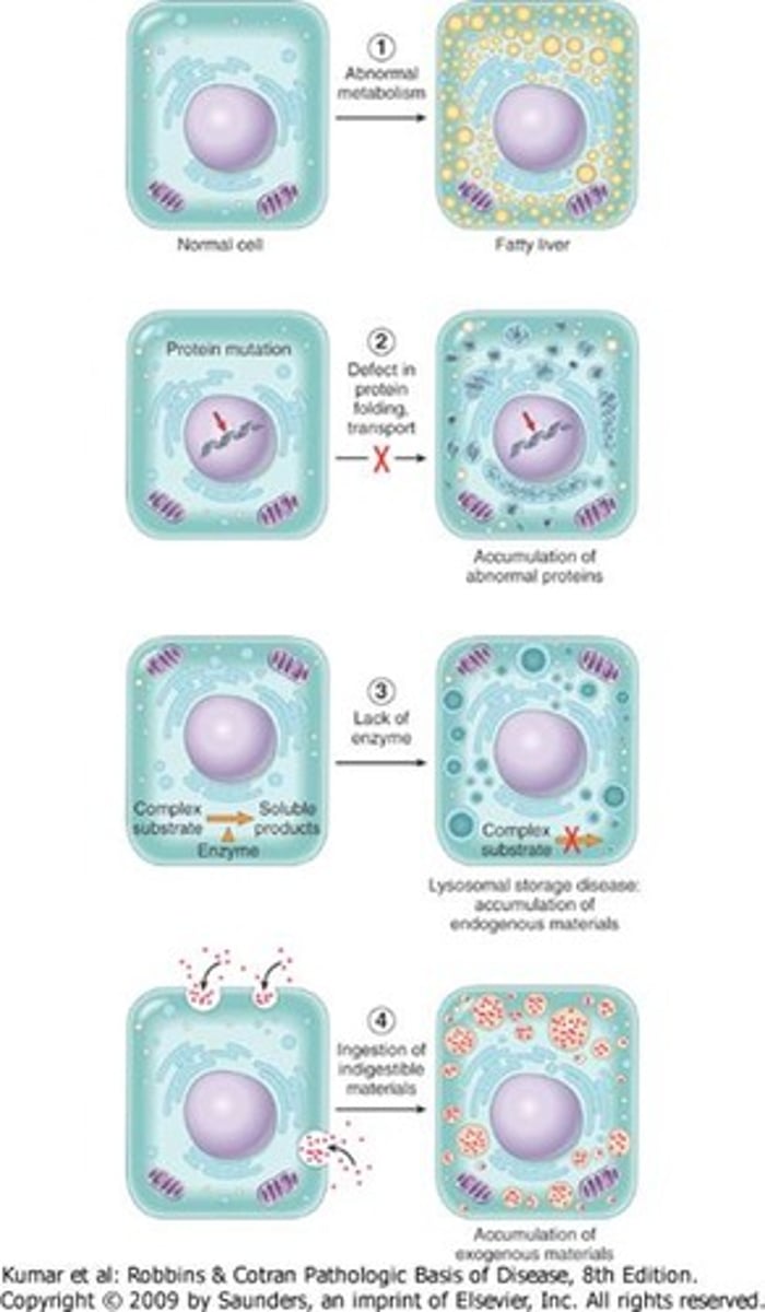
Mechanisms of intracellular accumulation
- A normal substance accumulates at an increased rate (abnormal metabolism) e.g., fatty liver
- A normal substance accumulates due to lack of enzymatic breakdown e.g., lysosomal storage disease (LSD)
- Inability to breakdown phagocytized particles
- Defect in protein folding, transport
Give some examples of common intracellular accumulations
- Water, electrolytes e.g., cerebral edema
- Lipids e.g., fatty liver
- Carbohydrates
- Proteins
- Pigments
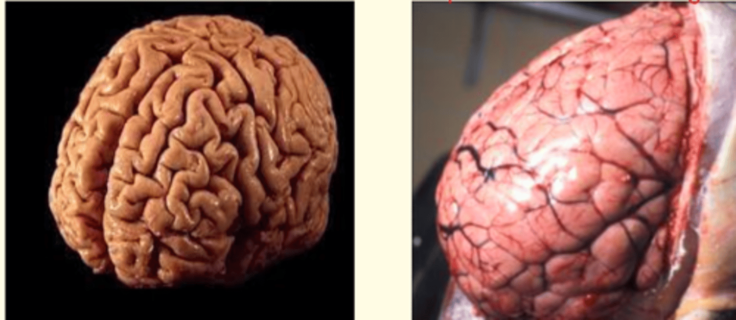
What is shown in this image?
Abnormal accumulation of fluid in the brain (hydropic swelling)
- Raised ICP
- Swollen sulci and gyri
- Cerebral edema
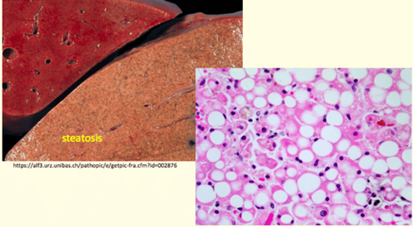
Steatosis
Abnormal condition of fat accumulation (increased fat at the cellular level often affecting the liver)
Endogenous pigment accumulation causes
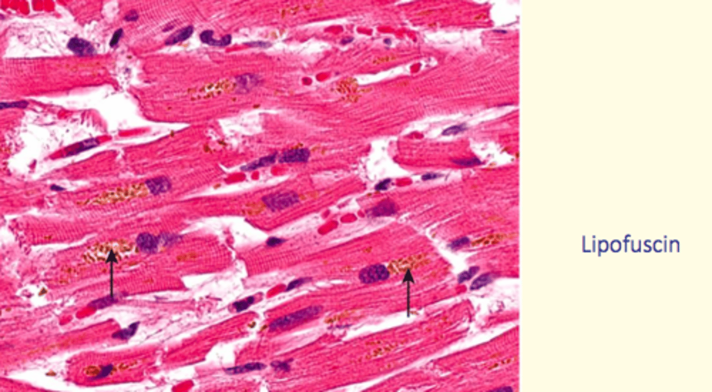
Lipofuscin
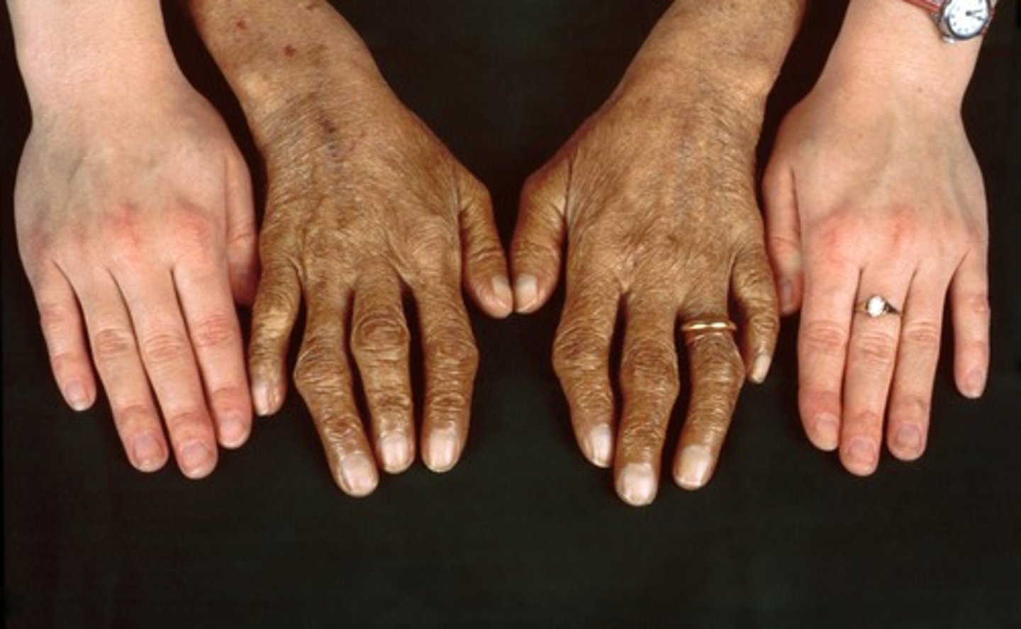
Haemosiderin (iron accumulation)
Hereditary haemochromatosis
Lipofuscin endogenous pigment
Age Pigment
Lysosomes with degradation products (residual body)
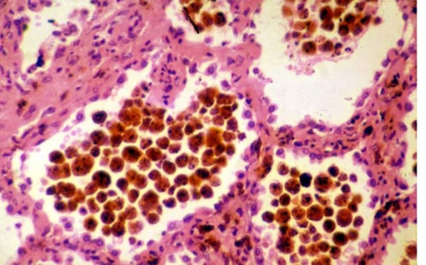
Haemosiderin endogenous pigment
Iron pigment from breakdown of blood or ingestion of iron
Golden brown pigment seen in macrophages
Bruising
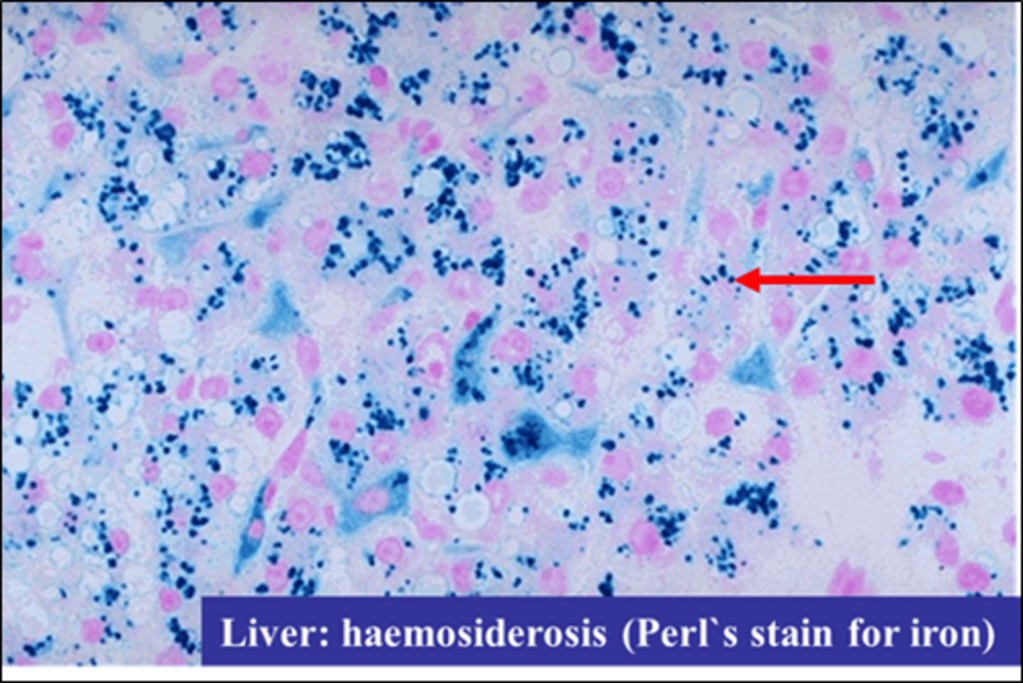
Haemosiderosis
Iron overload
An overload of iron in the body resulting from repeated blood transfusions. Hemosiderosis occurs most often in patients with thalassemia.
Abnormal deposit of hemosiderin
Hereditary haemochromatosis
Hereditary - autosomal recessive
Excessive absorption of iron from GI tract
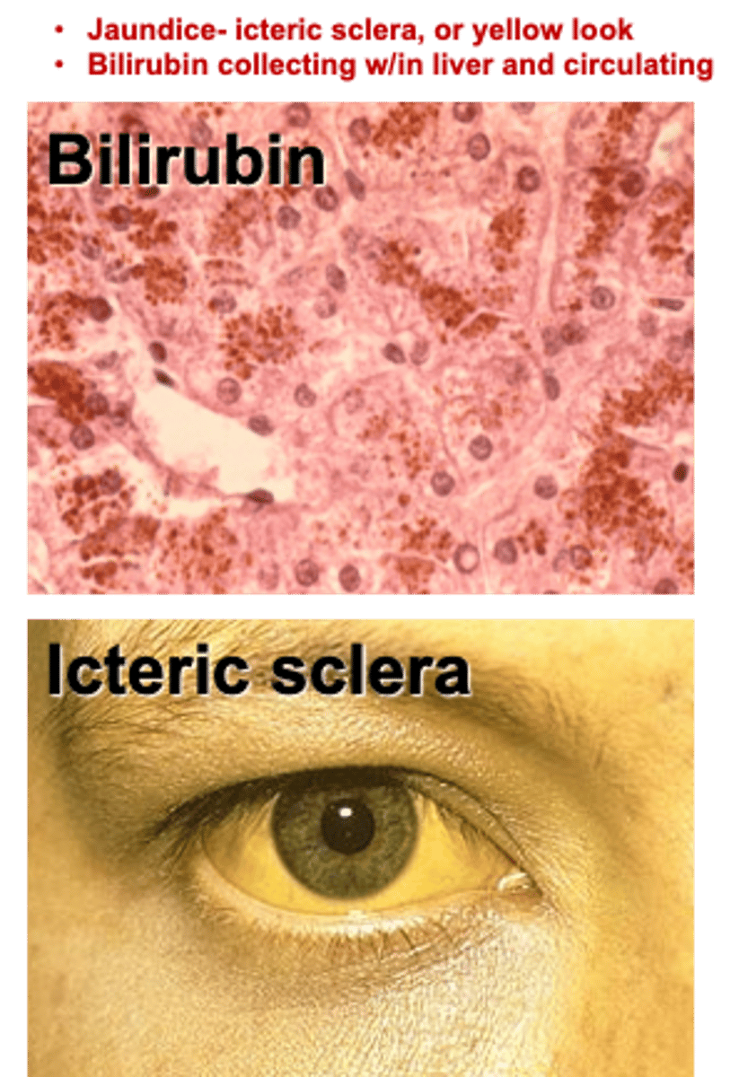
Bilirubin can accumulate in what pathologies?
Liver disease
Hemolytic anemia
Bilirubin endogenous pigment accumulation
Dystrophic calcifications (depositions)
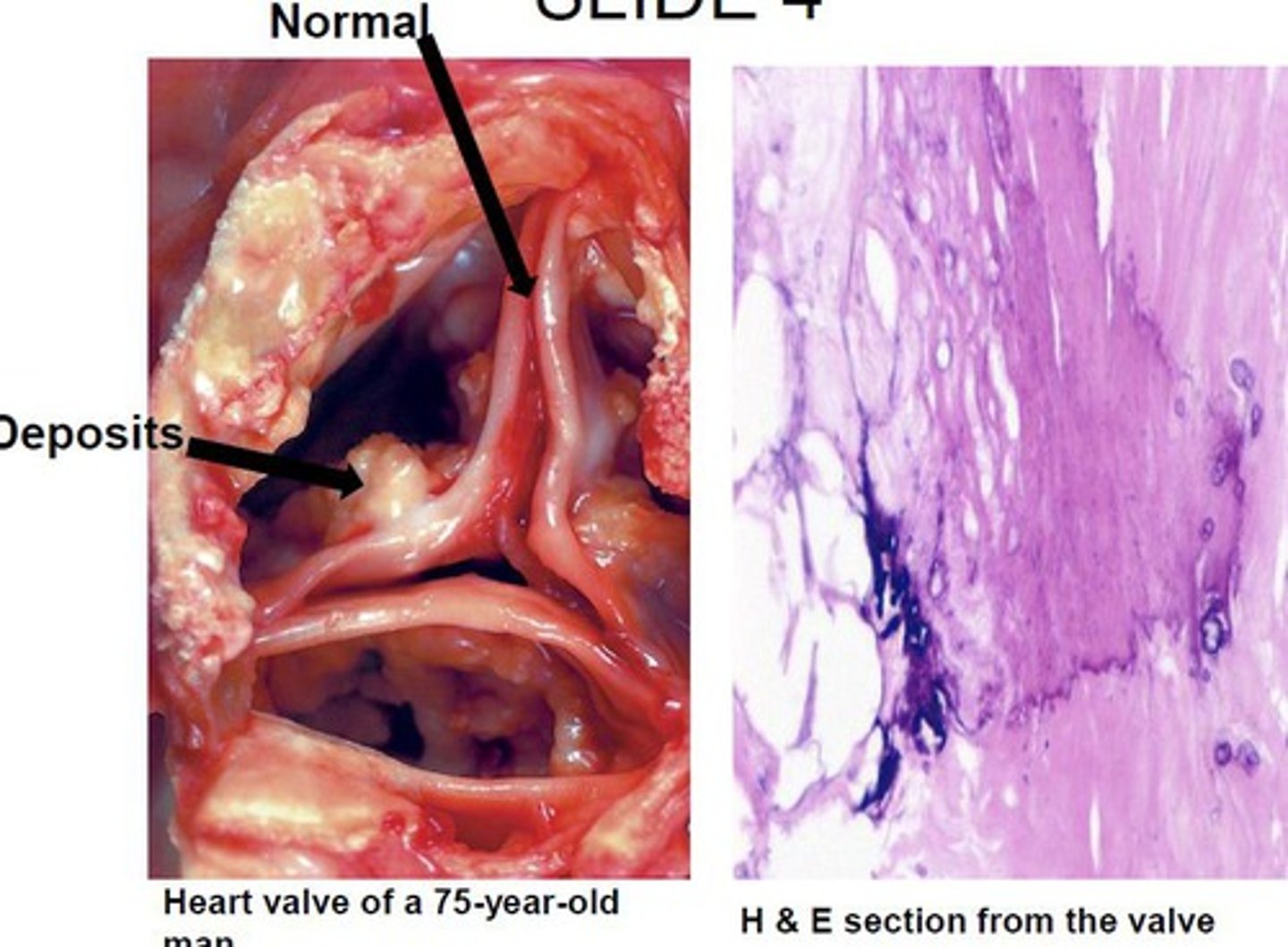
Pathological calcifications causes
- Parathyroid overactivity e.g., tumour/hyperplasia
- Malignant tumours e.g., breast/lung/bone
Other causes of calcification
- Vitamin D overdosage
- Paget's disease
- Prolonged immobilisation
Cellular ageing
Telomeres shorten
Infarcts in the spleen are usually haemorrhagic
True or false?
False

Infarcts in the lung are usually haemorrhagic
True or false?
True
Infarcts in the brain heal by gliosis
True or false?
True
Gliosis is the main reparative mechanism in the CNS
Infarcts usually result from ischaemia
True or false?
True
Infarcts are a manifestation of apoptosis
True or false?
False
Free radicals damage cells by cross-linking proteins
True or false?
True
Free radicals damage cells by breaking strands of DNA
True or false?
True
Free radicals damage cells by oxidising membrane lipids
True or false?
True
Lipid peroxidation
Free radicals damage cells by activating cell surface receptors
True or false?
False
Free radicals damage cells by activating cytoplasmic receptors
True or false?
False
There are no specific cytoplasmic receptors for free radicals
Apoptosis is involved in limb modelling in embryogenesis
True or false?
True
Apoptosis involves active transcription of genes
True or false?
True
Apoptosis is a physiological process
True or false?
True
Apoptosis is seen in the liver in hepatitis
True or false?
True
In reversible cell injury due to oxygen deprivation... ATP levels fall
True or false?
True

In reversible cell injury due to oxygen deprivation... ribosomes are detached from the endoplasmic reticulum
True or false?
True

In reversible cell injury due to oxygen deprivation... pyknosis occurs
True or false?
False
This occurs during irreversible cell injury only (image)
In reversible cell injury due to oxygen deprivation... mitochondria swell
True or false?
True
In reversible cell injury due to oxygen deprivation... there is lysosomal disruption
True or false?
False
Lysosomal disruption is a feature of irreversible cell injury. Apoptosis is programmed individual cell death
Myocardium would undergo irreversible cell injury within 60 minutes of complete cessation of blood supply
True or false?
True
Motor neurones would undergo irreversible cell injury within 60 minutes of complete cessation of blood supply
True or false?
True