Unit 2 Free Response
1/13
There's no tags or description
Looks like no tags are added yet.
Name | Mastery | Learn | Test | Matching | Spaced | Call with Kai | Chat |
|---|
No analytics yet
Send a link to your students to track their progress
14 Terms
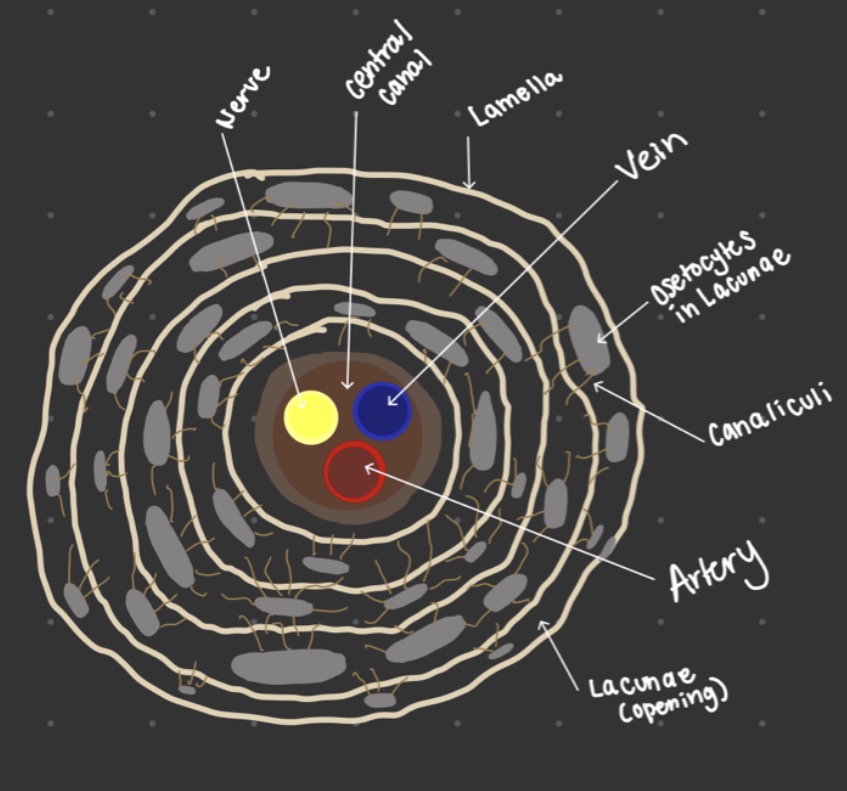
Be able to draw an osteon and label all components.
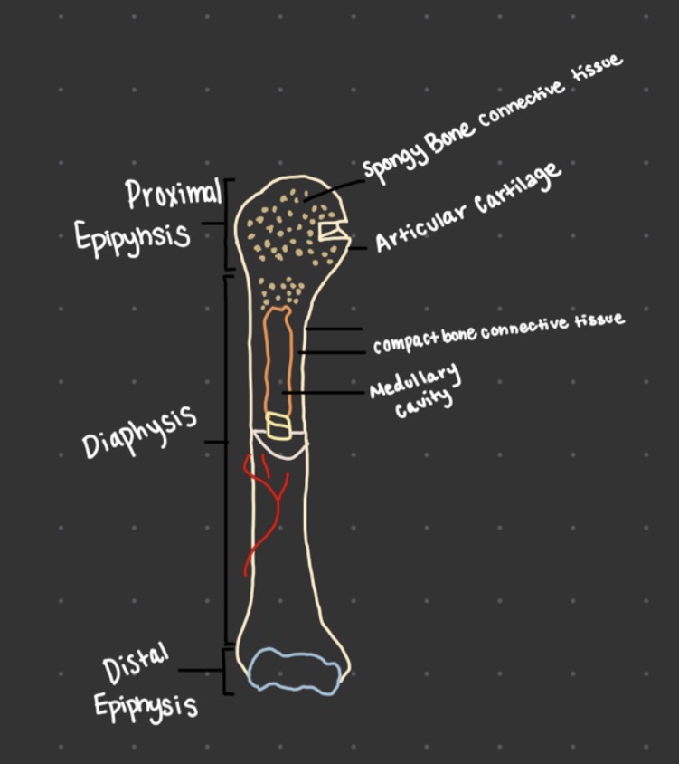
Be able to draw a long bone and label all components.
What are the types of marrow cavities?
red and yellow bone marrow
What is the composition of red marrow? What is its function? Location?
Composed of blood connective tissue
produces red and white blood cells
found in Ephysis of long bones
What is the composition of yellow marrow? What is its function? Location?
composed of adipose connective tissue
function is long term energy storage
found in medullary canal
Explain how intramembranous ossification occurs.
cluster around blood vessels
secrete a collagen and calcium matrix
calcium salts crystallize
trabeculare become denser and forms a thin outer layer of compact bone CT
Remodeling occurs via ostelasts
original sac becomes the periosteum (membrane that surrounds the bone)
Explain how endochondral ossification occurs.
Hyaline cartilage CT “template” forms first
formation of the primary ossification center in the diaphysis
Osteoblasts are going to produce spongy bone CT in the primary ossification center
Osteoblasts make compact bone CT beneath the periosteum (or around spongy bone CT)
Osteoclasts break down spongy bone CT forming the medullary canal
Osteoblasts make spongy bone CT in the secondary ossification center in the epiphysis
Epiphyseal growth plate is formed between the diaphysis and the epiphysis
Compare and contrast cartilage and bone tissue. Make sure to include location of tissue, mature cell type, location of mature cell, matrix composition, strength, flexibility, and vascularization.
Cartilage tissue
a) Location: nose, ears, joints, trachea, rib cage, vertebrae
b) Mature cell type: chondrocytes
c) Location of mature cell: chondrocytes in lacunae
d) Matrix comp: collagen fibers, elastic fibers
e) Moderately strong
f) Flexible
g) Avascular
bone tissue
a) Bones of the skeleton
b) Osteocyte
c) Osteocytes in lacunae
d) Collagen fibers
e) Very strong
f) Vascular
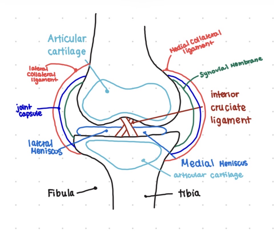
Be able to draw a knee joint and label all components. Know the histology and function of all components.
Articular Cartilage: Hyaline cartilage
Medial & Lateral Meniscus: Fibrocartilage
Tiba & Fibula: Bone tissue
Ligaments: Dense regular connective tissue
Joint capsule: Dense regular connective
Synovial Membrane: Areolar connective tissue
What are the connective tissues that surround muscle?
Epimysium
perimysium
endomysium
What is epimysium? What is its histology?
Superficial fascia that surrounds the whole muscle
dense reg CT
What is the perimysium? What is its histology?
Internal fascia the surround bundles of parallel fiber; forms fascicles
dense reg ct
What is the endomysium? What is its histology?
Surrounds each individual fiber (muscle cell)
areolar ct
What is a muscle agonist? Synergist? Antagonist?
Agonist (prime mover) - main muscle that generates a particular movement (action)
Synergist: helper muscle to the prime mover
Antagonist: opposite movement to the prime mover; reverse original movement