Exam 3 Study Guide - Abdominal Cavity Part 2 for Biology
 Call Kai
 Learn
Learn Practice Test
 Spaced Repetition
 Match
Match Flashcards
 Knowt Play
1/117

There's no tags or description
Looks like no tags are added yet.
Name | Mastery | Learn | Test | Matching | Spaced | Call with Kai | Chat |
|---|
No analytics yet
Send a link to your students to track their progress
118 Terms
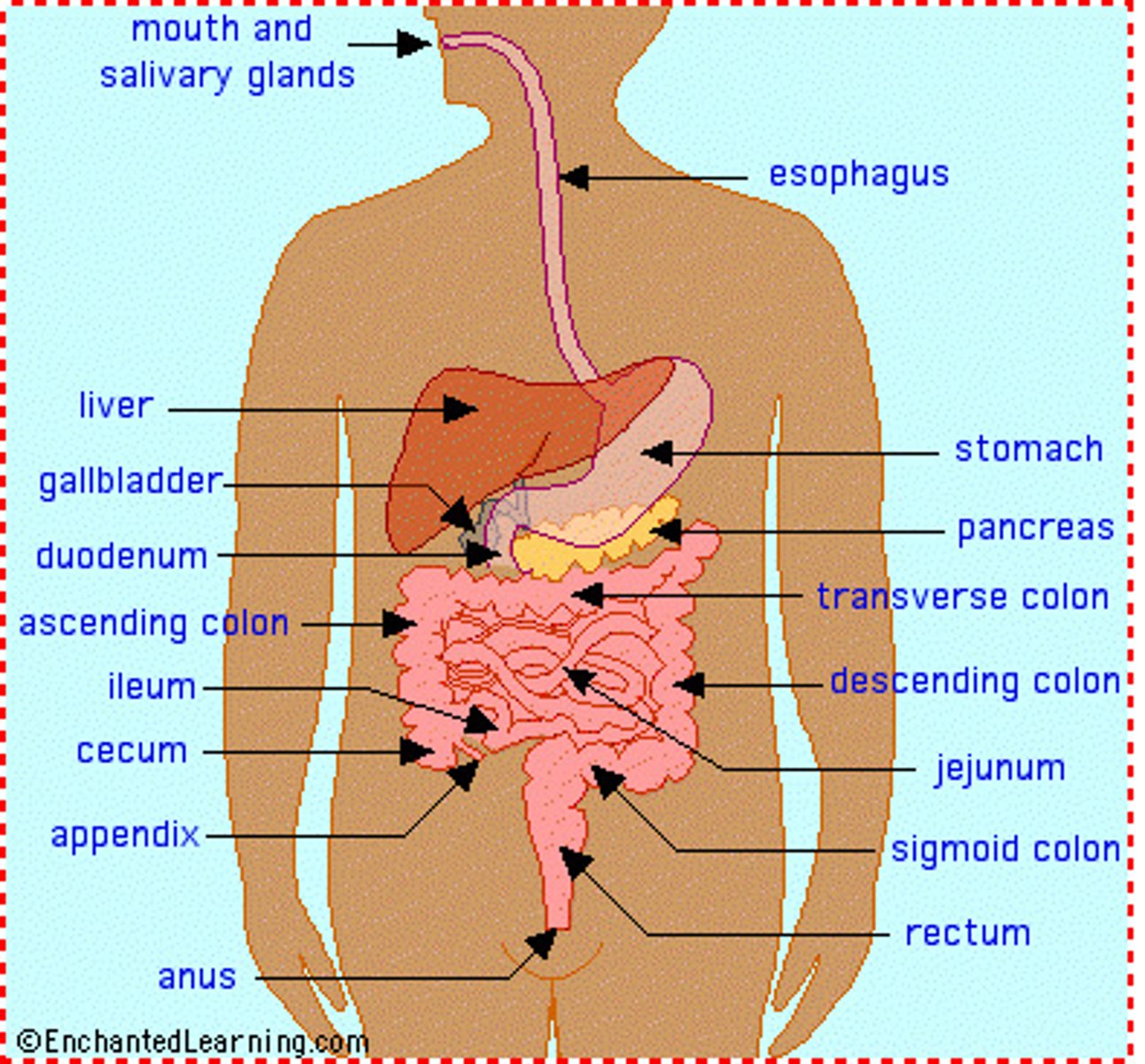
GI components
-mouth
-pharynx
-esophagus
-stomach
-small intestine
-colon
-rectum and anus
-gallbladder
-liver
-pancreas
Oral Cavity consists of
⚫ Teeth
⚫ Hard/soft palates
⚫ Tongue
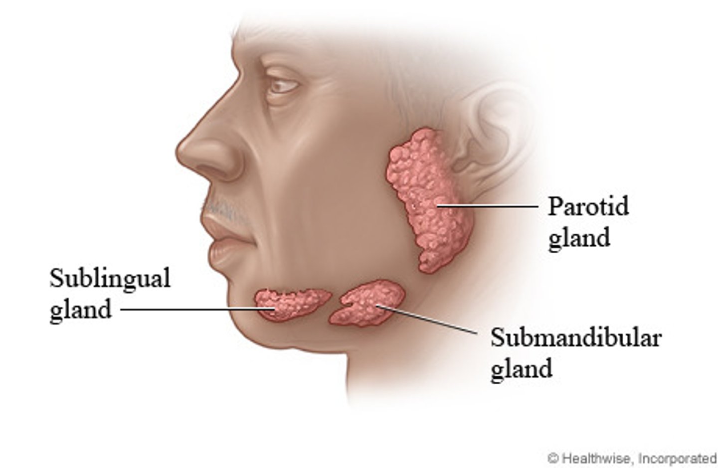
⚫ Salivary glands (3)
Three salivary glands
parotid (innervated by CN VII), submandibular, sublingual
Pharynx - three parts
nasopharynx, oropharynx, laryngopharynx
Pharynx muscles
External and internal
External muscles of the pharynx
CONSTRICTORS
Internal muscles of the pharynx
LEVATORS
Innervation of the pharynx external and internal muscle layers
pharyngeal plexus
# of teeth in adults
32 - variation with number of wisdom teeth
Teeth in children
Deciduous or milk Teeth (20 in total)
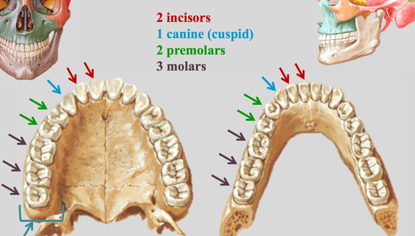
Dental formula
2:1:2:3
2 incisors
1 canine
2 premolars
3 molars
EX: 4 incisors on the top, 8 in total
this equation accounts for 1/4 of the teeth (1/2 of of the upper for example)
Esophagus landmarks (length)
C6-T10 vertebrae
Normal anatomical constrictions of the esophagus (3)
1. Junctions of the pharynx and esophagus (cricopharyngeous muscle)
2. Where the aorta and the L bronchus cross over the esophagus
3. Passage of esophagus through the diaphragm
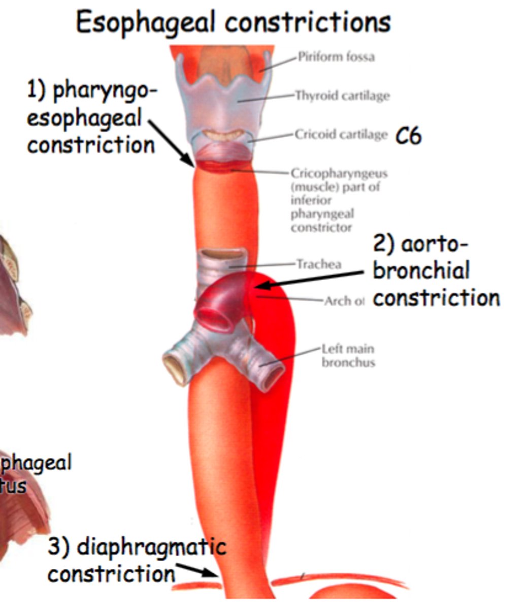
Normally the esophagus is
flat and opens up with food
Gastroesophageal Reflux Disease
Lower esophageal sphincter LES (@ diaphragm) doesn't function properly (not staying closed when it should be) and allows stomach contents to flow upwards and into the esophagus
- feels horrible and worse when laying down - need to sleep upright
pyrosis
heartburn; burning sensation in upper abdomen due to reflux of gastric acid
Bronchoaortic constriction
Normal anatomical constriction
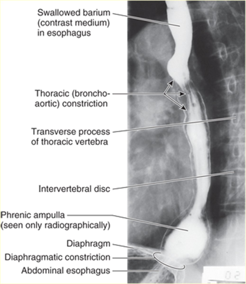
Diaphragmatic constriction
Constriction at the esophageal hiatus - Normal anatomical constrictions

Esophagus arterial supply
Inferior thyroid artery (upper portion)
L gastric artery
Esophagus venous drainage
azygous v
Esophagus innervation
esophageal plexus
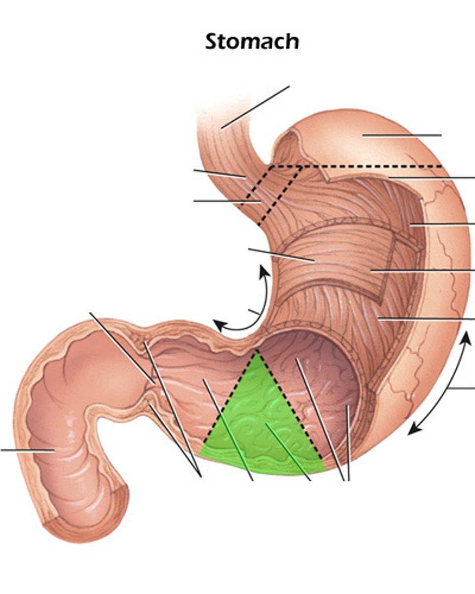
Portions of the stomach
cardiac, fundus, body, pylorus
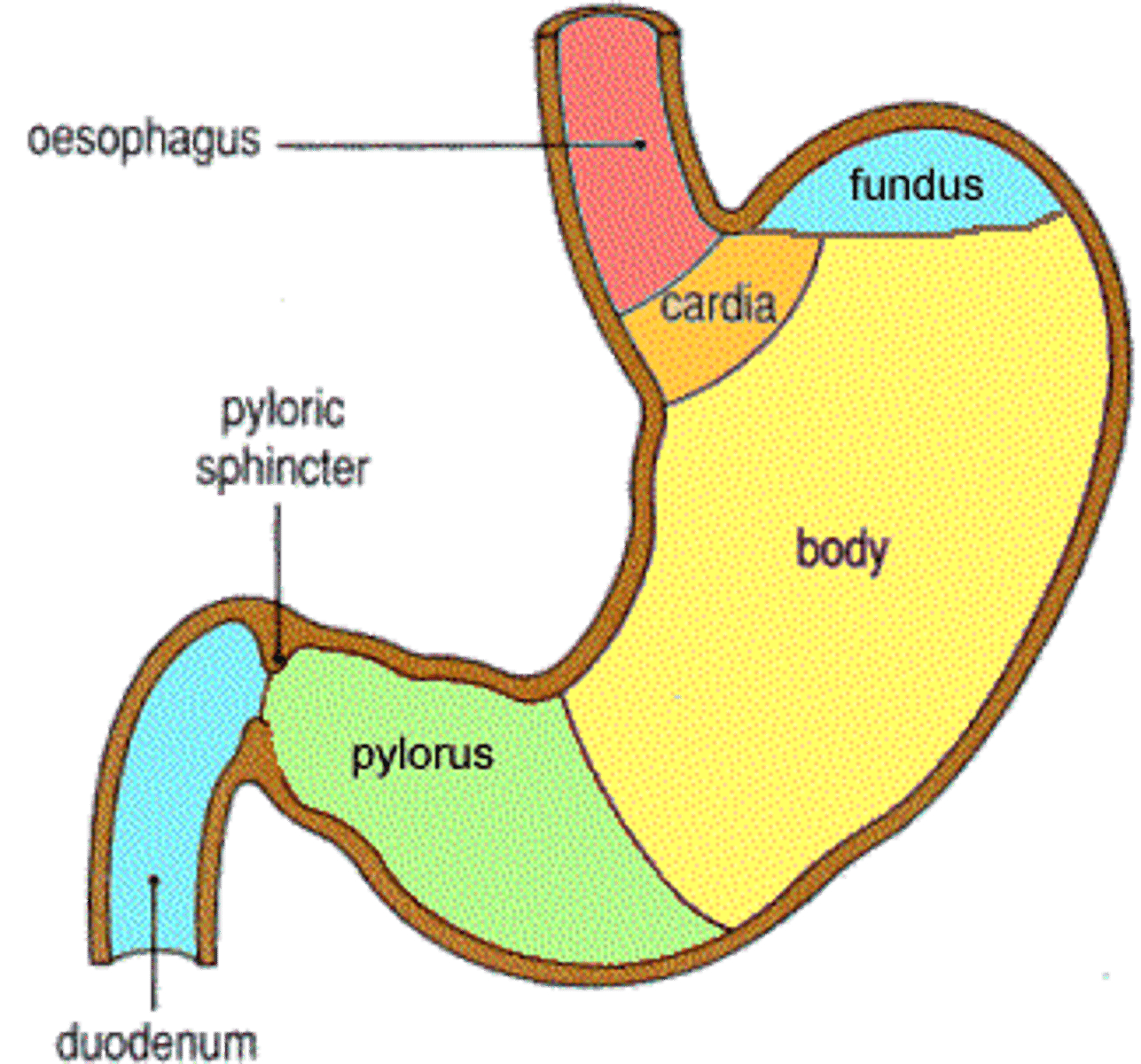
Curvatures of the stomach
greater and lesser
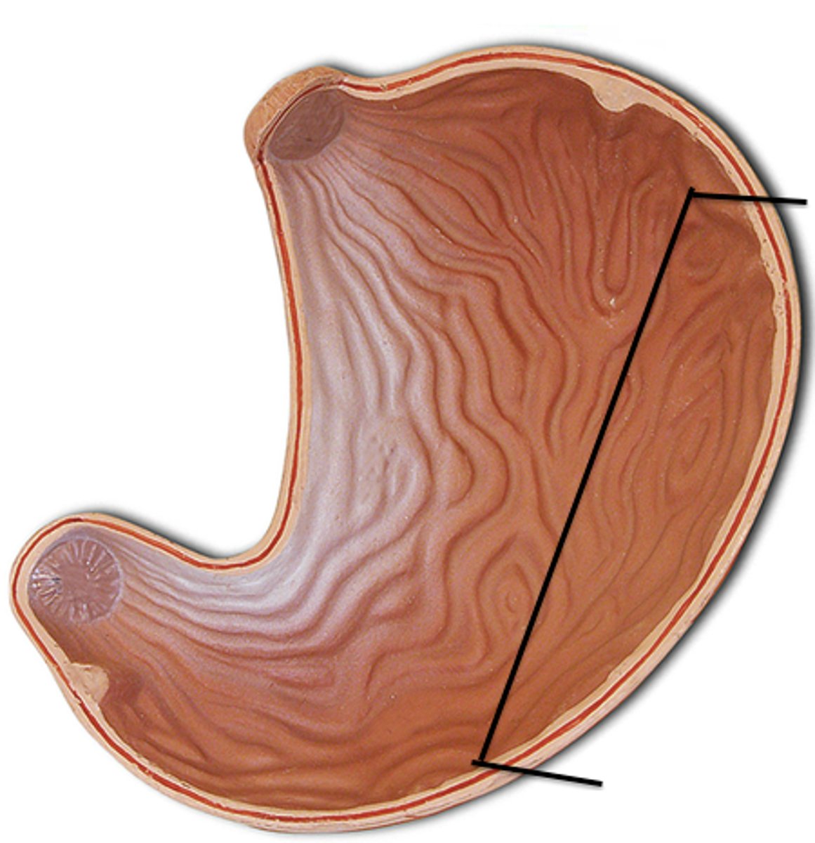
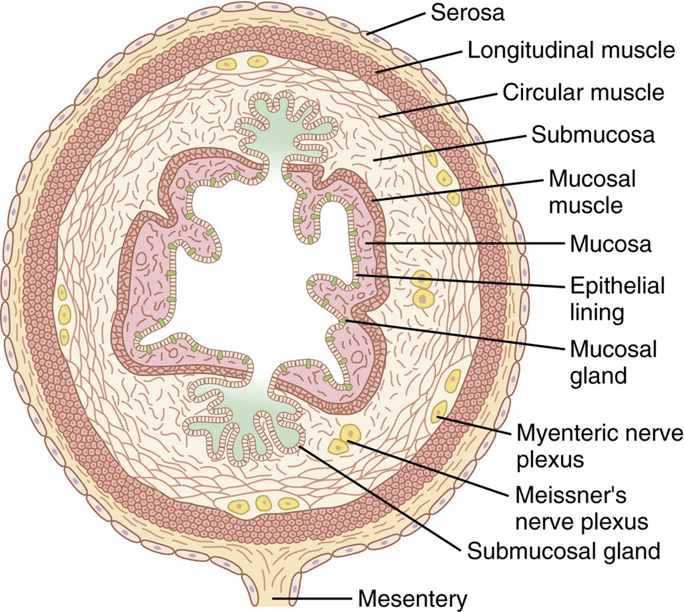
Mucosa of the stomach (4)
serosa, muscularis externa, submucosa, mucosa
Cells of the stomach (4)
Parietal cells (HCL), G cells (Gastrin), Chief cells (pepsinogen), ECL (histamine)
function of the stomach
storage, churn food
how much digestion is done in the stomach
20% of the protein digestion
3 layers of the stomach muscular wall & functions
1. longitudinal - regulate in length (outer)
2. circular - regulate diameter (middle)
3. Oblique - runs diagonally and helps churn the food (inner)

Hiatal Hernia
Protrusion of a part of the stomach thru esophageal hiatus in diaphragm - causing the fundus portion and potentially the cardiac to slide through the opening
mostly fundal region that slides through
-the esophageal hiatus opens more so stomach goes through
sliding is the most common in what
Middle age females - Hiatal Hernia
increasing the abdominal pressure can increase the size of the opening and allow for the sliding of the stomach through the esophageal hiatus
child birth and pressure change can increase the size of the hiatus
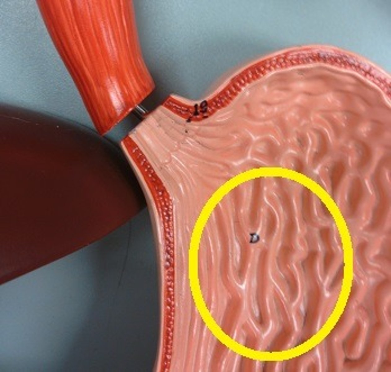
Rugae
folds in the stomach
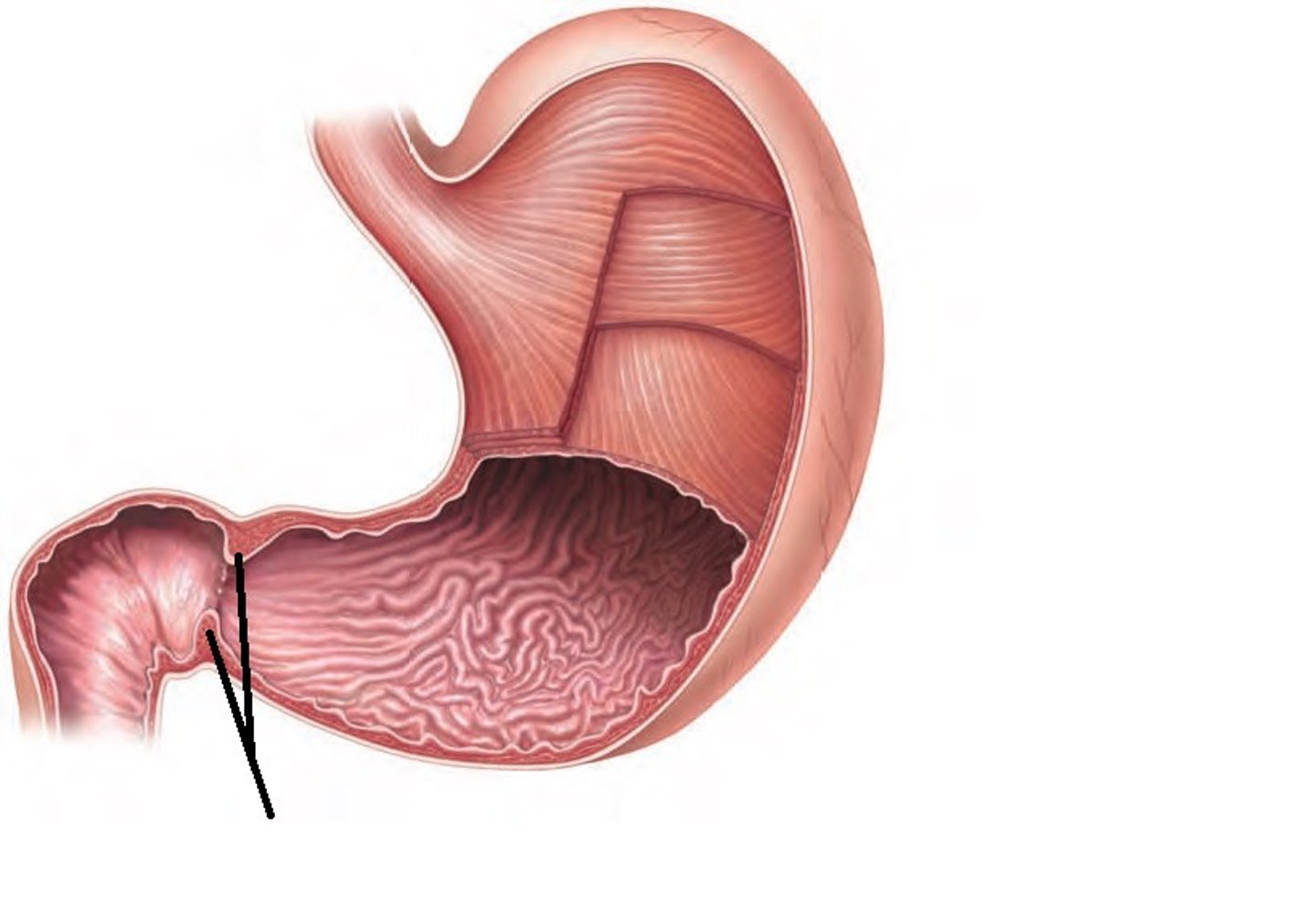
pyloric sphincter
Controls passage of food from stomach to small intestine
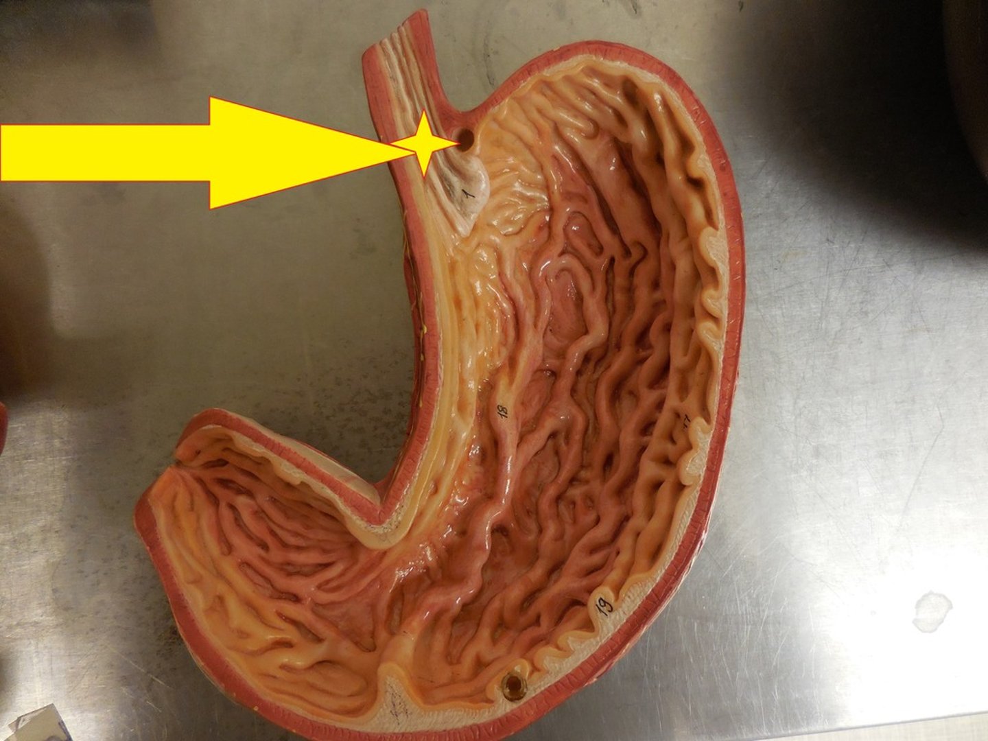
cardial orifice
pyloric canal
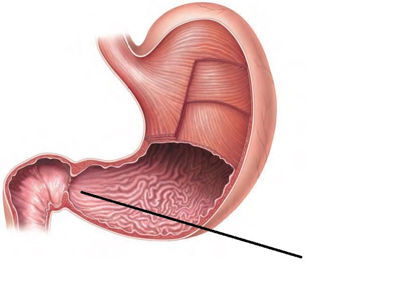
pyloric antrum
Stomach arterial supply
- celiac artery/trunk (mostly Left gastric artery)
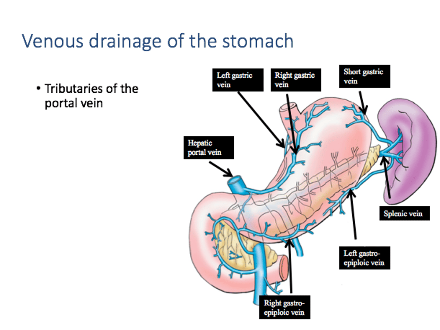
Stomach venous drainage
R/L gastric VV
Superior mesenteric V
splenic V
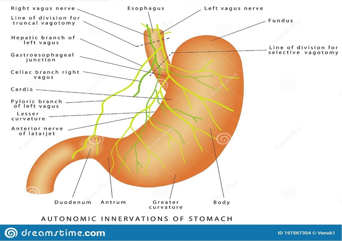
innervation of the stomach
celiac sympathetic plexus and the R/L Vagus N
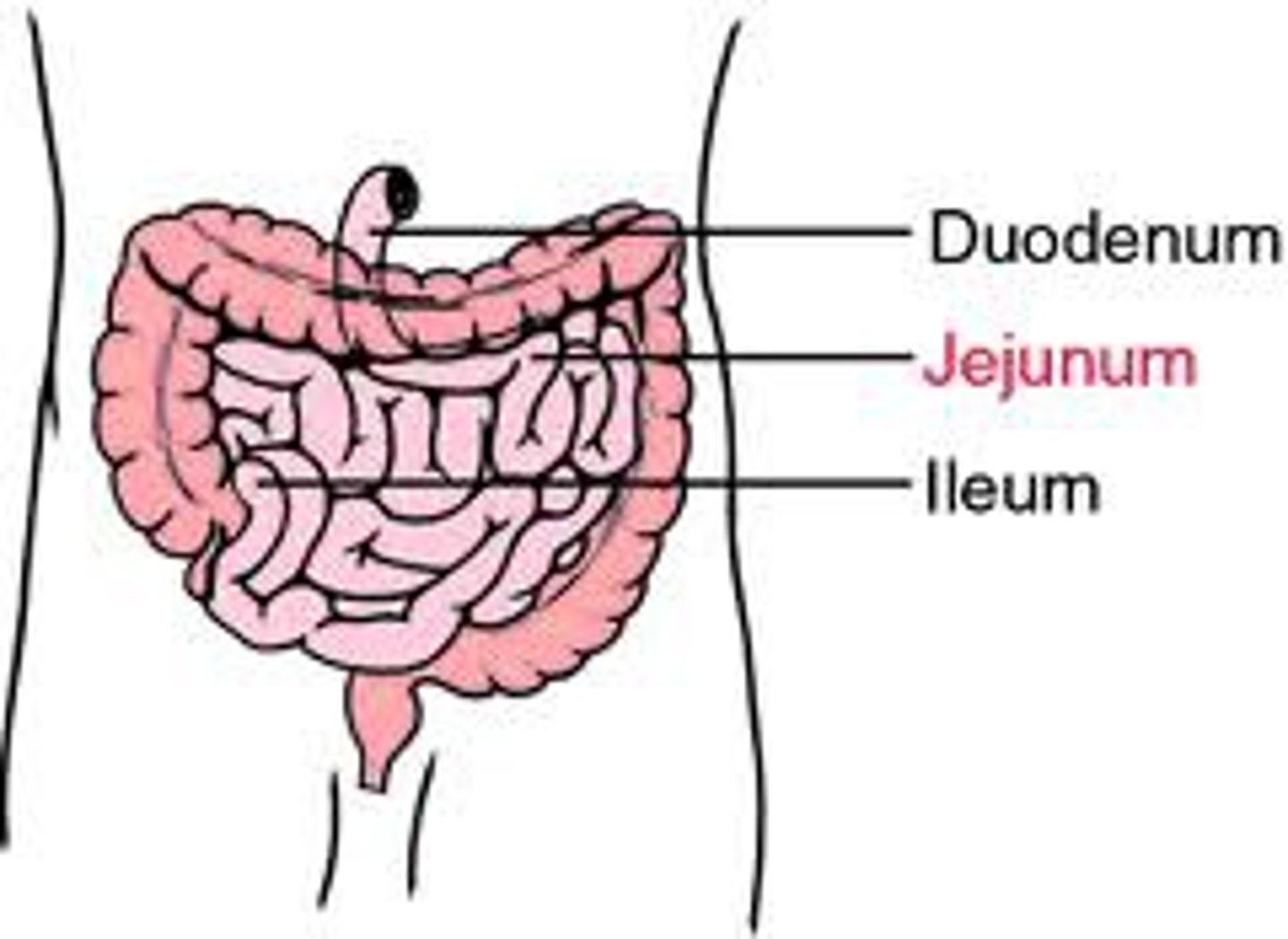
Small Intestine
⚫ 3 portions that can be differentiated by location & anatomy
Duodenum
jejunum
ileum
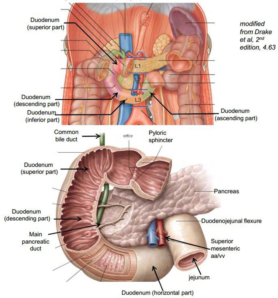
features of the duodenum
10"-12" (inches)
mostly retroperitoneal
C-shaped organ
small: made up of 4 parts
- superior
- descending
- horizontal
- ascending
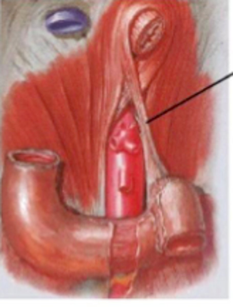
ligament of trietz
junction associated with duodenum and jejunum - connecting to the diaphragm
Features of the jejunum
8' long (feet)
WIDE
THICK WALLED
RED
more mesenteric vessel arcade (longer and less number)

features of the ileum
12' long- end at ileocecal junction
MORE FAT
MORE PYERS PATCHES
decrease mesenteric vessel arcade (more number but shorter)
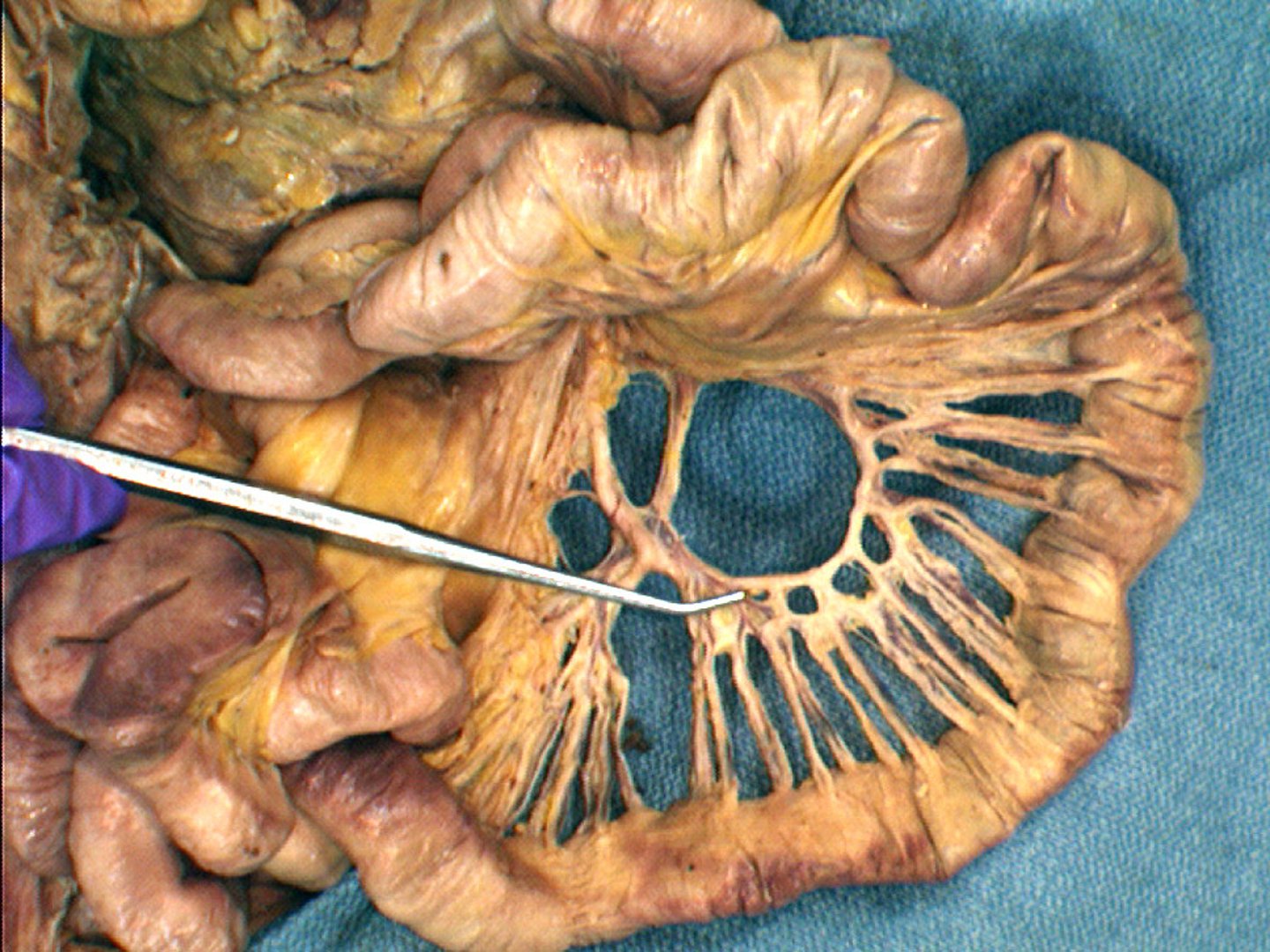
arterial arcades
loops of arteries around the jejunum and ileum
how to distinguish the small intestine regions
1. Location
2. structure (jejunum is wider, thicker, redder)
3. mesenteric vessel arcade (increased in jejunum)
4. lymphoid tissues (jejunum has fewer peyers patches)
5. fat deposition (less in jejunum)
Villi and function
finger like projections that function to increase surface area for absorption and absorb fats
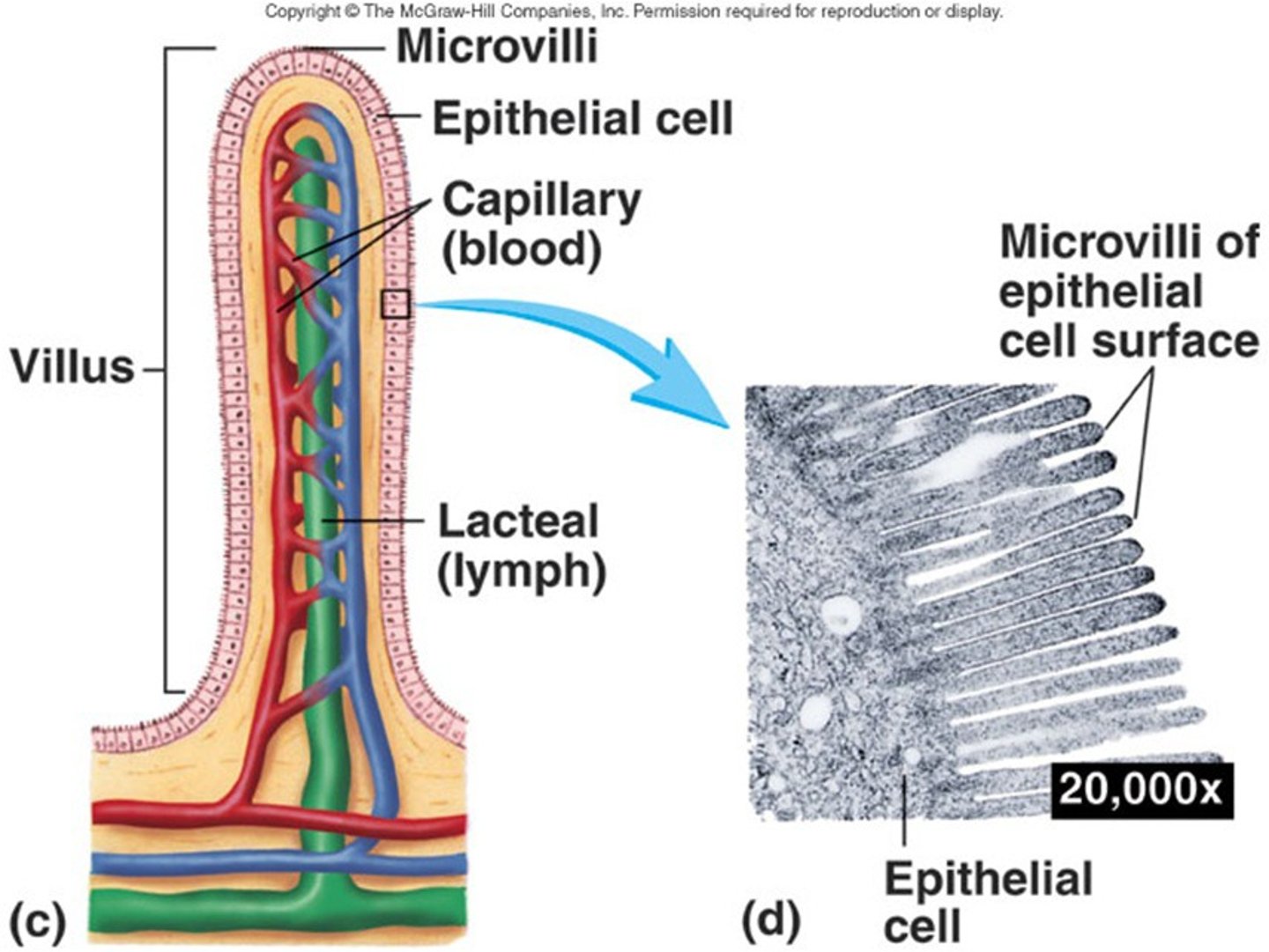
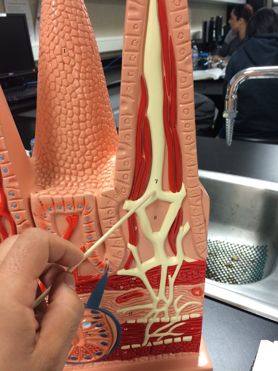
central lacteal
a wide lymph capillary that absorbs fats
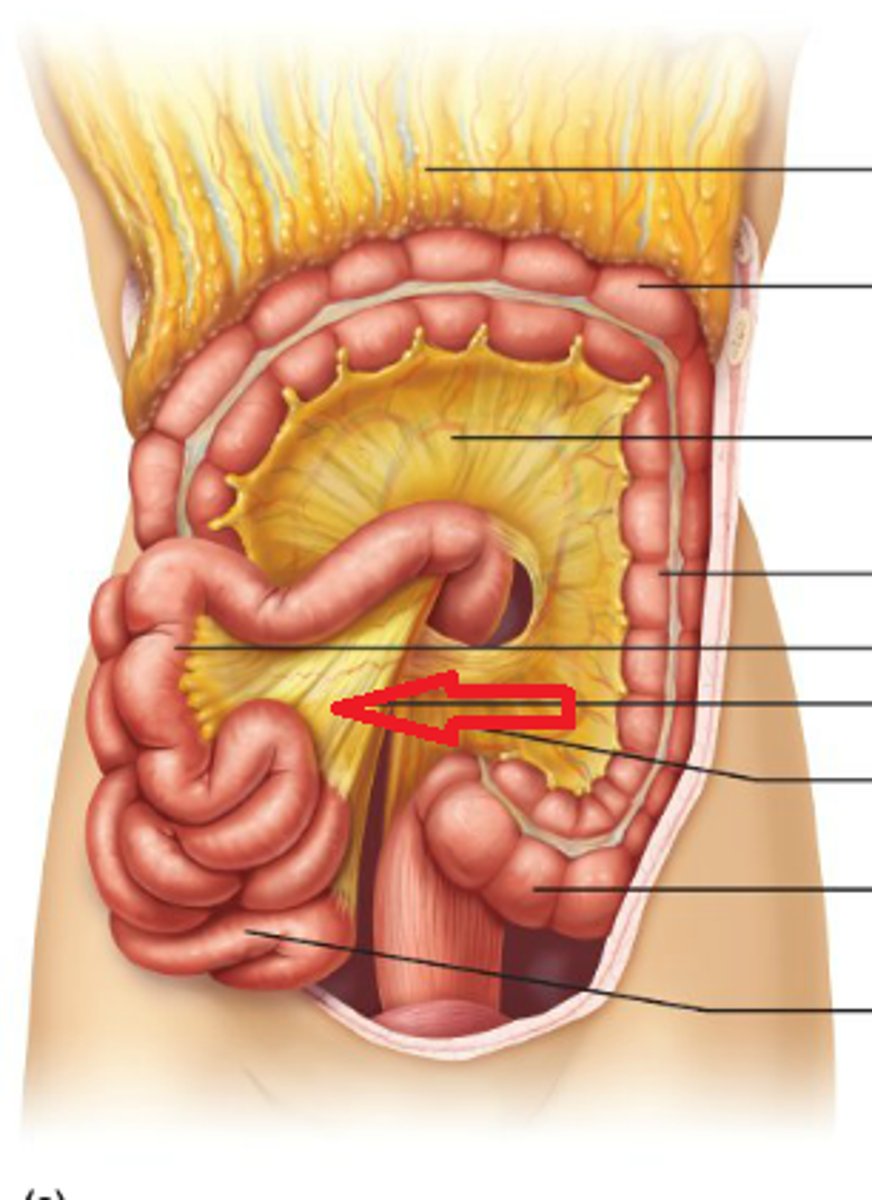
mesentery
ileocecal valve
valve between the ileum of the small intestine and the cecum of the large intestine
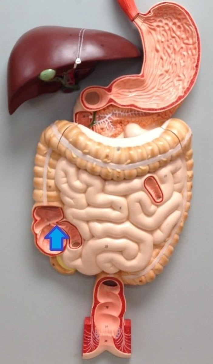
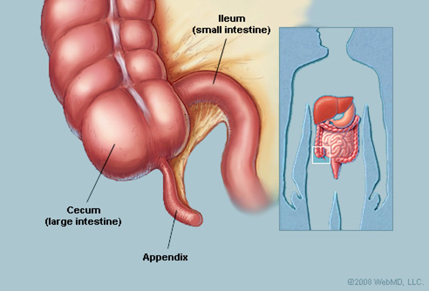
cecum
a pouch connected to the junction of the small and large intestines.

appendix
A small, fingerlike extension of the cecum; contains a mass of white blood cells that contribute to immunity.
Small intestine arterial supply
Superior mesenteric artery
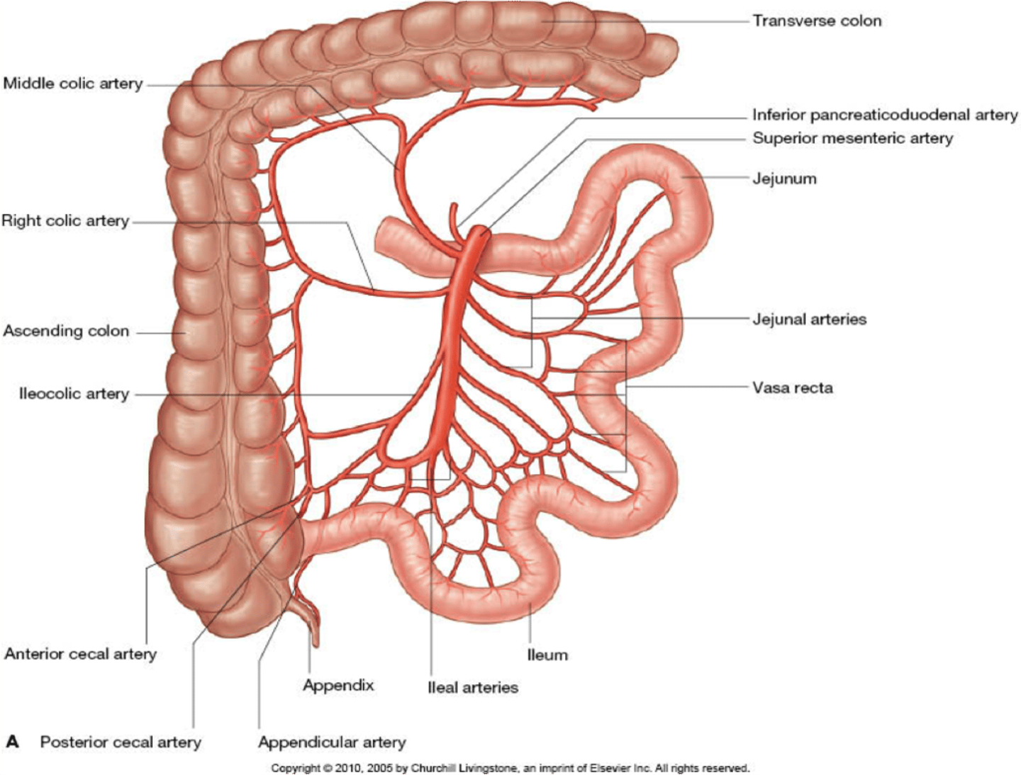
small intestine venous drainage
superior mesenteric vein

innervation of the small intestine
superior mesenteric plexus (sympathetic afferents) and the vagus n
Functions of the large intestine
transport
storage
water reabsorption ****
compaction
Parts of the large intestine
5' long (feet)
Cecum & apendix
1. ascending (r side)
2. transverse
3. descending (L side)
4. sigmoid - turns into the rectum at S3
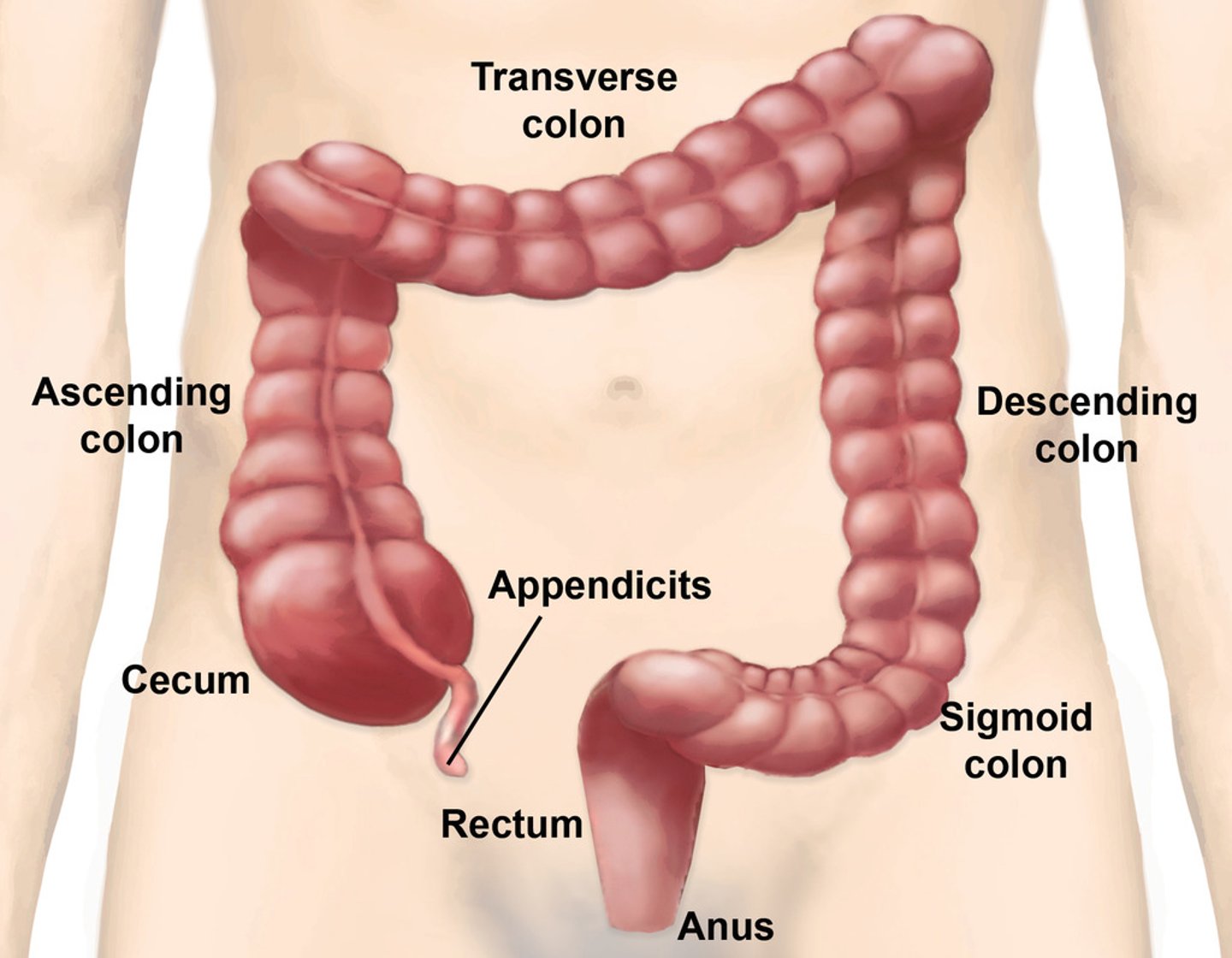
haustra
sacs that are only in large intestine because longitudinal muscle is shorter than gut tube
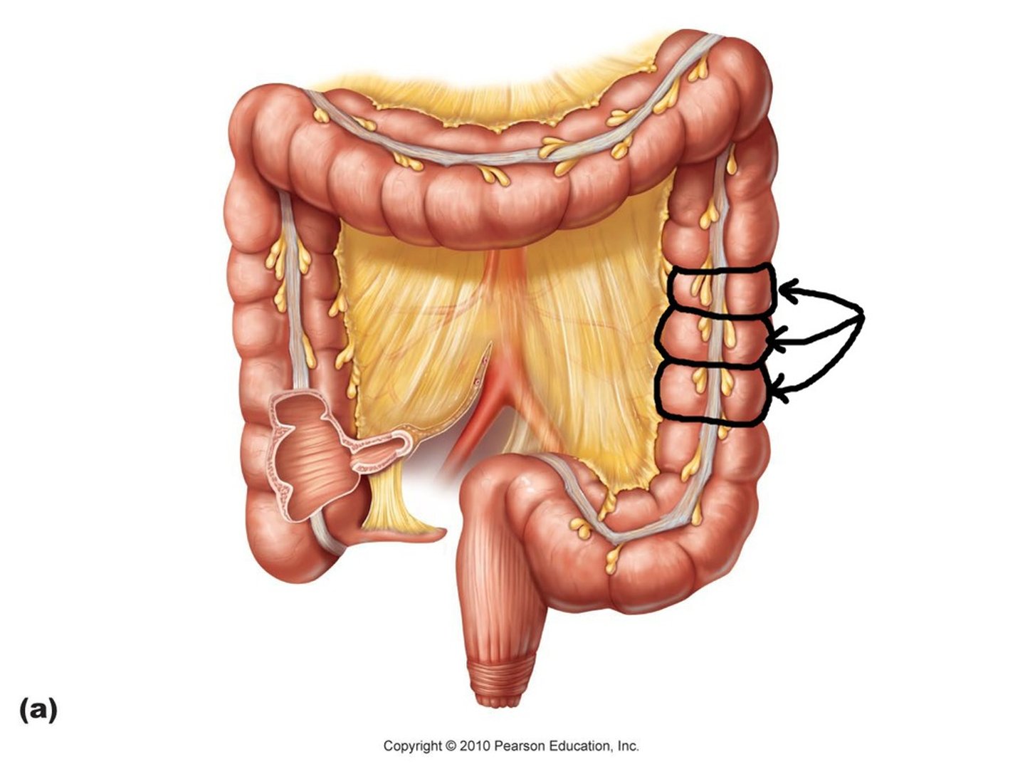
teniae coli
3 longitudinal ribbon of smooth muscle
additional layer of muscle
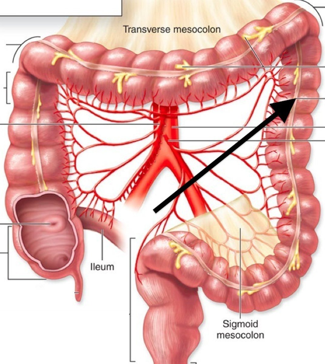
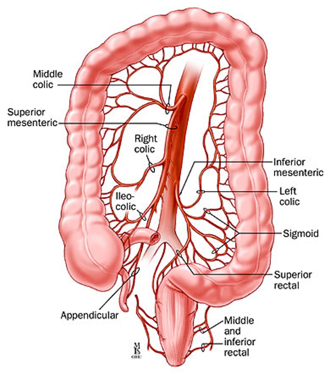
Large intestine arterial supply
superior and inferior mesenteric arteries
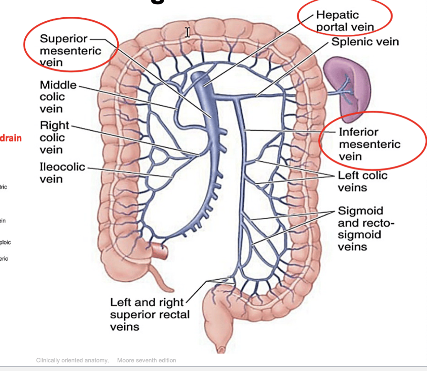
large intestine venous drainage
superior and inferior mesenteric veins
innervation of the large intestine
superior and inferior mesenteric plexus (sympathetic afferents) and the R/L vagus nerves
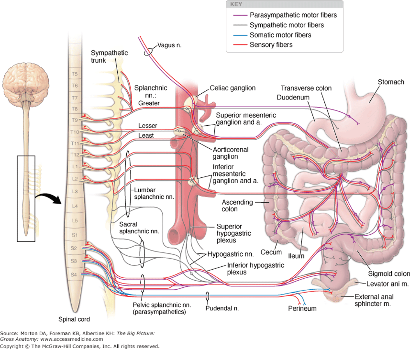
Omentum
Double-layered extension or fold of peritoneum that
passes from stomach & duodenum to adjacent organs in abdominal cavity or to the abdominal wall
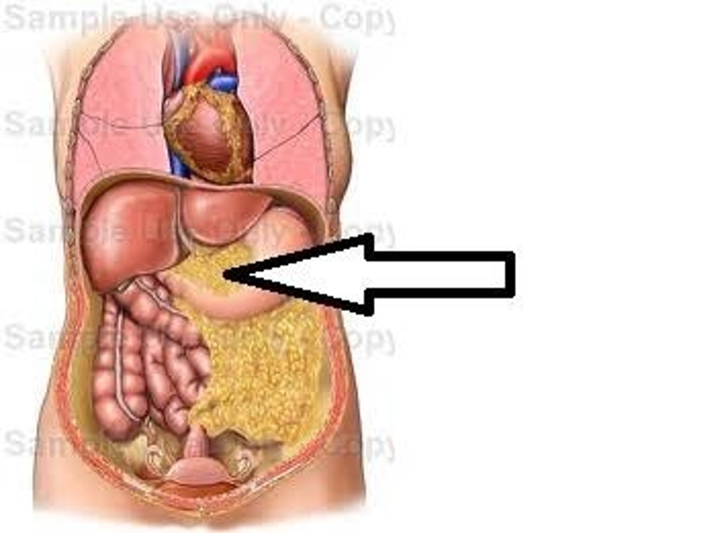
two types of omentum to note
1. greater omentum
2. lesser omentum
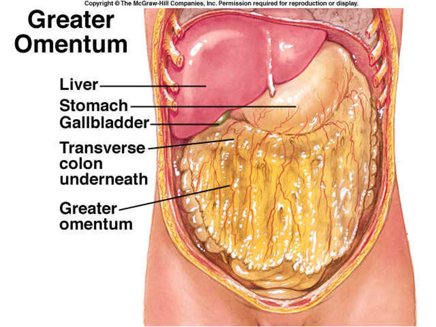
Greater Omentum - characteristics
giant fatty apron
1. 4 layers apron - double-layered omentum that is folded on itself
2. spans the stomach to the transverse colon
3. mostly fat
functions of the greater omentum
1. prevents parietal and visceral peritoneum from adhering to each other
2. forms a cushion
3. insulation (maintain body heat)
4. "police man" - protective function that will cause it to slightly move
3 ligaments of the greater omentum**
1. gastrophrenic
2. gastrosplenic
3. gastrocolic
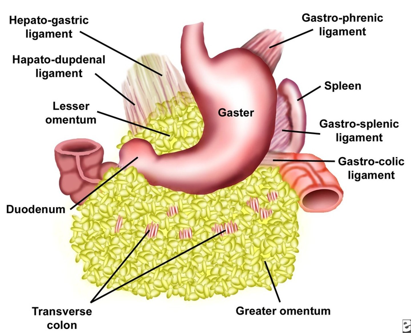
characteristics of the lesser omentum
spans the curvature of the stomach to the liver
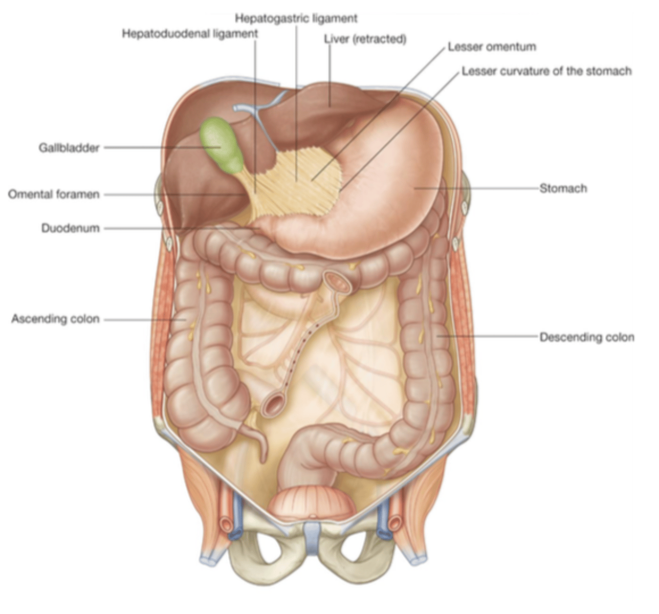
functions of the lesser omentum
anchor organs
2 ligaments of the lesser omentum
1. gastrohepatic
2. hepatoduodenal
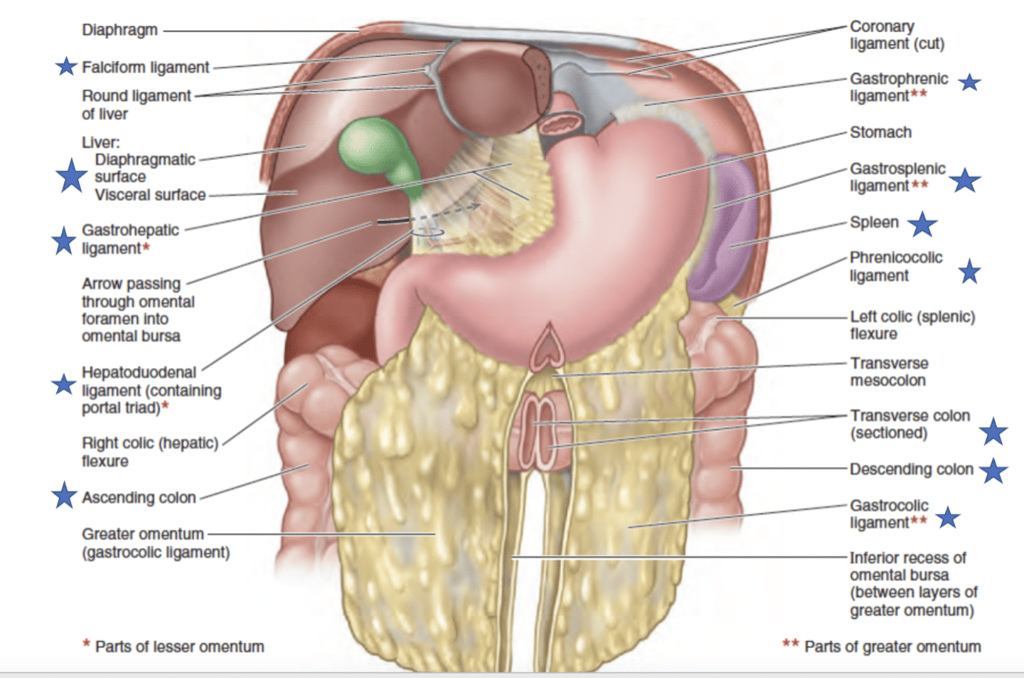
what does the hepatoduodenal ligament contain
the portal triad: hepatic portal vein, hepatic artery, bile duct
location of liver
-location: most of epigastrium and hypochondrium
Characteristics of the liver (R & L Sides)
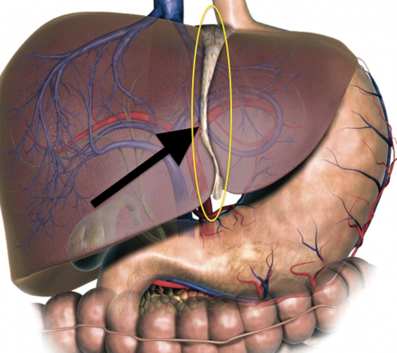
L and R sides are functionally independent
-divided by the falciform ligament
- contains its own blood supply from hepatic artery and portal vein
-contains its own venous and biliary drainage
porta hepatis
transverse fissure on the visceral surface of the liver
located between the quadrate and caudate lobes
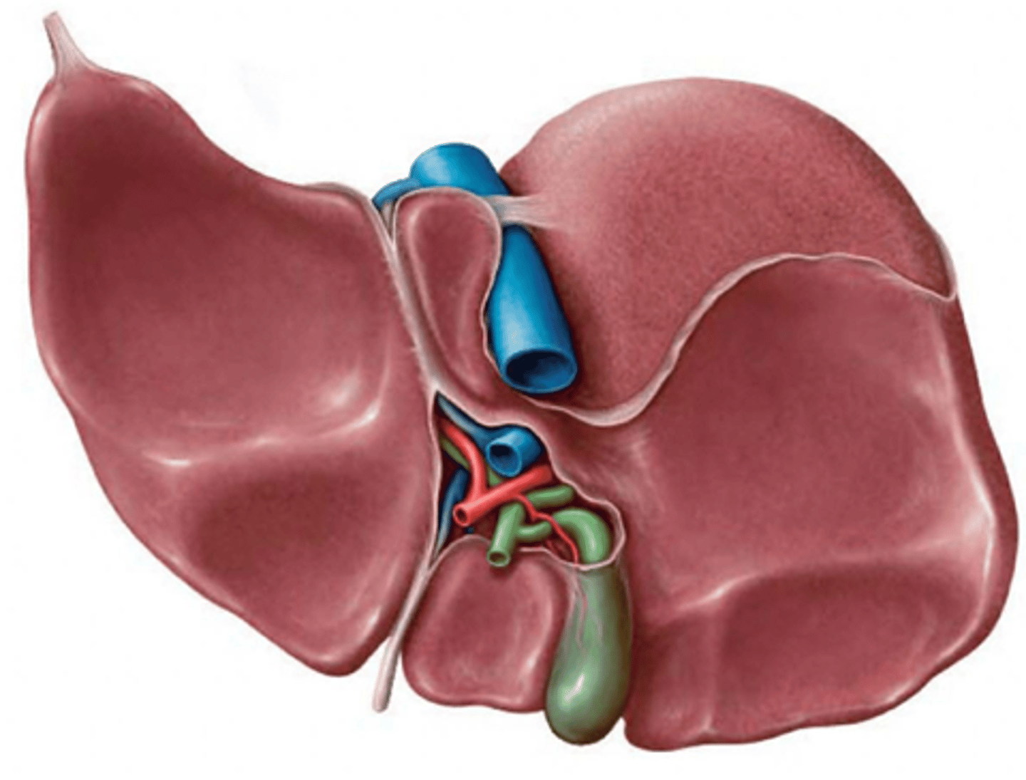
quadrate lobe
medial segment of the right lobe of the liver
gall bladder right next to it
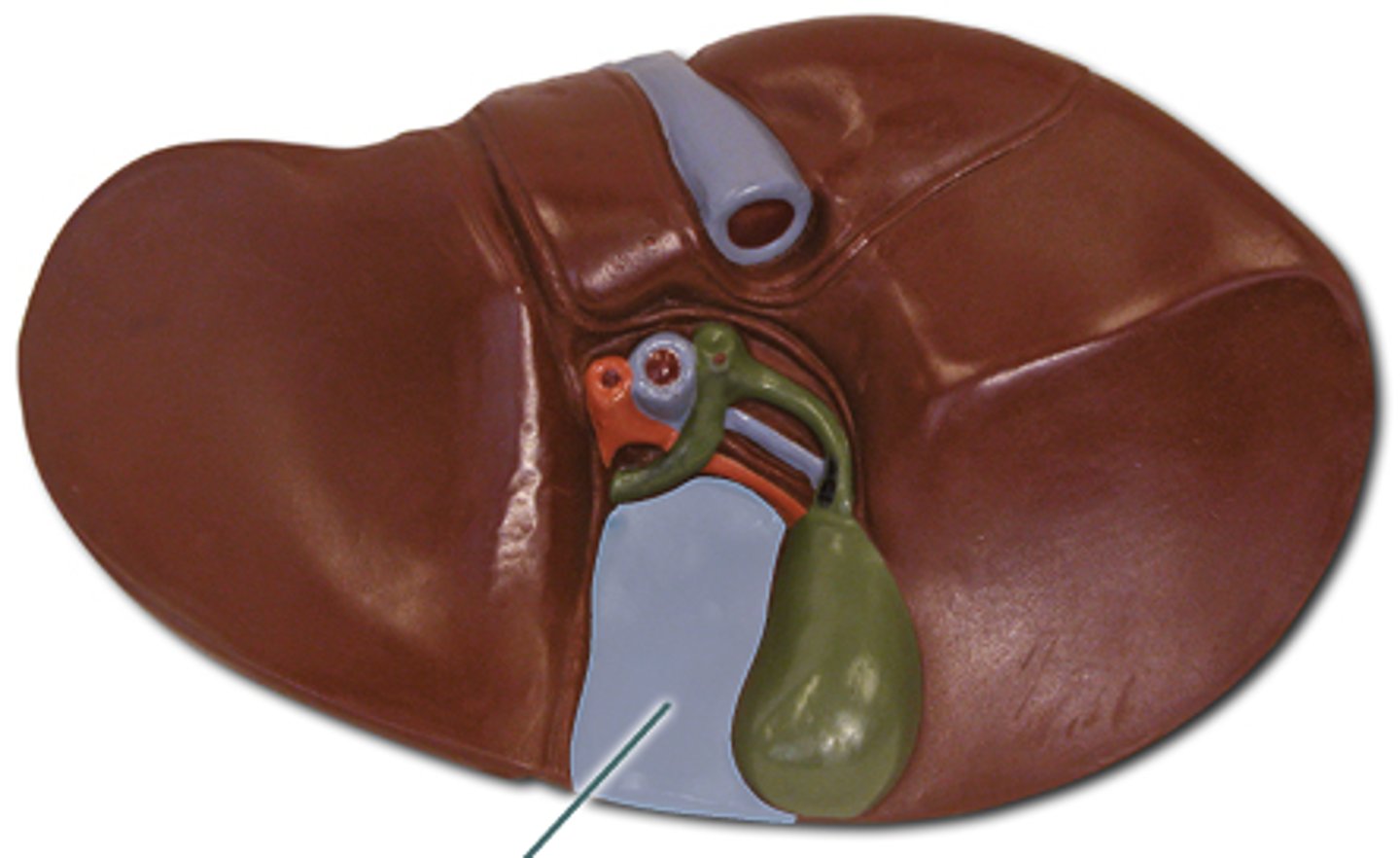
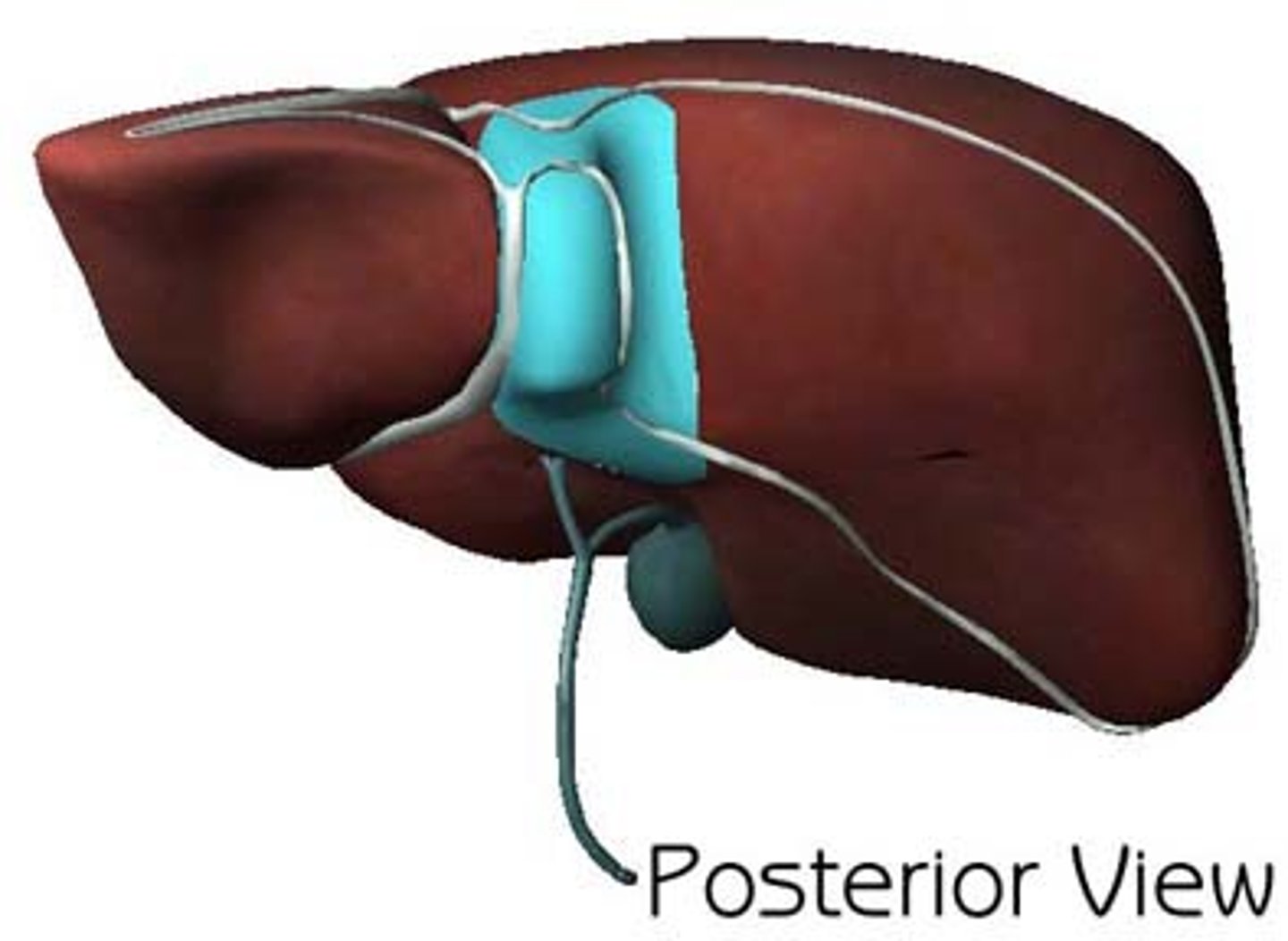
caudate lobe
smallest lobe of the liver situated on the posterosuperior surface of the right lobe the liver
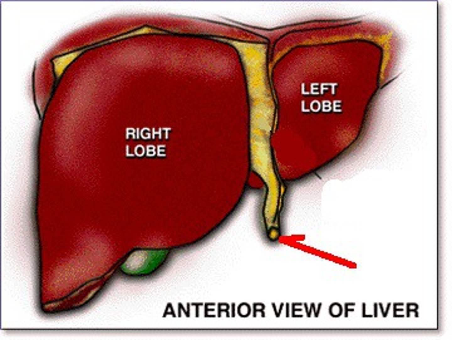
round ligament
The liver receives blood from 2 sources
portal vein (70%)
- from GI vessels to liver sinusoids (nutrient rich & Oxygen poor)
hepatic artery (30%)
- branch from celiac artery (Oxygen rich)
at the Porta hepatis both divide into L/R portions
portal vein & hepatic artery
porta hepatis gives passage to 5 VAN
hepatic portal vein, hepatic artery, hepatic ducts,
hepatic nerves (plexus) and the lymph vessels
The Gallbladder location
in the hollow between the R and L lobe of the liver (by the quadrate lobe)

Where is bile produced
Bile is produced in the liver.
where is bile released after production
into the R and L hepatic ducts
After bile is released into R and L hepatic ducts, it can either
-flow into common bile duct
-be stored in gall bladder
Where is bile stored
highly concentrated in the gallbladder
can store about 50ml
how much bile do we make in a day
1 L
what are the two fates of bile after production
released into R and L hepatic ducts, and it can either be stored, or be released into the common bile duct
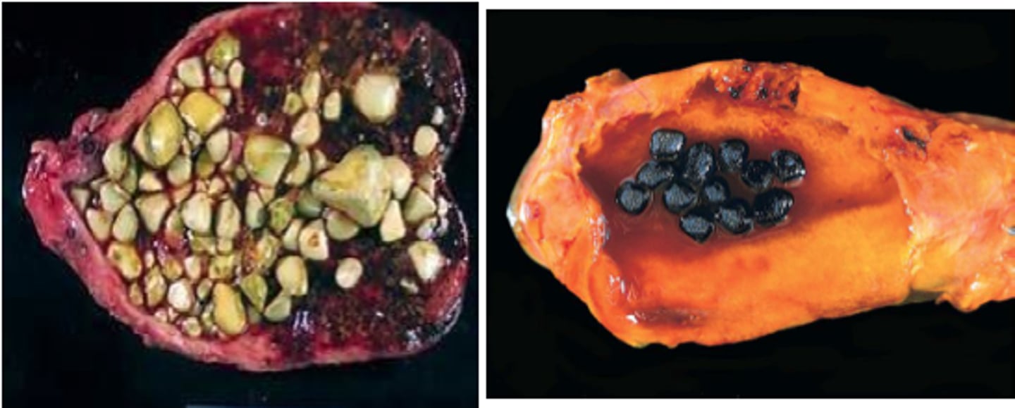
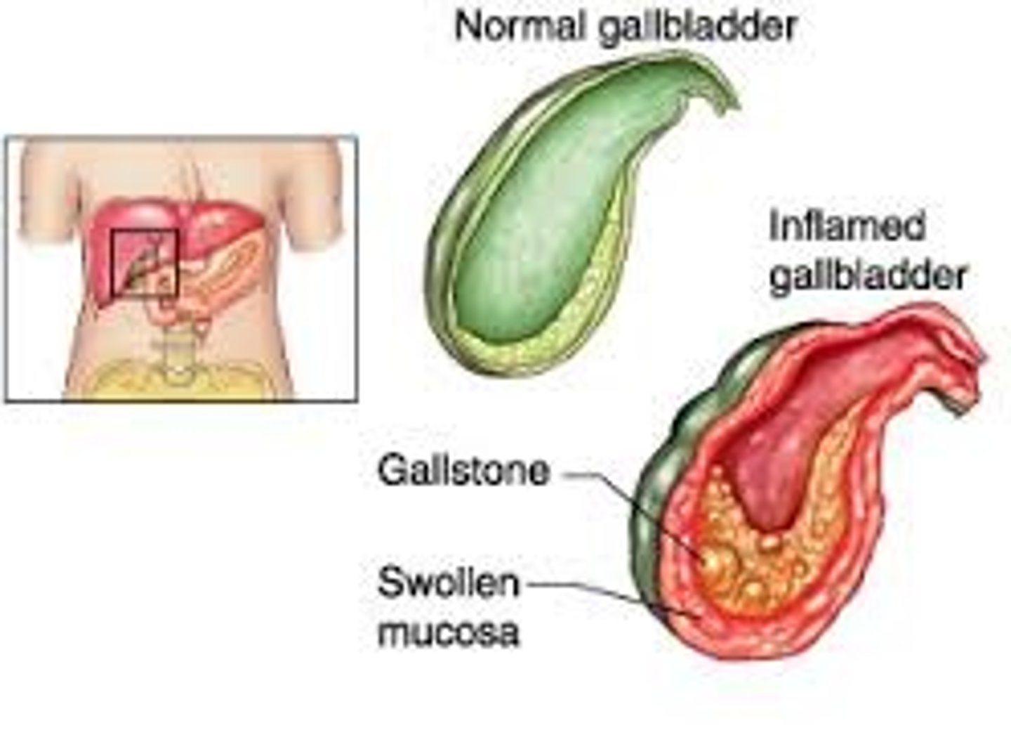
gall stones
Cholesterol rocks formed in the bile, blocks the bile ducts
cholecystitis
inflammation of the gallbladder; usually associated with gallstones - can be bacterial
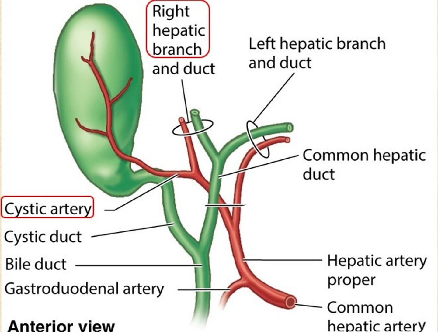
arterial supply of the gallbladder
cystic artery (branch of hepatic artery)
Venous drainage of the gallbladder
cystic vein (lead to portal vein)
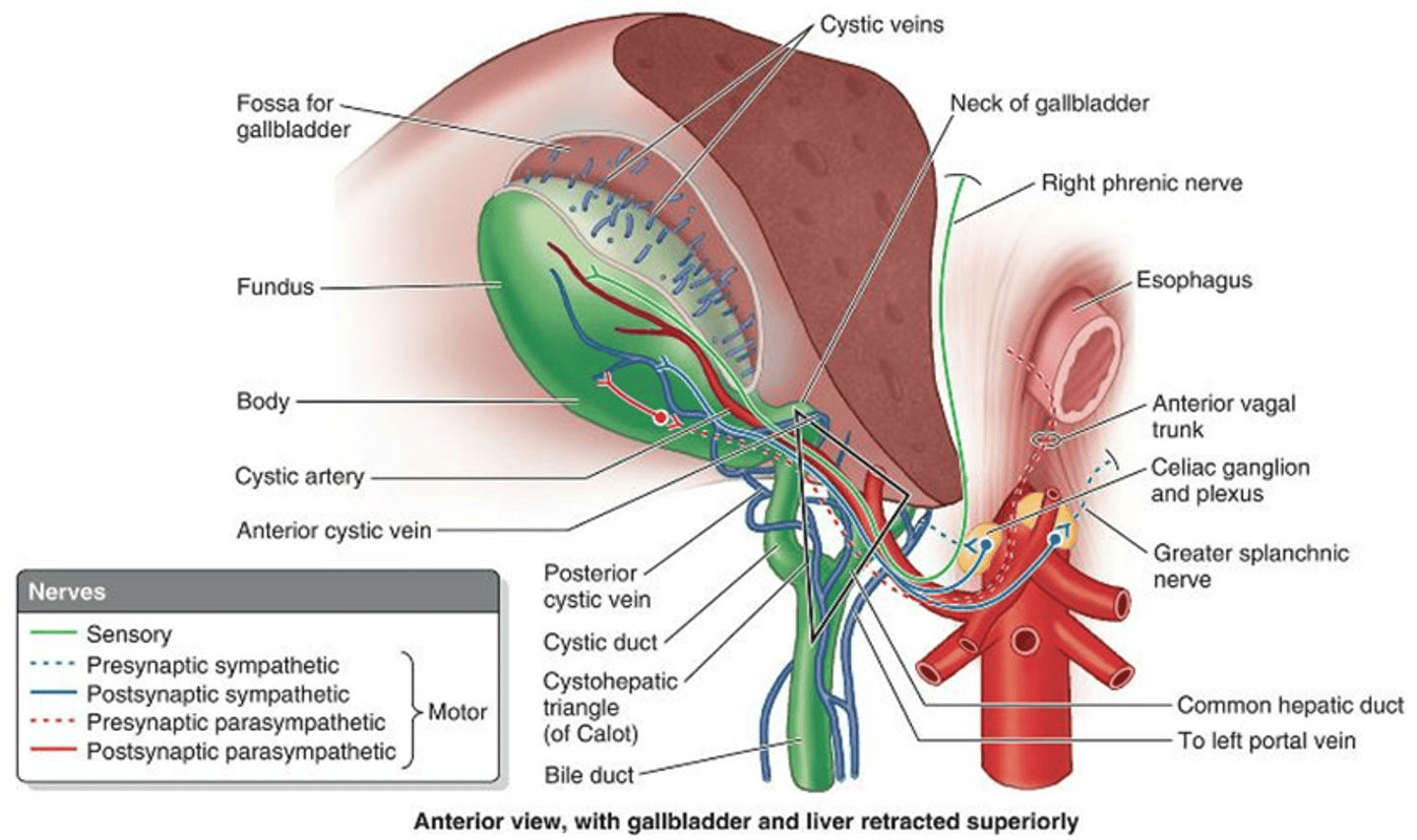
innovation of the gallbladder
celiac plexus
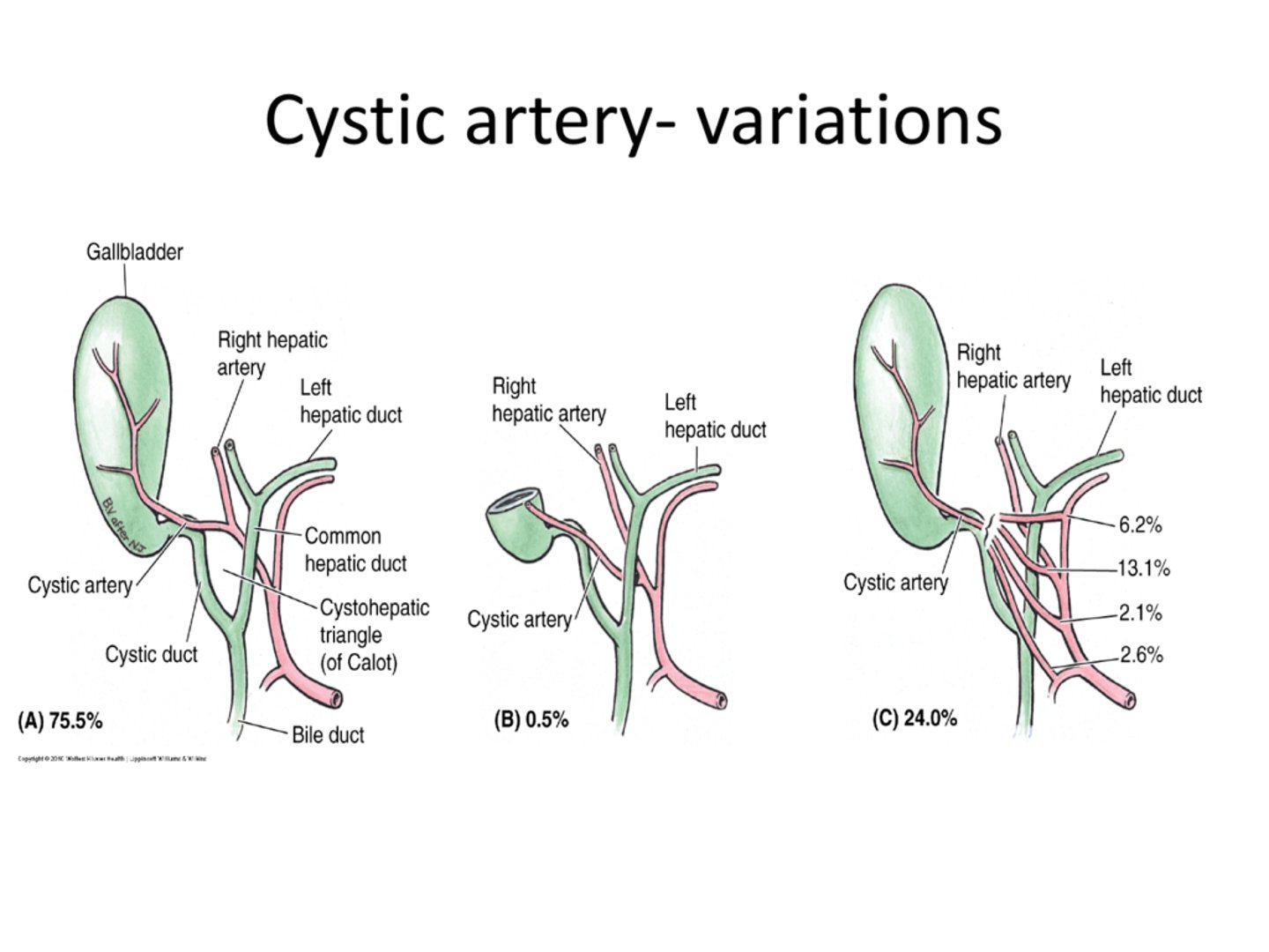
variations in the blood flow orientation of gallbladder
75% are typical with 24% making up other variant important for surgery!
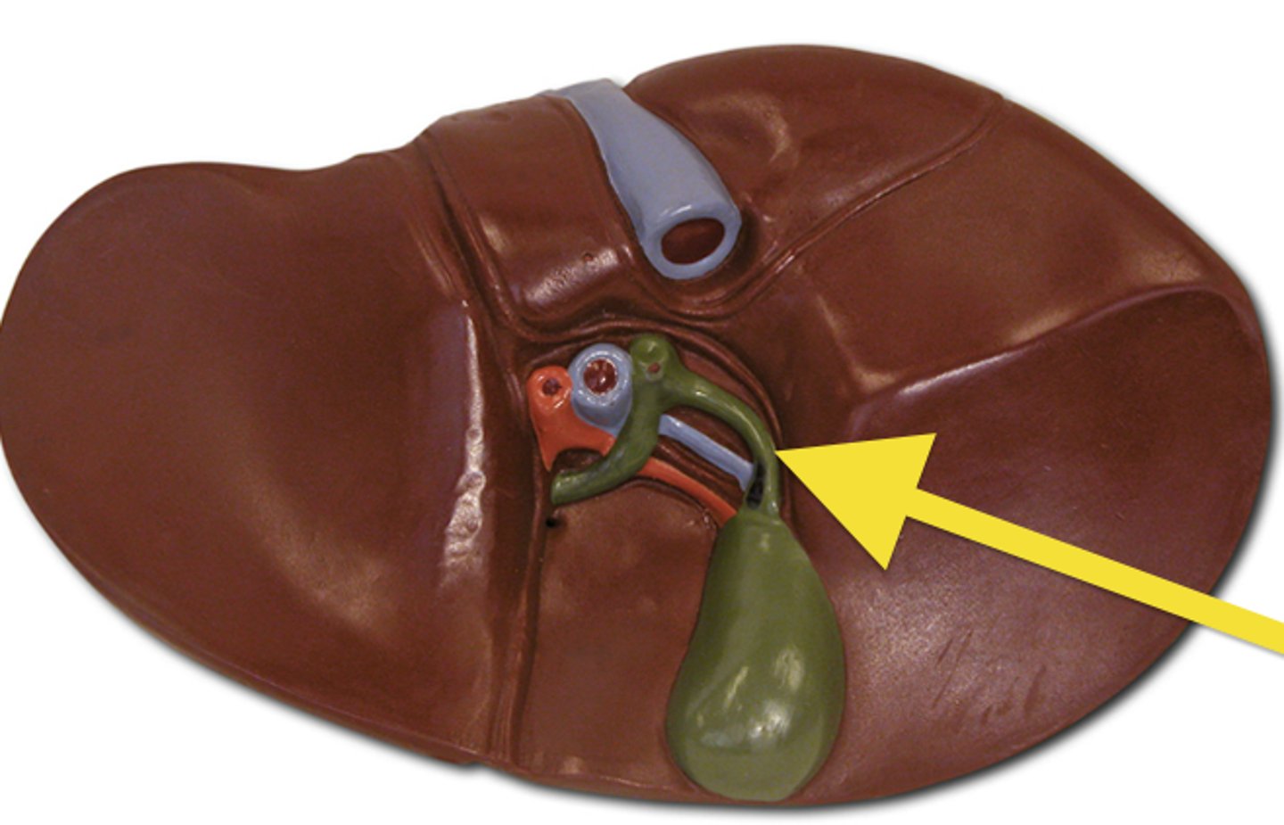
cystic duct
Duct leading from the gallbladder to the common bile duct; carries bile
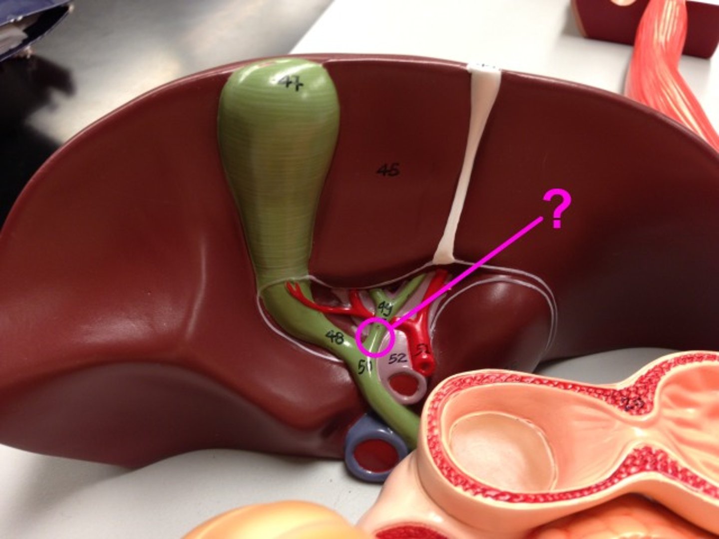
common hepatic duct
large bile duct leading from liver (right and left hepatic ducts); joins with the cystic duct to form the common bile duct
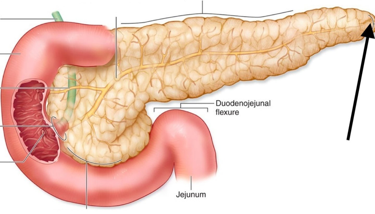
Pancreas is located
retroperitoneal, behind the stomach, "cradled in the arms of the duodenum)
2 major functions of the pancreas
1. exocrine
2. endocrine
exocrine function of the pancreas
produce enzymes and HCO3-
travels through the pancreatic duct

endocrine function of the pancreas
pancreatic islets secrete insulin (B cells) and glucagon (A cells) via the bloodstream

pancreatic duct
conducts pancreatic juice from the pancreas to the small intestine
letter= H

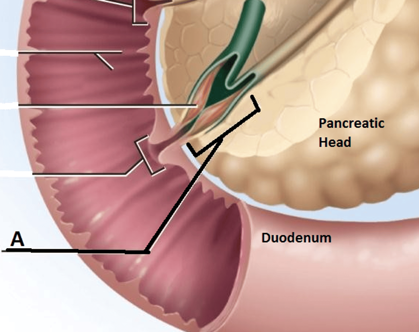
ampulla of vater
small opening in the duodenum in which the pancreatic and common bile duct enter to release secretions
guarded by the sphincter of oddi