Post-Lab Quiz 2 - 232 Lab
1/12
There's no tags or description
Looks like no tags are added yet.
Name | Mastery | Learn | Test | Matching | Spaced | Call with Kai |
|---|
No analytics yet
Send a link to your students to track their progress
13 Terms
Describe the composition of the 3 layers of a blood vessel wall and state the function of each
Tunica intima: endothelium lines lumen and provides a smooth surface for blood flow; releases N2O gas which relaxes tunica media
Tunica media: circular smooth muscle fibers supported by elastic fibers; perform vasoconstriction and vasodilation
Tunica external: outermost layer (collagen): attaches vessels to other structures
very large vessels have their own blood supply called vasa vasorum
Compare and contrast the structures and functions of arteries, veins, and capillaries including lumen diameter, tunica thickness, valves, and blood flow
Arteries:
Small lumen diameter
Thicker tunica media
Systemic: carry oxygenated blood
Pulmonary: carry deoxygenated blood
NO VALVES
Veins:
Large lumen diameter
Thinner tunica media
Systematic: carry deoxygenated blood
Pulmonary: carry oxygenated blood
CONTAIN VALVES
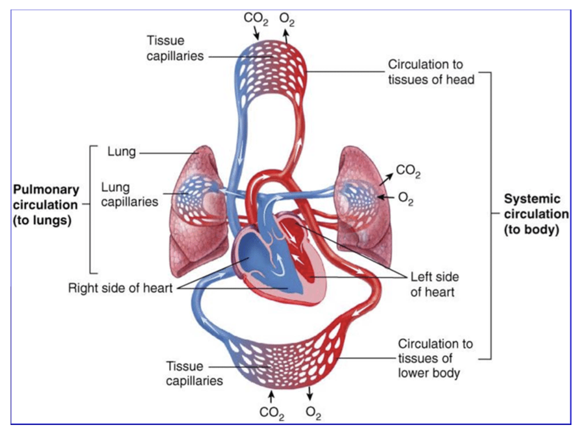
Compare and contrast pulmonary circulation and systemic circulation; trace blood flow of each
Systemic circulation: transports blood from the left side of the heart to the body for nutrient and gas exchanges and back to the right side of the heart
Supplies: oxygenated blood
Returns: deoxygenated blood
Pulmonary circulation: moves blood from the right side of the heart to the lungs for gas exchange and back to the left side of the heart
Supplies: deoxygenated blood
Returns: oxygenated blood
Know whether a vessel varries oxygenated or deoxygenated blood
Define vasoconstriction and vasodilation and explain how they relate to blood vessel lumen
Vasoconstriction: a reduction in blood vessel diameter, reduces blood flow, and increases pressure
Vasodilation: an increase in blood vessel diameter, increase in blood flow, decrease in pressure
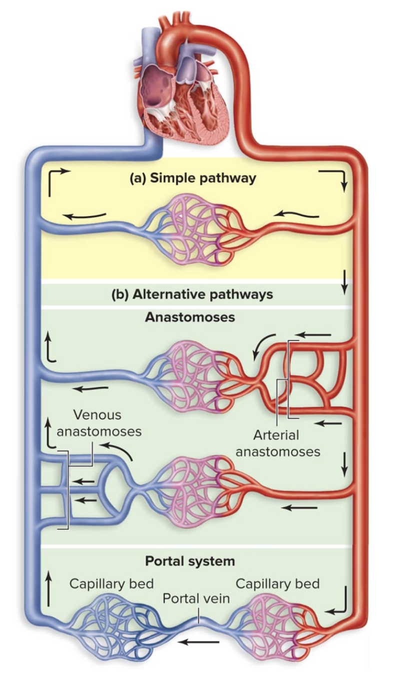
Compare and contrast end vessels and vessel anatomoses
End vessel: a simple pathway where one vessel takes blood to a tissue or organ
Anatomoses: when two or more vessels converge to take blood to a tissue or organ
Trace blood flow through vessels listed for Blood Vessels - Lab 2
Identify the vessel(s) blood just traveled from and the vesel(s) it will enter next; be prepared to identify more than one vessel if possible
Identify the organ, region, or anatomical structure a blood vessel services
BODY
→ Superior vena cava and Inferior vena cava →
RIGHT ATRIA
→ Tricuspid valve →
RIGHT VENTRICLE
→ Pulmonary valve →
PULMONARY TRUNK
→ Left and Right Pulmonary Arteries →
LUNGS
→ Left and Right Pulmonary Veins →
LEFT ATRIA
→ Bicuspid Valve →
LEFT VETRICLE
→ Aortic Valve →
AORTA
Trace Blood Flow:
Describe the major anatomical features of the heart including its two sides, 4 chambers, 4 valves, attached great vessels, and the structures listed below
Orient and identify major features of the heart from anterior, posterior, superior, and inferior orientations when you give sagittal, frontal, and transverse cross sections
Trace blood flow through the heart
Explain coronary circulation; identify coronary arteries and veins, and the regions of the heart they serve
Describe the structure of cardiac muscle, including intercalated discs, and explain how it differs from skeletal and smooth muscle