Male Reproductive Anatomy
 Call Kai
 Learn
Learn Practice Test
 Spaced Repetition
 Match
Match Flashcards
 Knowt Play
1/25

There's no tags or description
Looks like no tags are added yet.
Name | Mastery | Learn | Test | Matching | Spaced | Call with Kai | Chat |
|---|
No analytics yet
Send a link to your students to track their progress
26 Terms
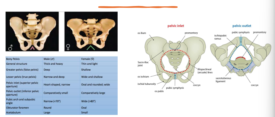
Male vs female pelvises
Male sexual anatomy
Primary sex organ: testes → sperm + testosterone
Secondary sex organs: ducts, glands + penis → transport/support sperm
Functions: (SECSH)
Spermatogenesis
Hormone secretion
Semen production
Ejaculation
Copulation
External genitalia
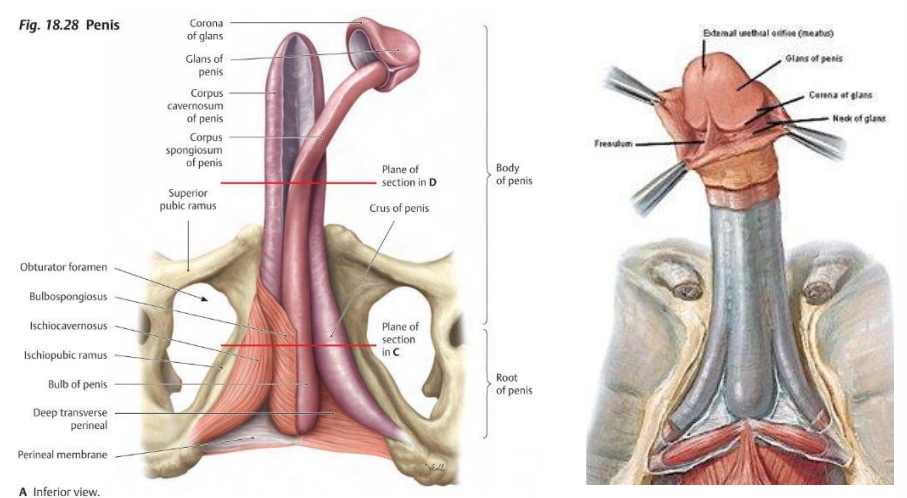
Penis
Root: 2 crura → attach to inferior pubic rami
Bulb: expansion of shaft at base
Body: shaft
Glans: distal end
Erectile tissue – “2 for erection, 1 for protection”
2 corpora cavernosa → dorsal → erection
1 corpus spongiosum → ventral → contains urethra
Protects + keeps urethra open during ejaculation
Forms glans (“acorn”)
Scrotum
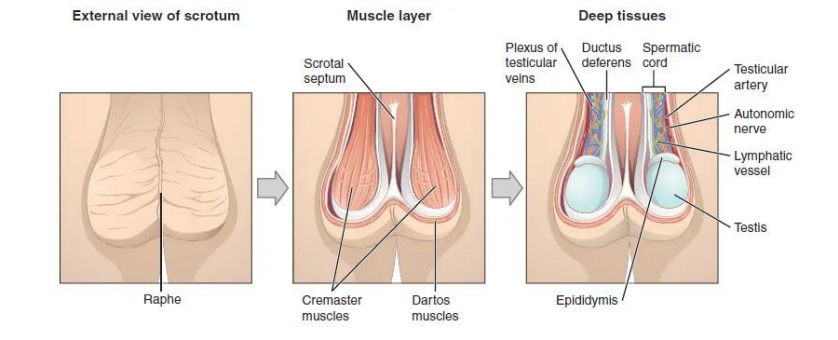
Skin-covered sac → divided by median septum
Thermoregulation: 2–3°C below body temp
Dartos muscle: wrinkles skin
Cremaster muscle: elevates testes
Homologous structures
Homologous = same embryonic origin, but may develop differently and have different functions.
**know the table

Cross section anatomy of the penis
Erectile Tissues
2 corpora cavernosa
1 corpus spongiosum → contains urethra
Tunica albuginea → fibrous covering around erectile tissue
Blood supply
Deep + dorsal penile arteries ← internal pudendal artery
Innervation
Pudendal nerve → somatic/sensory
Pelvic splanchnic nerves → autonomic

Perineum
Diamond-shaped region between thighs, ↓ pelvic diaphragm
Boundaries: anterior = pubic symphysis; posterior = coccyx; lateral = ischial tuberosities
Roof: pelvic diaphragm
Floor: skin + superficial fascia
Divided into 2 triangles:
Urogenital
Anal
Anterior Urogenital Triangle
Contents
Root of penis
Scrotum
Penile/spongy urethra
Bulbourethral glands + membranous urethra → deep perineal pouch
Muscles
Bulbospongiosus
Ischiocavernosus
Superficial transverse perineal

Penis pics
Scrotum
Sac of loose skin, fascia + smooth muscle, divided into 2 pouches by a septum
Function: temperature regulation of testes
Contents:
Testes
Epididymis
Spermatic cord → vas deferens, blood vessels + nerves
Layers — “Some Damn Englishman Called It The Testes”
Skin
Dartos muscle/fascia
External spermatic fascia
Cremaster muscle/fascia
Internal spermatic fascia
Tunica vaginalis → serous membrane surrounding testes
Tunica albuginea → dense, white capsule
Scrotum pics
Temperature regulation of the testes
Temperature Regulation of Testes
Dartos muscle: smooth muscle lining of scrotum
Cold/arousal → elevates testes
Wrinkles skin → ↓ surface area → ↓ heat loss
Cremaster muscle: in spermatic cord; slip of internal abdominal muscle
Cold → elevates testes
Pampiniform plexus: counter-current heat exchange
Testicular artery + vein
Cool venous blood cools arterial blood travelling to testes
Easy mnemonic: Cool Down Please!
Cremaster → elevates testes
Dartos → wrinkles skin
Pampiniform → cools arterial blood
Testes
Testes
Paired, oval glands
Left usually lower than right
Covered by:
Tunica vaginalis
Tunica albuginea
Develop in abdomen → descend through inguinal canal just before birth
Internal structure:
Septa → divide testes into 200–300 lobules
Each lobule → 2–3 seminiferous tubules
Seminiferous tubules → spermatogenesis
Rete testis → efferent ductules (12-20)→ epididymis
Blood supply: testicular artery
Venous drainage: pampiniform plexus

Seminiferous Tubules
Seminiferous Tubules
Contain:
Sperm-forming cells
Sertoli cells
Form blood-testis barrier
Support/nourish developing sperm
Secrete inhibin → ↓ spermatogenesis
Produce fluid + control sperm release into lumen
Leydig cells (interstitium)
Secrete testosterone
“S = Support, L = Libido”

Epididymis
Epididymis
Single, coiled duct posterolateral to testis
Head → receives efferent ductules
Body
Tail
Site of sperm maturation + storage
Smooth muscle → aids sperm movement

Vas deferens (Ductus Deferens)
Long, muscular duct → carries sperm from epididymis → urethra
Ampulla → dilated portion
Passes through spermatic cord
Joins seminal vesicle duct → forms ejaculatory duct in pelvic cavity
One per testicle

Accessory Glands
Seminal Vesicles (2)
Elongated outgrowths of vas deferens
Posterior to bladder, between bladder + rectum
Secrete clear, fructose-rich alkaline fluid + prostaglandins, ATP & coagulating enzymes
Nutrition for sperm → supports optimal function
Prostate (1, unpaired)
Walnut-sized, inferior to bladder
Posterior to pubic symphysis, anterior to rectum
Continuous with bladder neck
Surrounds urethra
Ejaculatory ducts pass through posterior aspect → open into prostatic urethra
Secretes milky prostatic fluid (enzymes + citrate) → kills bacteria
Bulbourethral (Cowper’s) Glands (2)
Small, pea-sized mucous glands, inferior to prostate
Lateral to membranous urethra
Open into spongy urethra at root of penis
Secrete alkaline pre-ejaculate (mucus-like)
Lubrication
Urethral neutralisation
Accessory glands pic

Semen
Sperm + seminal fluid from accessory glands
Composition:
60% seminal vesicles
30% prostate
5% bulbourethral glands
Slightly alkaline, milky appearance
Typical ejaculate: 2.5–5 mL
Normal sperm count: 50–150 million/mL
<20 million/mL → sterile (per lecture)
Duct System
Vas deferens (2)
Thick, muscular wall → propels sperm during ejaculation
Joins seminal vesicle duct → ejaculatory duct
Ejaculatory ducts (2)
Pass through prostate → prostatic urethra
Urethra
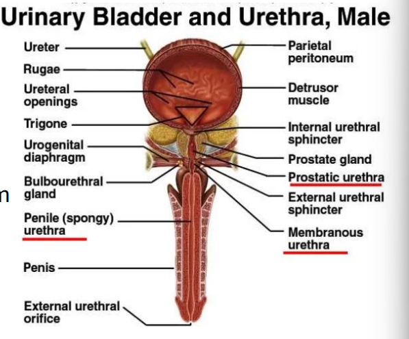
Prostatic → Membranous → Spongy/Penile
Prostatic urethra
Receives ejaculatory ducts
Membranous urethra
Shortest + least protected
Common injury site in pelvic trauma
Runs through urogenital diaphragm
Penile/Spongy urethra
Within corpus spongiosum
Opens at external urethral meatus
Urethra pic
Inguinal canal
Short, oblique passage in lower anterior abdominal wall (~4 cm)
Spermatic cord passes through
Extends deep inguinal ring → superficial inguinal ring
Potential site of inguinal hernia
Herniation: abnormal exit of tissue/organ through the wall of the cavity where it normally lies
Spermatic Cord
Spermatic Cord
Passes through inguinal canal
Contains:
Vas deferens
Testicular artery
Pampiniform plexus → heat exchange
Cremaster muscle
Lymphatics + nerves
Clinical: inguinal hernia + torsion of the cord
Spermatogenesis
Production of sperm in the seminiferous tubules
Spermatogonia → mature spermatozoa through:
Mitosis
Meiosis
Spermiogenesis

How semen exits
Semen Exit: Erection → Emission → Ejaculation
1. Erection
Sexual stimulation → parasympathetic reflex
Arterioles dilate → ↑ blood flow → veins compressed → blood trapped
Penile blood sinuses engorge → erection
Bladder sphincter closes
2. Emission
Sympathetic reflex
Fluids propelled through vas deferens → seminal vesicles → ejaculatory ducts → bulb of penis
Prostatic fluid enters urethra
3. Ejaculation
Skeletal muscle contractions → squeeze semen through urethra
Smooth muscle at bladder neck prevents reflux into bladder
Pathway of sperm
SEVEN-UP — Sperm Pathway
S → Seminiferous tubules
E → Epididymis
V → Vas deferens
E → Ejaculatory duct
N → Nothing
U → Urethra
P → Penis
Blood Supply & Innervation
Arterial supply
Testes: testicular artery ← abdominal aorta
Penis: internal pudendal artery → dorsal + deep penile arteries
Venous drainage
Testes → pampiniform plexus → testicular vein
Right → IVC
Left → left renal vein
Innervation
Pudendal nerve → sensory/motor to most perineal muscles
Autonomic: pelvic splanchnics + hypogastric plexus
Genitofemoral nerve → cremaster reflex
Clinical considerations and Male timeline
Clinical Considerations
Vasectomy: cut vas deferens → sterilisation; potentially reversible
Prostate enlargement (e.g. carcinoma, prostatitis) → difficulty urinating
Varicocele: dilated pampiniform veins
→ infertility risk
→ testicular atrophy + ↓ sperm production
Male Timeline
Infancy: Erections begin
11–14 yrs: Secondary sex characteristics appear
13–16 yrs: Sperm produced in adult amounts (puberty onwards)
Late teens: Peak sexual urges
Throughout life: If healthy → sex drive + ability to fertilise an ovum