Cytology
 Call Kai
 Learn
Learn Practice Test
 Spaced Repetition
 Match
Match Flashcards
 Knowt Play
1/92

There's no tags or description
Looks like no tags are added yet.
Name | Mastery | Learn | Test | Matching | Spaced | Call with Kai | Chat |
|---|
No analytics yet
Send a link to your students to track their progress
93 Terms
Describe TWO rules for blood sample collection
Fast animals for 6 - 12hr prior to prevent lipaemia
→ Artificial elevation of Hb, TP, AST and ALT
Avoid haemolysis of sample
→ Interferes with TP, lipase, bilirubin, urea, AST, ALT, ALP, phosphorus and potassium
Fill out the following table
Colour top | Anticoagulant | Mechanism | Use |
Red | |||
Purple | |||
Grey | |||
Blue | |||
Green |
Colour top | Anticoagulant | Mechanism | Use |
Red | None (plain tube) | Clotted blood is spun down to obtain SERUM |
|
Purple | EDTA | EDTA chelates calcium (binds Ca2+ necessary for secondary haemostasis) | Haematology #1 |
Grey | Potassium oxalate/sodium fluoride | Oxalate = Anticoagulant Fluoride = Prevents glucose utilisation | Glucose assessment |
Blue | Sodium citrate | Sodium citrate chelates calcium | Coagulation panel #1 |
Green | Lithium heparin | Inhibits thrombin formation |
|
Serum vs. plasma
Serum = Liquid part of blood that remains AFTER the blood has clotted (contains NO fibrinogen)
Plasma = Liquid part of blood that has NOT clotted (contains fibrinogen and other clotting factors) using an anticoagulant (eg. EDTA)
Fill out the following table
Colour | Cause |
Clear | |
Yellow | |
Milky white | |
Red |
Colour | Case |
Clear | NORMAL cat and dog |
Yellow |
|
Milky white | Lipaemia |
Red | Haemolysis |

Blood Smear Preparation
When to prepare
Method
6 Common errors
When: ASAP after blood collection (cells break down after removal from the body)
Ideally use fresh blood with no anticoagulant
EDTA most common, but may cause artefactual changes
Method:
Spreader slide = Polished/ground edge slide to allow distribution of blood cells
Errors:
Drop size too big or small
Not allowing drop of blood to spread along edge of spread slide before smearing
Not enough or too much pressure on spread slide
Spread too fast/slow
Removing spread slide too early
Ensure adequate angle of spread slide (too steep = short smear, too flat = long smear)
Cytology
Definition
7 Advantages
5 Disadvantages
4 Reasons to perform cytology in-clinic
Definition: Harvesting cells or fluid for microscopic examination of INDIVIDUAL cells
+ve:
Quick
Cheap
Samples collected without GA
Relatively non-invasive
Assessment of tissue AND fluid (cannot assess fluids with histology)
Some interpretation in-clinic
Superior for round cells
-ve:
No appreciation of lesion architecture or its margins (cannot stage a tumour)
Small sample that is not always representative (multiple samples required)
Difficult to distinguish between neoplasia or hyperplasia (cannot rule out malignancy 100%)
NOT ideal for mesenchymal lesions (difficult to distinguish fibroblasts in granulation tissue from neoplastic mesenchymal cells due to lack of architecture)
Results are as good as the person performing it (refer samples when in doubt)
In-Clinic:
Clinician must have basic skills to ensure specimen is adequate to submit to the lab
Allows for faster examination = faster prognosis = faster treatment (advantageous for patient)
Local extension of the clinical exam
Clinic revenue
Describe THREE methods of cytology collection
Equipment
Method
Indications
ONE: FNA
Equipment: 5 - 10mL syringe, 23 - 25G needle and slides
Methods:
FNI
Needle ONLY (no syringe) is placed into the mass and redirected serval times to obtain cells in the needle/hub
Indication: Delicate cells (eg. lymphocytes) or bloody tissue (eg. spleen)
FNA
Negative pressure technique which aims to dislodge cells and draw into the needle hub
Needle attached to syringe is inserted into the mass and negative pressure is created while redirecting several times
Indication: Firm or fibrous mass
Indications:
5cm SC mass
Fluid in abdomen
Enlarged liver
TWO: IMPRESSION SMEAR
Equipment: Slides ONLY
Method:
Cut mass and gently dab the cut surface or swab lesion dry to remove blood and exudate
Gently dab cut surface ono slide
Indication: Biopsies of solid mass or lesions at necropsy (eg. intestinal mass)

THREE: SCRAPING
Equipment: Scalpel and slides
Method:
Cut fresh surface of mass
Blot sample dry with paper towel or clean ulcerated surface with saline and dry to remove exudate
Scrape surface gently with scalpel at a 45˚ angle OR until blood oozes from the ulcer if on live animal
Slide edge of scalpel blade at 45˚ along the glass slide making a trunk
Use tip of scalpel blade to make stems ("trunk and stem pattern") which contain a single layer of cells for examination
Indications:
Mesenchymal tumour excisional biopsies
Shallow ulcerated lesions (difficult to insert needle for FNA) eg. SCC in cats
Lesions at necropsy

3 ways to smear a thin fluid sample
Smear like blood smear
Concentrate sample before smearing by centrifuging to remove supernatant (fluid)
Make fluid stop prep (suddenly stop blood smear to create concentrated line of cells)

5 Tips for preparing slides
ALWAYS prepare multiple samples as not all are likely representative of the lesion
Label frosted edge with pencil as stains will dissolve pen or marker
Only stain 1 or 2 smears for examination before sending rest (unstained) to lab
Store adequately
Do NOT store slides in fridge (water condensation destroys cells)
Dried smears do NOT require immediate staining
Place smears in plastic slide holders to send to lab
Keep slides away from formalin (separate packaging) as it destroys cells in cytological samples
ALWAYS provide history and be precise with the description of the lesion location (eg. thoracic lesion vs. SC thoracic lesion)
Describe the 5 steps of the diff quik staining technique
Ensure slide is air-dried
Fixative (methanol) = 6 dips and dab off excess (fixes cells to slide)
Pink = 6 dips and tap off excess onto paper towel to prevent dilution of purple and make it last longer
Purple/blue = 12 dips
Rinse off excess stain with water after and allow to dry before examining (can use hair drier to accelerate drying process)
5 Recommendations for diff quik staining
Ensure you dip as movement of dye over the cells encourages uptake
Can go back and re-dip into pink or purple stain if not enough on initial examination
Variable staining intensity is expected due to difference in cell thickness in different areas
ALWAYS replace lids to prevent evaporation of stains due to alcohol content
Separate stains for ear smears to prevent yeast and bacterial growth in stains
Position of condenser for:
Stained slide
Unstained slide (eg. urine sediment)
Stained slide: Condenser directly under stage
Unstained slide: Condenser as far away from stage as possible
Label the following slide artefacts
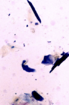 |  | 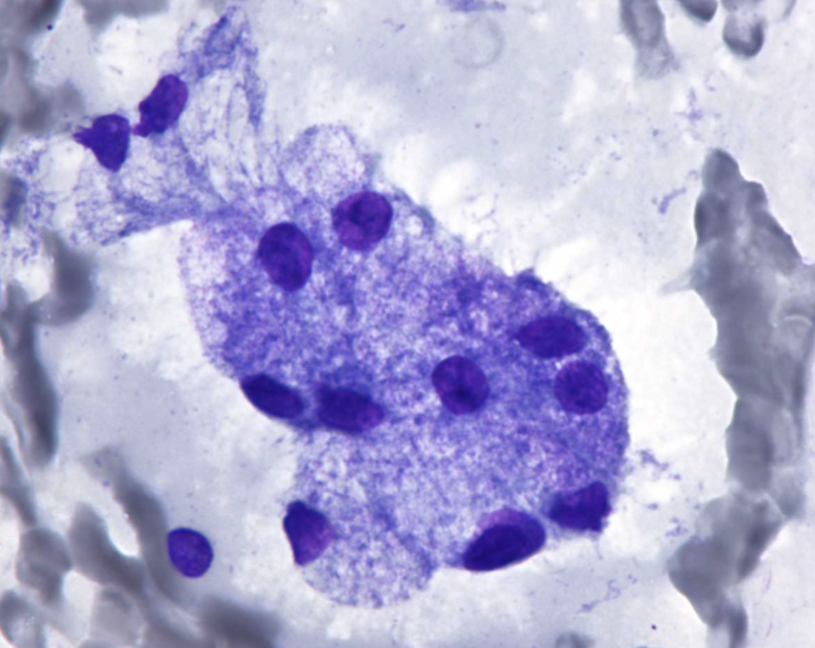 |
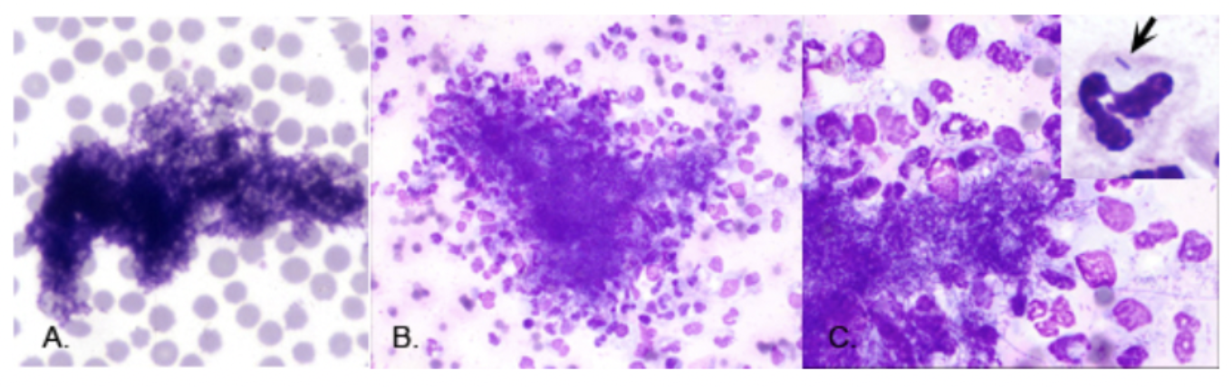 |  |  |
Squames = Keratinised epithelial cells from the skin of patient or fingerprints of operator
| Glove Powder (L) = Round/oval structures with which refractile and crystalline with a clear outer edge and a small dark dog in the centre
R = Cryptococcus = Larger purple centre that is more variable in size and NOT refractile | Salivary Gland Epithelial Cells = Collection of salivary glands when aspirating submandibular lymph nodes
|
Stain Precipitate (L) = Highly variable sized/shaped chunks of dark purple precipitate which is coarsely granulated
Bacteria (M and R) = Uniform in shape | U/S Gel = Magenta and granular mass that obscured cells | Muscle Fibres = Collection of muscle fibres when aspirating SC
|
What is this?
Appearance
Misinterpreted as?

Nuclear Debris
Appearance: Purple strands of nuclear material due to unavoidable damage of cells during preparation
Misinterpretation: Fungal hyphae
How to determine contamination vs. bacterial infection
Assess for inflammatory response to confirm infection
Organisms within cytoplasm of neutrophils or macrophages are unlikely to be contaminants
CSI-TeaM approach for examination of cytology samples
Are there useful cells present?
Is the staining adequate?
Inflammatory vs. non-inflammatory (neoplastic)?
What is the cell type present?
Is it malignant or benign?
ONE: ARE THERE USEFUL CELLS PRESENT?
Method
Ideal
4 Indications to resample
Method: Assess in 4x objective
Look a the feathered edges where cells tend to aggregate
Take note of cellular areas of interest to examine in higher power
Ideal: Areas of intact NON-HAEMATOGENOUS cells which are well-spread out into a single layer (should see cell definition)
Resample:
ONLY blood
No cells or too few to examine
Too thick (many cells, but not smeared adequately)
Ruptured cells during smearing ALL PRODUCE NON-DIAGNOSTIC RESULTS

TWO: IS THE STAINING ADEQUATE?
Method
Ideal
Method: Assess on low or medium power
Ideal:
Varying shades of pinks, purples and blues (not just all pink or all purple)
Clear differentiation between cytoplasm vs. nucleus vs. background

THREE: INFLAMMATORY OR NON-INFLAMMATORY?
Method
3 Steps after confirming inflammation
4 Types of inflammation
4 Causes of non-inflammatory sample
Method: High power 40x to assess cellular detail
Inflammation:
Describe (type of cells)
Identify organisms (± septic)
Treatment Do NOT continue with steps 4 and 5 (reserved for non-inflammatory lesions)
Types of Inflammation:
Neutrophilic >85% = Suppurative (purulent)
Septic = Suppurative inflammation with bacteria present (within degenerate neutrophils)
Mononuclear
Histiocytic = Macrophages (chronic inflammation)
Granulomatous = Epitheliod macrophages and multinucleated giant cells (mycobacteriosis, fungi, parasites, foreign body reaction)
Lymphocytic/plasmacytic (allergy, immune reaction, viral infection, chronic inflammation)
± Eosinophilic >10% (hypersensitivity, parasite migration)
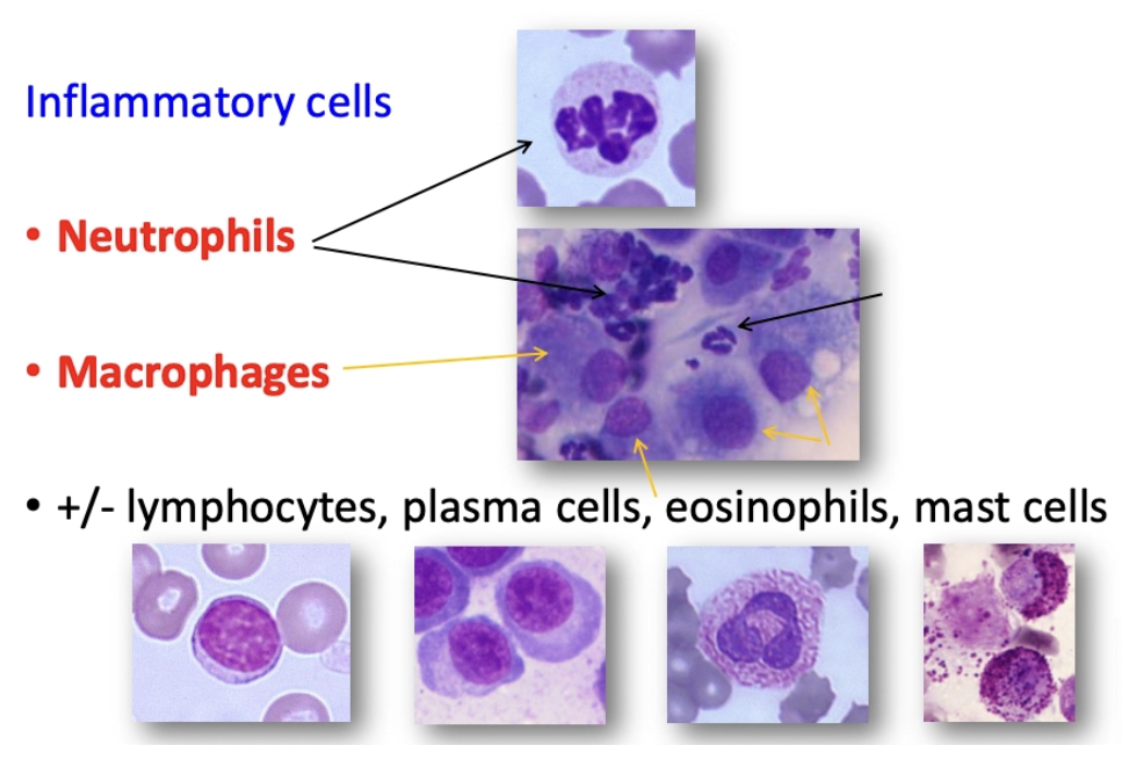
Non-Inflammatory:
Neoplasia
Hyperplasia
Cysts
Calcium deposits
What are these? What do they mean?

Naked Nuclei = Nucleus with no cytoplasm and appears larger than normal (as do their nucleoli) which may be interpreted as malignancy and should NEVER be interpreted
Does the presence of inflammatory cells exclude the possibility of underlying neoplastic lesion? Why?
No
Many neoplasms have inflammation due to secondary infection, response to necrosis within the neoplasm or paraneoplastic response
Blood contaminated cells will also have neutrophils NORMALLY
Degenerate neutrophils
Abnormal looking neutrophils with degraded nuclei ± pyknosis (small), karyorrhexis (fragmentation) or karyolysis (fading) which is most common in septic environments

FOUR: WHAT CELL TYPE IS PRESENT?
3 Cell types
How to differentiate
Types:
Epithelial
Mesenchymal
Round
Differentiate:
Size
Shape
Cell borders
Clustering/clumping
Cellularity of FNA
Name the Cell Type
Size
Clustering/clumping?
Cellularity of FNA
Morphological appearance
Cell borders
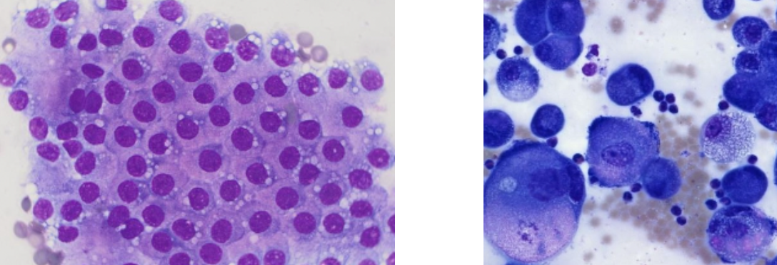
Epithelial Cells
Size: Large cells
Clustering: Usually cohesive and STICK together in sheets or clusters (can be single too)
Cellularity: Exfoliate easily so FNA or impression smears yield large numbers of cells for an epithelial lesion
Appearance: Oval to angular cells with round nuclei and ABUNDANT cytoplasm
Cell Borders: Distinct cell junctions between attached cells
Name the Cell Type
Size
Clustering/clumping?
Cellularity of FNA
Morphological appearance
Cell borders

Mesenchymal Cells
Size: Medium to large cells
Clustering: Usually individual cells
Cellularity: Exfoliate poorly so FNA contains very few cells → Scraping
Appearance: Spindle cells with cytoplasmic tails BUT may also appear plump
Cell Borders: Indistinct cell borders which fade into the background
Bound by bright pink ECM produced by the cells
Round cells
Size
Clustering/clumping?
Cellularity of FNA
Morphological appearance
Cell borders
Size: Small to medium cells (usually similar to RBCs)
Clustering: Usually single BUT may look like they are sticking
Cellularity: Exfoliate well on FNA
Appearance: Small, circular cells with round nuclei
Cell Borders: Distinct cytoplasmic borders
List the 5 (6) types of round cells
Lymphocytes
Plasma cells
Mast cells
Histiocytes
TVT
± Melanocytes
Name the Round Cell
Appearance
3 DDx

Lymphocyte
Appearance:
Small, round cell
Large nucleus which fills almost the entire cell
Small rim of basophilic cytoplasm
DDx:
Normal lymphoid tissue
Reactive lymphoid tissue
Lymphoma
Name the Round Cell
Appearance
2 DDx

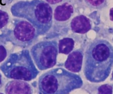
Plasma Cell
Appearance:
Eccentric nucleus (to one side)
Moderate amount of DARKLY-stained basophilic cytoplasm (darker than lymphocytes)
Often with pale area within cytoplasm = Golgi apparatus
DDx:
Plasma cell myeloma
Cutaneous plasmacytoma
Name the Round Cell
Appearance
DDx

Mast Cell
Appearance:
Central to eccentric, round nucleus
Abundant pale to clear cytoplasm
PURPLE GRANULES
DDx: MCT
Name the Round Cell
Appearance
2 DDx

Histiocyte
Appearance:
"Fried egg" appearance
Central to eccentric nucleus
ABUNDANT clear to PALE basophilic cytoplasm
Potentially with darker area of basophilia (opposite of plasma cell)
DDx:
Histiocytoma
Histiocytic sarcoma
Name the Round Cell
Appearance
Significance

Transmissible Venereal Tumour
Appearance: Histiocyte but vacuolated
Significance: NOT in NZ
Usually found around the perineal area
Name the Round Cell
Appearance
DDx

Melanocyte
Appearance:
Green/black granules in cytoplasm
Moderate amount of basophilic cytoplasm
DDx: Melanoma (care with amelanotic melanoma = no granules)
Label the Following Round Cells
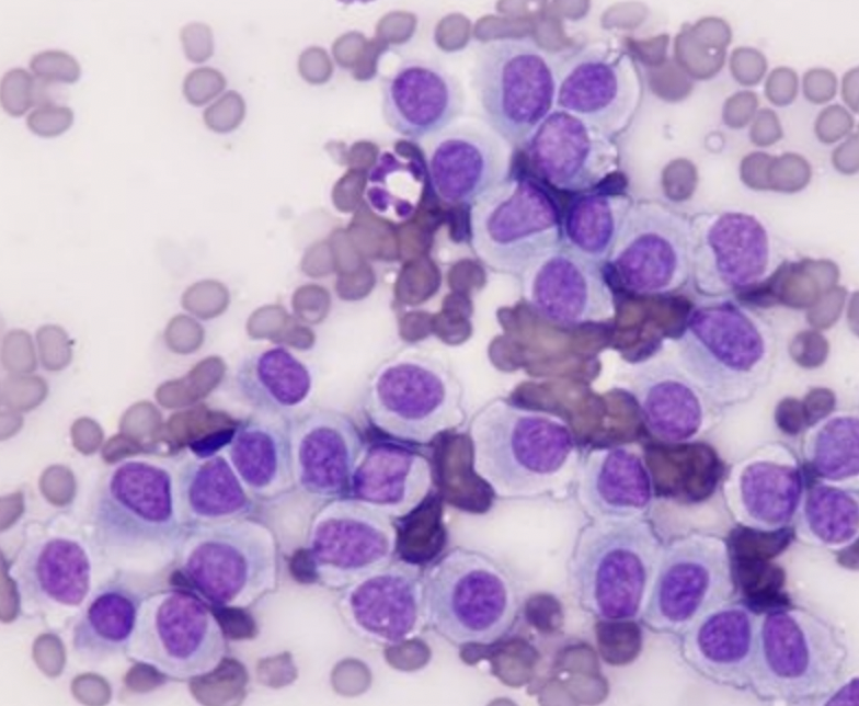 |  | 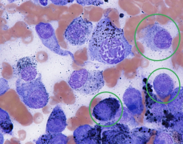 |
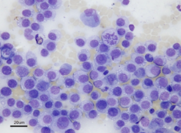 |  |  |
Histiocytes | Mast Cells | Melanocytes |
TVT | Lymphocytes | Plasma Cells |
FIVE: IS IT MALIGNANT OR BENIGN?
Criteria of malignancy
4 Whole cell features
7 Nuclear features
3 Exceptions
Criteria of Malignancy: ≥3 criteria (more variation → more malignant)
Whole Cell Features:
Pleomorphism = Variability of the same cell type (anisocytosis, variable shape and high nucleus:cytoplasmic ratio)
Disorganisation of cells within clusters
Hypercellularity of smears (more exfoliating cells)
Occasional very large cells
Nuclear Features
Macronuclei
Anisokaryosis
Multinucleation
Macronucleoli (same size as RBC)
Anisonucleoliosis (variation in nucleoli size and shape)
Increased mitotic figures
Abnormal mitotic figures
Exceptions:
Lymphoma (less variable than normal lymphoid tissue)
Thyroid carcinoma (uniform cells BUT malignant)
Apocrine gland adenocarcinoma (uniform cells but malignant)
Describe what is wrong with the following samples
 |  |  |
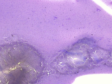 |  |  |
Formalin = RBCs become green and background is murky | Splattergram = Material is expelled but NOT spread Cells are too close for examination | Squished Cells = Rough handling → Naked nuclei with nuclear debris |
Slide Too Thick = Need to spread | Squished Cells = Rough handling → Naked nuclei with nuclear debris | Too Pink = Dip again into purple stain |
Statement for:
Are there adequate cells present?
2. Is the stain adequate?
Inflammatory vs. non-inflammatory (when it is non-inflammatory but there are neutrophils present)
Cells: Although there is blood contaminating the sample, there are sufficient intact cells which are spread out into a thin enough layer to obtain enough single cells to see cell definition
Staining: Yes. There is variation in colour and there is a clear difference between the nucleus, cytoplasm and background
Non-Inflammatory: Although there are neutrophils present, because they are the most abundant WBC in blood, and red cells are observed in the slide, the neutrophils likely they came from contamination of the sample with blood
List the only FOUR structures observed on an ear smear
Skin cells (squamous epithelial cells) ± melanin granules
Yeast (Malassezia spp.)
Bacteria (cocci and rods)
Neutrophils
Label the Following Image

White Arrow = Melanin pigment within keratocytes
Common in dark-skinned animals
Red Arrow = Bacteria (cocci)
Green Arrow = Degenerate neutrophils
Label the Following Image

Grey Arrow = Keratin bars/scrolls (rolled up keratinised epithelial cells)
Red Arrow = Bacteria
Label the Following Image
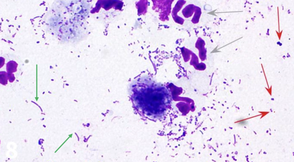
Grey Arrow = Neutrophils
Red Arrow = Cocci
Green Arrow = Rods
Label the Following Image

Yellow Arrow = Melanin granules
Red Arrows = Yeast
Large dark blue structures = Epithelial cells
How to differentiate yeast from bacteria on ear smear
Yeasts are MUCH larger than bacteria 40x = easily see yeast while bacterial presence is questionable
100x = BOTH (yeasts are obvious and bacteria more clearly visible)
Yeast = Footprints
Effusion
Definition
Normal
Definition: Accumulation of fluid in the body cavity produced by mesothelial cells of the visceral and parietal serosal lining
Normal: <1mL for lubrication
What is Starling's law?
Defines the retention of fluid in vessels which is determined by FPnet (net filtration pressure)
FPnet = HSP - COP (hydrostatic pressure - colloid osmotic pressure)
Describe the 5 steps of fluid sample collection
Clip site and surgically prep area for aseptic technique
FNA with 18 - 22G needle
Place fluid into an EDTA tube to prevent clot formation
Prepare fluid sample that is NOT from the EDTA tube (EDTA is bactericidal)
Make smears like a blood smear OR stop prep OR centrifuge to remove supernatant
4 Step of preliminary fluid analysis after sample collection
Assess gross appearance before and after centrifuge (colour and turbidity)
Cytology and cell counts to assess cellularity
TP with refractometer (centrifuge to measure protein of supernatant first)
± Culture and sensitivity
What are these? What is their significance?


Reactive Mesothelial Cells = Mesothelial cells lining the pleural, peritoneum and pericardium become reactive (produce more fluid), → Plump up and divide and hence often have:
TWO nuclei but are NOT neoplastic
Pink fringe/tutu
Difficult to differentiate from neoplastic mesothelial cells
Fill out the Following Table on Classifications of Effusions
Feature | Transudate | Modified Transudate | Exudates |
Colour and consistency | |||
TP | |||
Nucleated cell count (TNCC) | |||
Specific gravity | |||
Cells | |||
Mechanism | |||
Cause | |||
Clot |
Feature | Transudate | Modified Transudate | Exudates |
Colour and consistency | Clear and watery | Vague intermediate | Turbid (cloudy) and thick |
TP | Low (<25g/L) | Moderate (25 - 50 - 70g/L) | High (>30g/L) |
Nucleated cell count (TNCC) | Low (<0.5 x 10^9/L) | Moderate (0.5 - 7 x 10^9/L) | High (>3 x 10^9/L) |
Specific gravity | Low (<1.017) | Moderate (1.017 - 1.025) | High (>1.025) |
Cells | Normal cell (mainly mononuclear, occasional non-degenerate neutrophils ± mesothelial cells) | Reactive mesothelial cells, macrophages, non-degenerate neutrophils, lymphocytes | Septic: Large number of degenerate neutrophils ± bacteria |
Mechanism | Low COP or increased HP (non-inflammatory) | Increased HP or impaired lymphatic drainage | Increased vascular permeability |
Cause | Severe hypoalbuminaemia (liver failure) and early heart failure |
| Inflammatory ± septic (bacteria) |
Clot | No | Mixed | Yes (high fibrinogen) |
List 6 effusions which do not fit into the categories well
Haemorrhage (frank blood)
Chylous effusions
Ruptured abdominal organs (eg. uroperitoneum or bile peritonitis)
Equine abdominal paracentesis
Feline infectious peritonitis
Neoplastic effusion
Haemorrhagic Effusion
Appearance
2 alternative DDx and differentiation
Types of cells present >few hours old and greater than 2 days old
Appearance: Red or serosanguinous colour → Brown
DDx: ALSO due to contamination at sampling or accidental splenic aspiration (would have platelets and clumping)
Cells:
< Few hours = Macrophages with RBCs in cytoplasm
>2d = Macrophages with haemosiderin
Chylous Effusion
Appearance
MoA
Typical location
Cells present (4)
4 DDx
3 Diagnostic methods
Appearance: Milky white effusion (may be pink with blood)
MoA: Leakage of lymphatic vessels carrying fat
Location: Thorax #1
Cells:
Small lymphocytes
Macrophages (with lipid vacuoles)
Neutrophils
Fat droplets
DDx:
Heart disease in cats (cardiomegaly prevents emptying of thoracic ducts)
Neoplasia (eg. lymphoma or thymoma)
Lymphatic rupture
Lung torsion
Diagnosis:
Centrifuge sample (remains white if chylous)
Measure TAG content compared to serum (chylous TAG > 2 - 3x serum TAG)
Refrigerate sample overnight (fat rises to top and solidifies)

CASE 1: 1yr X-breed
History: Dog presented with pendulous abdomen. Abdominal radiology confirmed fluid in the abdomen

Question: CSI-TM approach and provide and interpretation/diagnosis
CSI-TM:
Useful cells present? Yes (lots of intact cells present, thinly enough spread to see cell definition and not just blood)
Adequate staining? Yes (can see cytoplasm vs. nucleus and background is clear)
Inflammatory vs. non-inflammatory? Inflammatory
Lots of neutrophils, some degenerate
Occasional intracellular bacteria (rod-shaped)
Interpretation: Septic, suppurative peritonitis
CASE 2: 6yr ME GSD
History: Presented for scooting and difficulty defaecating
PE: Mass on rectal palpation


Question: CSI-TM approach and provide and interpretation/diagnosis
CSI-TM:
Useful cells? Yes
Lots of intact cells that are thin enough to see cell definition
Adequate staining? Yes (can see difference between nucleus and background)
Inflammatory or non-inflammatory? Non-inflammatory (no neutrophils and macrophages)
Cell type? Epithelial (cell clustering and abundant cytoplasm)
Malignant or benign? Benign
Interpretation: Benign epithelial mass (perianal adenoma)
CASE 3: 14yr DSH cat
History: Multiple small (<1cm) skin masses over entire body
PE: Splenomegaly and hepatomegaly

Question: CSI-TM approach and provide and interpretation/diagnosis
CSI-TM:
Useful cells? Yes
Adequate staining? Yes
Inflammatory or non-inflammatory? Non-inflammatory
Neutrophils present due to blood contamination (RBCs present)
Cell type? Round cells (mast cells)
Interpretation: Mast cell tumour
CASE 4: 9yr DSH
History: Cat presented for coughing and lethargy
PE: Pleural fluid found
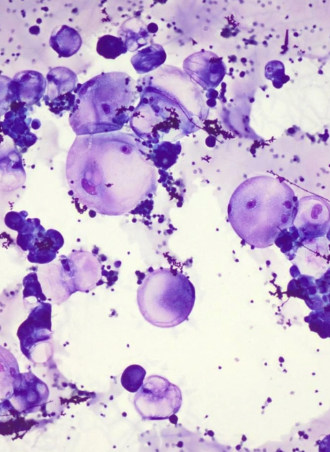
Question: CSI-TM approach and provide and interpretation/diagnosis
CSI-TM:
Useful cells? Yes
Adequate staining? Yes
Inflammatory or non-inflammatory? Non-inflammatory
Cell type? Epithelial cells (clustering)
Signet ring = Epithelial cells swell and have a chunk at the top
Malignant or benign? Malignant (highly variable, lots of balloon cells, anisocytosis, anisokaryosis = variable nuclear cells, signet rings)
Interpretation: Carcinoma
CASE 5: 9yr Rottweiler
History: Presented 3-legged lame (non-weight bearing) on right forelimb
PE: Extreme pain of right shoulder
Radiographs: Lysis of proximal humerus

Question: CSI-TM approach and provide and interpretation/diagnosis
CSI-TM:
Useful cells? Yes
Adequate staining? Yes
Inflammatory or non-inflammatory? Non-inflammatory
Cell type? Mesenchymal (tails, not clustering, fairly large)
Malignant or benign? Malignant (highly variable, lots of balloon cells, anisocytosis, macronucleoli, angular nucleoli)
Interpretation: Sarcoma (osteosarcoma)
CASE 6: 11yr cross-breed dog
History: Large abdominal wall mass
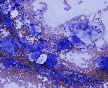
Question: CSI-TM approach and provide and interpretation/diagnosis
CSI-TM:
Useful cells? Yes
Adequate staining? Yes
Inflammatory or non-inflammatory? Both
Neutrophils and occasionally macrophages
Cell type? Epithelial (clumping)
Malignant or benign? Malignant (highly variable, balloon cells, anisocytosis and anisokaryosis)
Interpretation: Carcinoma with inflammation
CASE 7: Horse with colic
PE: Abdominal fluid aspirated and shown pre- and post-centrifugation
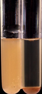


Question: CSI-TM approach and provide and interpretation/diagnosis
CSI-TM:
Useful cells? Yes
Grass in peritoneal cavity = Intestinal rupture
Adequate staining? Yes (not a lot of variation in colour, but only single cell type)
Inflammatory or non-inflammatory? Inflammatory
Neutrophils, some degenerate
Some intracellular bacteria = septic
Interpretation: Septic peritonitis
CASE 8: 11yr Cat
History: Lethargy and an extended abdomen
PE: Ascites


Question: CSI-TM approach
CSI-TM:
Useful cells? Yes
Adequate staining? Yes
Inflammatory or non-inflammatory? Non-inflammatory
Cell type? Epithelial (clumping)
LARGE compared to neutrophils (round cells would be similar size to leukocytes
Malignant or benign? Malignant
Anisokaryosis
Anisocytosis
Anisonucleoliosis (macronucleoli as big as RBCs)
CASE 9: 7m Burmese
History: Extreme lethargy
PE: Abdominal fluid
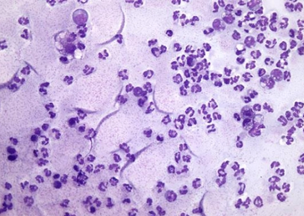
Question: CSI-TM approach and provide and interpretation/diagnosis
CSI-TM:
Useful cells? Yes
Adequate staining? Yes
Inflammatory or non-inflammatory? Inflammatory
Neutrophils with some macrophages
Organisms? None
High protein fluid = Purple background
Fingernail clippings = Protein folds
Interpretation: Mixed inflammation in abdomen (FIP)
CASE 10: 13yr MC Corgi
History: Skin mass on flank

Question: CSI-TM approach and provide and interpretation/diagnosis
CSI-TM:
Useful cells? Yes
Adequate staining? Yes
Inflammatory or non-inflammatory? Non-inflammatory
Cell type? Round cells = Plasma cells
Interpretation: Plasmacytoma
CASE 11: 8yr FS Labrador
History: Skin mass on HL
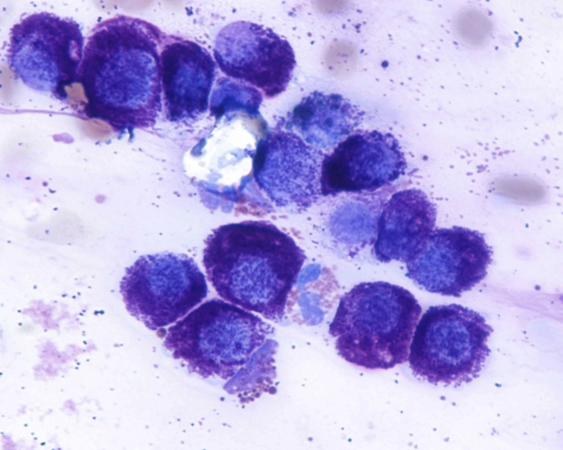
Question: CSI-TM approach and provide and interpretation/diagnosis
CSI-TM:
Useful cells? Yes
Adequate staining? Yes
Inflammatory or non-inflammatory? Non-inflammatory with single ruptured eosinophil (must be >10% to be eosinophilic inflammation)
Cell type? Round cell = Mast cell
Interpretation: Mast cell tumour
CASE 12: 4yr Rottweiler
History: Swollen stifle and dog was in a fight a few days ago

Question: CSI-TM approach and provide and interpretation/diagnosis
CSI-TM:
Useful cells? Yes
Adequate staining? Yes
Inflammatory or non-inflammatory? Inflammatory
Unknown if bacteria present (requires 100x)
Neutrophils in lines = Wind rowing (joints have highly proteinaceous background due to high viscosity)
Interpretation: Suppurative arthritis (immune-mediated OR septic)
CASE 13: 10yr Dalmatian
History: Skin mass in preputial area

Question: CSI-TM approach and provide and interpretation/diagnosis
CSI-TM:
Useful cells? Yes
Adequate staining? Yes
Inflammatory or non-inflammatory? Non-inflammatory
Cell type? Round cell = Melanocyte
Interpretation: Melanoma
CASE 14:
History: Skin mass on lateral wall of abdomen, near thigh

Question: CSI-TM approach and provide and interpretation/diagnosis
CSI-TM:
Useful cells? Yes
Adequate staining? Yes
Inflammatory or non-inflammatory? Non-inflammatory (neutrophils = blood contamination)
Cell type? Mesenchymal
Malignant or benign? Malignant (anisocytosis, anisokaryosis, anisonucleoliosis)
Interpretation: Sarcoma
CASE 15: Dog
History: Abdominal mass
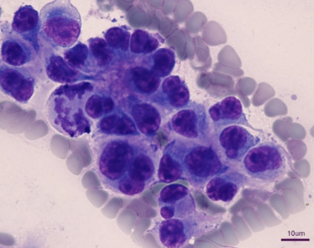
Question: CSI-TM approach and provide and interpretation/diagnosis
CSI-TM:
Useful cells? Yes
Adequate staining? Yes
Inflammatory or non-inflammatory? Non-inflammatory
Cell type? Epithelial
Malignant or benign? Malignant (anisocytosis, abnormal mitotic figure, anisokaryosis, anisonucleoliosis)
Interpretation: Carcinoma
CASE 16: Cat
History: Fluid in thorax
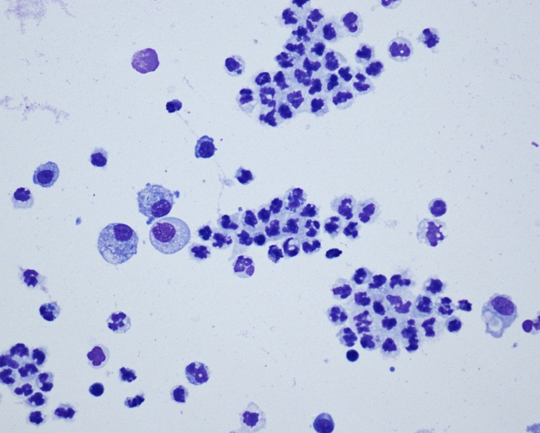

Question: CSI-TM approach and provide and interpretation/diagnosis
CSI-TM:
Useful cells? Yes
Adequate staining? Yes
Inflammatory or non-inflammatory? Inflammatory with intracellular rods
Interpretation: Septic pyothorax
CASE 17: 1.5yr Poodle
History: Mass on muzzle

Question: CSI-TM approach and provide and interpretation/diagnosis
CSI-TM:
Useful cells? Yes
Adequate staining? Yes
Inflammatory or non-inflammatory? Non-inflammatory
Cell type? Round cell = Histiocytes
Interpretation: Histiocytoma
CASE 18: Dog
History: Intra-abdominal mass
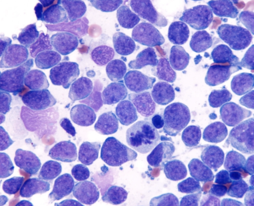
Question: CSI-TM approach and provide and interpretation/diagnosis
CSI-TM:
Useful cells? Yes
Adequate staining? Yes
Inflammatory or non-inflammatory? Non-inflammatory
Cell type? Round cell = Lymphocytes
Interpretation: Lymphoma
CASE 19: Cat
History: Fluid in thorax

Question: CSI-TM approach and provide and interpretation/diagnosis
CSI-TM:
Useful cells? Yes
Adequate staining? Yes
Inflammatory or non-inflammatory? Inflammatory with intracellular rods
Interpretation: Septic pyothorax
3 Indications of fresh/frozen samples for PM
Infectious/toxic causes
Bacterial culture
PCR
Toxicology
Direct vs. Indirect Testing
Definition
3 Examples of each
Direct Testing = Identifies presence of agent itself
Examples:
Bacterial or fungal culture → Promotes growth/replication of infectious agent for identifications
PCR → Extract, amplify and sequence DNA of infectious agent from sample
Serology Ag test → Add Ab to sample which binds with the Ag to form an Ag-Ab complex which can be visualised
Indirect Testing = Identifies animal's response to agent (i.e. Ab) vs. agent itself
Examples:
IFA
ELISA
MAT
Serology
Definition
MoA
4 Types
Qualitative vs. quantitative
MoA
Definition: Examining serum/other bodily fluids (milk, CSF) for presence of Ab/Ag of an infectious agent (occasionally of a hormone, enzyme like pancreatic lipase or auto-Ab)
MoA: Ag binds specific Ab → Ab-Ag complex formed → Indicators bind complex to visualise reaction and ID positive result
Indicators: Coloured indicator dyes, fluorescent markers or agglutinating particles (eg. latex)
Intensity of reaction is proportional to amount of Ab/Ag present in sample
Types:
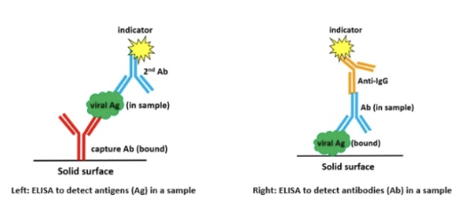
ELISA (Enzyme-linked immunosorbent assay) = Qualitative
Ag (plates/wells coated with Ab which Ag in sample binds to)
Ab (plates/wells coated with Ag for Ab in sample to bind to)
2nd Ab added after sample which is linked to a chromogenic indicator molecule → Sandwich
Positive result: Colour change
RIM (Rapid immuno-migration tests) = Qualitative
Type of ELISA test in cassette form (aka. SNAP or POC tests)
eg. FeLV Ag, parvovirus Ag and FIV Ab

Agglutination tests (LAT and MAT) = Qualitative (titres)
Use the tendency of Ag to clump together (agglutinate) in the presence of specific Ab in suspension
LAT = Latex particles bind Ag-Ab complexes when present → Detectable mat which floats to surface (eg. Toxoplasma gondii Ab)
MAT = Examine with dark-field microscopy for Ab-Ag complex formation and agglutination (eg. Leptospiral Ab)

IFA and TIA (Immunofluorescence assay and turbidimetric immunoassay) = Qualitative (titres)
eg. Failure of passive transfer in foals
Titres
Definition
2 Example tests that measure titres
Paired titres
Definition: Way to express concentration (quantity) of particular Ab in sample
Determined by serially diluting sample and testing each → Final titre = Highest dilution that still produces positive test reaction
Higher denominator for final titre → More Ab present in sample (eg. 1/512 is a stronger positive result than 1/32 → 1/512 more likely to indicate active of current infection)
Tests:
Agglutination tests (LAT and MAT)
IFA
Paired Titres: TWO samples taken 2 - 4 weeks apart to determine if recent/current infection is present (4-fold rise in Ab titres)

2 Disease vaccinations that influences SNAP results
Cats vaccinated against FIV → Positive Ab result
Dogs vaccinated against parvovirus in last 7 - 10 days → Weak positive Ag result
5 Causes of low titre (<1/800) for Leptospirosis
Recent infection (titre still rising)
Recent vaccination
Chronic infection (subclinical)
Convalescent titre (dropping after previous infection)
Maternal antibody in calves → Distinguish from recent infection with paired titres
Serum sample from with with 1/200 titre for Leptospira hardjo and 1/1600 titre for Leptospira pomona Which serovar is the likely cause of leptospirosis?
L. pomona is the likely cause of current infection and L. hardjo titre likely due to previous vaccination
PCR
5 Advantages
3 Disadvantages
4 Samples to collect
+ve:
Small sample volumes (often swab sufficient) necessary (vs. > 1 - 2mL of blood for serology)
High Sn and Sp
Infectious agent does NOT need to be alive (VS. bacterial/fungal culture)
Can detect early in disease course (do not have to wait for Ab to rise)
Cost effective screening for some diseases and performed on samples pooled from multiple animals
-ve:
May need multiple PCR tests for different infectious agents to make diagnosis → $$
Quality of sample, primers and protocol influences Sp and Sn
False positive possible (eg. tiny remnant of Toxoplasma in sheep sample → positive PCR despite NOT being cause of abortion)
Samples: Depends on disease being tested (check with lab first)
Sterile swabs
Fluids samples
MCF = EDTA vs. BVD = Serum
Faeces (GIT disease)
Stomach contents of aborted foetus
What type of virus is tested on FIP PCR?
FeCV (some tests distinguish between FeCV and FIPV, but the normal FeCV should not be present in effusion anyway)
Parvovirus Diagnosis
Test
3 Disadvantages
Test: SNAP RIM for Ag in faeces
-ve:
False positive due to recent vaccination with modified live vaccine in last 7 - 10 days
False negative due to transient shedding
False negative due to faecal Ab present binds the virus
FPT in Foals
2 Tests (+ sample)
When should blood be collected?
Tests: Horses do NOT have high levels of GGT in colostrum
IgG (Ab) TIA test at diagnostic lab = Serum
Quantitative and higher DSe
SNAP IgG (Ab) "foal side" test = Serum
Results within 7 minutes
Qualitative result (<400, 400 - 800 or >800mg/dL = Failure, partial failure and adequate passive transfer respectively)
Collection: 18 - 24hr post-birth
BVDV Testing
6 Tests (+ sample)
First BVD test to perform on a dairy herd + sample submitted
Results
Follow-up test
Tests:
Pooled Ab ELISA = Bulk milk tank/serum
Individual Ab ELISA = Individual milk/serum
Individual Ag ELISA = Serum or ear notch (ear notch only <35 days as maternal Ab from colostrum neutralise viral Ag in blood)
Individual Ag RIM/SNAP = Ear notch (2 - 3mm tissue)
Pooled PCR = Milk or serum
Individual PCR = Serum or ear notch
1st Test: Bulk milk tank ELISA Ab (or pooled serum) → Define BVD status of herd through S/P ratio
S/P > 0.75 = High number of seropositive cows indicates current or recent exposure to BVD due to presence of PI animal → Individual Ag ELISA for PI hunt
0.17 < S/P < 0.75 = Presence of transiently infected (TI) animals rather than PI animals → Assess bulk tank milk for BVD Ag to ID any unidentified PI animals in the milking herd for PI hunt
Cows infected with PI animal over fence line
Contact with PI animal that has since died/been culled
PI animal recently introduced and not yet had time to infect many other animals
PI present but extensive grazing results in low contact rates between animals
S/P < 0.17 = No PI animal

2 Test options for screening young stock for BVD for PI animals
Direct testing (serum or ear notch) for BVD virus with PCR or ELISA Ag +ve result = PI or TI → Retest 3 - 4 weeks later TWO +ve results → PI
Indirect testing (serum) for BVD Ab with ELISA ONLY recommended in unvaccinated animals with low chance of PI animal in the group as it relies on the fact that in herds with PI, many have high Ab titre
6 Reasons for a false negative test result
Ag tests in late stages of infection (eg. intermittent and reduced shedding as disease progresses)
Ab tests in early stages of infection (prior to effective seroconversion)
Immunocomplexing (endogenous Ab binds Ag which prevents it from being free to bind the test reagent Ab)
Wrong/bad sample tested (Ag or Ab in sample becomes degraded and will not react)
Test not performed correctly
Wrong test
2 Reasons for a false positive test result
Ab (and some Ag) tests with recent vaccination (many tests cannot distinguish between Ab produced in response to vaccination vs. infection)
PCR tests in general (very sensitive eg. using sample needle on multiple animals)
What steps would you recommend for a farm dog with suspected leptospirosis presenting acutely?
Given the acute presentation, I would try for a PCR on a urine sample first to detect Leptospirosis Ag. Ab are unlikely present in serum as there has not been sufficient time for seroconversion. PCR is not specific to a serovar but the disadvantage is false negatives due to intermittent shedding. If the test is negative, but I still highly suspect Leptospirosis, I would then do a serology for Ab with MAT. Because I must select a specific serovar, I would start with Hardjo and Pomona as this is a farm dog, and go from here (save owner cost). These tests must be repeated in 2 - 4 weeks to show a rising titre (>4-fold increase). As such, I would treat for leptospirosis while awaiting the 2 - 4 weeks to pass.
3 Potential causes for Leptospira copenhagenii titre of 1/50
Not infected (or recent vaccination)
Infected with different serovar
Infected with copenhagenii, but insufficient time for seroconversion → False negative