Scrotal anatomy and physiology
 Call Kai
 Learn
Learn Practice Test
 Spaced Repetition
 Match
Match Flashcards
 Knowt Play
1/38

There's no tags or description
Looks like no tags are added yet.
Name | Mastery | Learn | Test | Matching | Spaced | Call with Kai | Chat |
|---|
No analytics yet
Send a link to your students to track their progress
39 Terms
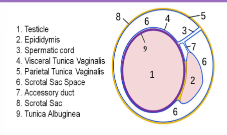
Testicle is surrounded by a dense fibrous capsule called:
Tunica albuginea
Septula arise from the tunica albuginea to form…
Mediastinum testis
Mediastinum testis
Creases an incomplete septum and provides support for testicular vessels
Each testicle contains approximately ______ seminiferous tubules that converge to form 20-30 larger ___________
840; Efferent ducts
The efferent ducts form channels called the RETE TESTIS that …
Carry the seminal fluid from the testicle to the epididymis
Average testical size
Length: 3-5cm
Width: 2-4cm
AP: 3cm
In pediatric pts, testicular volumes can be calculated using
Lambert formula = length x width x height x 0.71
Ellipsoid formula = length x width x height x 0.52
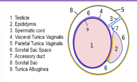
Tunica vaginalis
Extension of the peritoneum into the scrotal sac
Normally there is a small amount of fluid between layers
Visceral layer: Inner later covering the testes, epi and lower part of spermatic cord
Parietal layer: lines the walls of the scrotal pouch
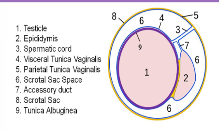
Tunica albuginea
Thin layer of tissue that is in direct contact with the testicular tissue
Folds of this layer form sections within the testicles called the mediastinum testis
The rete testis is a series of channels within mediastinum
Blood vessels and ductules enter/exit the testicle at the mediastinum
The visceral layer of the tunica vaginalis covers the
Tunica albuginea
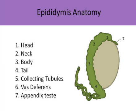
Epididymis
Curved lying posterior lateral to the testicle
Usually 6-7cm in length
Head (globus major) - formed by tubules from rete testis, 10-12cm width
Tubules converge into ductus epididymis which forms the body and tail (globus minor), 1-2mm width
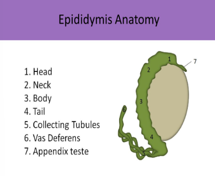
Appendix testis
Remnant of Mullerian duct
Small ovoid structure usually located between the superior pole and epididymal head
Spermatic cord
Holds the vas deferens, cremasteric, deferential and testicular arteries, pampiniform plexus of veins, lymph vessels, nerves
Scrotal wall
Skin - outermost layer
Dartos - muscle layer that forms scrotal raphe and separates the two testicles
Tunica vaginalis - Inner layer
Right and left gonadal arteries (testicular arteries)
Originate from anterior aorta to supply blood to the testicles
Normally low resistance, RI 0.5-0.75
Capsular artery courses along testicle periphery and produces branches called Centripetal arteries
Centripetal arteries
Course through the parenchyma to deliver O2 blood to tissues
These arteries are sampled with Doppler to assess testicular perfusion
Normally demonstrate low resistance, high flow volume
Deferential artery
Originates from the internal iliac artery
Feeds epi and vas deferens
High resistance, RI >0.75
Cremasteric artery
Originates from external iliac artery
Feeds peri-testicular tissues
High resistance, RI >0.75
Venous drainage
Pampiniform plexus is the network of small veins that merge to form the testicular vein
Right testicular vein drains directly into IVC
Left testicular vein drains into left renal vein
Testicles function
Exocrine = sperm production
Endocrine = Testosterone production
Epididymis function
Sperm maturation
Ejaculatory duct function
Sperm and seminal vesicle fluid
Urethra
Drainage channel for semen, sperM and urine
Seminal vesicle
Secretes alkaline component of semen and nutrients for sperM
Prostate gland
Secretes alkaline fluid into semen
Cowper gland
Secretes mucous for lubrication
Exam indications for testicular
Palpable mass (#1), pain, swelling, hydrocele, trauma, torsion, neoplasm
Use ____ for comparison views for echotexture and vascularity → keep setting same through exam for color
Dual screen
USA testicle
Homogenous, uniform, medium level echoes
Mediastinum testis seen as a linear echogenic band within testicle
Tunica albuginea not normally visualized
Doppler waveforms demonstrate antegrade, monophasic flow
Testicles normally have low vascular resistance and moderate antegrade diastolic flow
Epididymis USA
Normally isoechoic or mildly hyperechoic to testicle
Head is best evaluated in the long plane as a triangular or crescent shaped structure superior to the upper pole of the teste
Body and tail are usually located posterior and inferior to the head
Normal variants
Epididymal cysts (<4mm), epididymal calcs, minimal hydrocele, testicular cyst (2-3mm), testicular band, bilobed testicle, polyorchidism
What is the dense fibrous layer that is in direct contact with the testicle?
Tunica albuginea
What layer is an extension of the peritoneum
Tunica vaginalis
The mediastinum testes is formed by folds of the …
Tunica albuginea
Which of the following lies posterior lateral to the testicle?
Rete testis
Epididymis
Medial raphe
Dartos muscle
Epididymis
The ________ arteries encircle the testicle and the ____________ arteries penetrate the testicular parenchyma
Capsular; centripetal
The centripetal arteries normally demonstrate __________flow while the deferential arteries normally demonstrate _________
Low resistance; High resistance
The left testicular artery originated from the ____________ and the left testicular vein empties into __________
Aorta; Left renal vein
When evaluating a patient with testicular pain:
The US system settings should be established while scanning the UNAFFECTED side and remain unchanged when scanning the affected side