PROPAGATION OF INFORMATION
1/26
There's no tags or description
Looks like no tags are added yet.
Name | Mastery | Learn | Test | Matching | Spaced |
|---|
No study sessions yet.
27 Terms
What is myelin?
Myelin is a multi-layered, compacted membranous sheath that surrounds axons in the peripheral (PNS) and central nervous systems (CNS) to increase the conduction speed in the nervous system.
What is myelin composed of and how is it organised?
It is a multi-layered structure primarily composed of lipids and expresses unique proteins that facilitate its dense compaction.
Myelin is clustered into internodes which are found between the nodes of Ranvier gaps.
What are the cell types that give rise to myelin?
Schwann Cells:
PNS
provide myelination, with one cell typically myelinating a single axon (1:1 myelination).
Oligodendrocytes:
CNS
can myelinate multiple axons (50:1 myelination).
Identify five advantages that myelin confers to the neuron
Increased Conduction Velocity: Myelin enables saltatory conduction by changing the capacitance of the axon, allowing action potentials to jump from node to node, greatly increasing the speed of signal transmission.
Metabolic Efficiency: Myelination reduces the amount of energy required for nerve impulse conduction by minimizing the continuous depolarization along the axon, allowing for greater metabolic efficiency.
Allows for more compact nervous system
Compartmentalization of Axons: Periaxonal space is really important for transferring substrates from axon to oligodendrocyte
Transporters exist upon myelin and axon membranes, allowing for glucose and lactate to shuttle between the oligodendrocyte and the axon, giving the mitochondria in the axon energy in which to operate
Myelin can repair itself
What are neurotransmitters?
Neurotransmitters are chemical signals that are released from presynaptic nerve terminals into the synaptic cleft. This process facilitates communication between neurons.
What is the basic process for neurotransmitter release and neurotransmission?
Transmitter synthesized and stored (within vesicles within the nerve terminals
Action potential
Depolarisation and opening of VGCC
Ca2+ influx through channels
Ca2+ causes vesicles to fuse with presynaptic membrane
Transmitter is released into the synaptic cleft via exocytosis
Transmitter binds to receptors on postsynaptic membrane
Opening of postsynaptic channels
Post-synaptic current causes EPSP or IPSP changing membrane potential/excitability of the post-synaptic cell
Reuptake and/or breakdown of transmitter
What are the 6 criteria for neurotransmitters?
Synthesis: The chemical must be produced within a neuron.
Storage: The chemical must be found within a neuron
Release: When stimulated (depolarized) a neuron must release the chemical
Receptor: When the chemical is released it must act on a post-synaptic receptor and cause a biological effect
Inactivation: After the chemical is released there must be a mechanism for inactivation (uptake, degradation)
If chemical is applied on post-synaptic membrane, it should have the same effect as when released by a neuron.
What are the major types of neurotransmitter with an example of each?
Neurotransmitters can be categorized into two main types:
Classical (small molecule)
Amino Acids: Include Glutamate, GABA, and Glycine.
Monoamines: Include Noradrenaline, Dopamine, Adrenaline, and Serotonin (5-HT).
Acetylcholine (ACh): A major peripheral neurotransmitter.
Non-classical (large peptides)
Peptides: Include Substance P, Vasoactive intestinal peptide (VIP), Somatostatin, Neuropeptide Y (NPY), Enkephalin, and Kisspeptin.
Gases: Include Nitric oxide and Carbon monoxide.
Lipids: Anandamide is the primary example.
Describe the basic organisation of a nerve terminal
Nerve terminals contain multiple synaptic vesicles filled with neurotransmitters, and they also have mitochondria to meet the high energy demands necessary for neurotransmitter release.
What is the role of voltage gated calcium channels and what are the main types?
Voltage-gated calcium channels (VGCC) play a crucial role in neurotransmitter release enabling the influx of Ca2+ with membrane depolarization. The Ca2+ that enters binds to release proteins to cause vesicle fusion and the release of neurotransmitters into the synaptic cleft.
The main types are N-type (Cav2.2) and P/Q-type (Cav2.1) calcium channels
Explain quantal release of neurotransmitters
Transmitters are released in packets. The amount of neurotransmitter in 1 vesicle is called a quantum
Because miniature endplate potential (mEPP) is significantly less than threshold, normal neurotransmission results from the release of many vesicles simultaneously
How does the frequency of stimulation determine what type of neurotransmitter is released in co-transmitters?
Sometimes more than one neurotransmitter is released from a synaptic terminal.
Low frequency stimulation preferentially raises the Ca2+ concentration close to the membrane favouring the release of transmitter from small clear-core vesicles.
High frequency stimulation leads to more general increase in Ca2+ concentration causing the release of neuropeptides from large dense core vesicles as well as small molecule neurotransmitters.
What is the difference between co-release and co-transmission?
Co-release:
both neurotransmitters are packaged into the same set of synaptic vesicles. When the presynaptic terminal is depolarized it causes the release of both neurotransmitters.
Co-transmission:
requires transmitters to be packaged into distinct synaptic vesicles with differential release mediated by differential Ca2+ sensitivity. Alternatively, co-transmission can rely on spatial segregation of vesicle populations to different boutons to different synaptic targets.
What are the key types of synaptic proteins of a SNARE complex and how do they work?
Synaptobrevin, syntaxin, SNAP-25, synaptotagmin
The vesicle docks and synaptobrevin (found in vesicle membrane) binds to syntaxin and SNAP-25 (found on presynaptic plasma membrane), forming a snare complex
This causes the opening of the voltage gated calcium channels and calcium ions bind to the synaptotagmin (found on vesicle membrane) and catalyses membrane fusion for neurotransmitter release
Where are synaptic proteins found and what are their functions?
The surface of synaptic vesicles are densely covered in proteins, most of which act at one or more steps of the synaptic vesicle cycle and the function of some of these proteins are largely unknown.
Most of these proteins are important for vesicle recycling
What are the three models for vesicle recycling?
Kiss-and-Run Model: Vesicles temporarily fuse, after the neurotransmitter release, the fusion pore is closed and the vesicles are recovered.
Clathrin-Mediated Endocytosis: Collapse of vesicle into the membrane, then new vesicle formation elsewhere.
Ultrafast Endocytosis: Rapid internalization via exocytosis, vesicle delivery to endosome. Clathrin-mediated regeneration of synaptic vesicles occurs at the endosome.
What are the major elements of post-synaptic densities and identify their roles in synaptic transmission?
Membrane and cytoplasmic proteins clustered immediately opposite release sites or active zones at synapses
These proteins include:
Ligand gated ion channels
Anchoring proteins (scaffolding)
Cytoskeleton
Regulatory proteins
Known as density because appears dense in electron microscopy
What is the difference between the PSD core and the PSD pallium and what is typically found in each?
PSD core: more towards the surface of the postsynaptic cell, has high concentration of PSD-95
PSD pallium: deeper layer, containing a scaffold of Shank and Homer proteins
What is the primary function of most post synaptic density proteins?
Cell adhesion molecules (CAMs) that facilitate synaptic stability and signaling by linking receptors and scaffolding proteins.
What are the four post synaptic density proteins and identify their main function?
MAGUKs (membrane-associated guanylate kinases). The most abundant MAGUK at the mammalian post synaptic density is PSD-95
Scaffolding protein important for recruiting, trafficking, and stabilizing receptors like NMDA and AMPA receptors
GKAP (guanylate kinase-associated protein), also called PSD- 95- associated protein,
binds to Shank and PSD-95.
Shank
important scaffolding protein
Homer
Major function is to crosslink different proteins
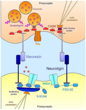
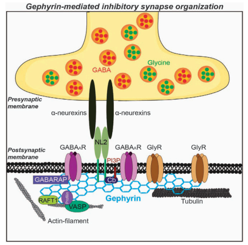
What are Neuroligins and Neurexins and what is their function?
Neuroligins are on postsynaptic cell membrane and are cell adhesion proteins that mediates the formation and maintenance of synapses between neurons
Neurexins are expressed on presynaptic membranes, bound to neuroligins to form that synapse and connect the presynaptic and postsynaptic structures.
What are the four types of neuroligins and which synapses are each expressed on?
Neuroligin 1 characteristically found in all glutamatergic synapses
Neuroligin 2 found preferentially in some inhibitory synapses (GABA)
Neuroligin 3 found in excitatory and inhibitory synapses
Neuroligin 4 found preferentially at glycinergic synapses in retina
What does PSD-95 bind to and with which domain?
PSD95 has 3 PDZ binding domains, as well as other domains
Neuroligins bind to PSD95 via 3rd PDZ domain
PSD95 binds AMPA glutamate receptors via 1st PDZ domain
Calcium-calmodulin protein kinase II (CaMKII)
Regulates NMDA and AMPA cycling
Neuronal nitric oxide synthase
Implicated in synaptic plasticity and neuroprotection
Shank proteins, indirectly via GKAP
Shanks can also bind directly to neuroligins
What is a key organising molecule that is specific for GABA and glycinergic inhibitory synapses?
Gephyrin
Anchors the receptors and important proteins to the post synaptic membrane
What are shanks?
Important scaffolding proteins that tether other immediate scaffolding proteins
Explain post-synaptic and pre-synaptic regulation via cell adhesion molecules (CAMs)
Post-synaptic specializations regulate pre-synaptic specializations via CAMs and vice versa
Bidirectional Communication:
Post-synaptic and pre-synaptic specializations influence each other through cell adhesion molecules (CAMs), which physically and functionally link the two sides of the synapse.
Explain the broad structure of the post-synaptic density
The postsynaptic density (PSD) is a dense cluster of proteins just beneath the post-synaptic membrane.
It contains dozens of molecules crucial for signal transduction and synaptic plasticity, including:
Glutamate receptors:
Ionotropic (e.g., NMDA-R)
Metabotropic (mGluR)
Receptor tyrosine kinases (RTKs): Involved in broader cell signalling.
Intracellular signalling molecules, especially CaMKII (a key enzyme in memory and synaptic strength).
These components are anchored and organized by scaffolding proteins, which help position receptors next to their associated signalling molecules for efficient function.