Psych 207 - Module 2: The Brain, Structure and Function
1/18
There's no tags or description
Looks like no tags are added yet.
Name | Mastery | Learn | Test | Matching | Spaced |
|---|
No study sessions yet.
19 Terms
Phylogenetic Division
Organizes the brain structures in terms of the order in which they are thought to have evolved
Major structures in the brain
Forebrain → main focus because this is where most cognitive processes happen
Midbrain → responsible for lower level non-cog functions (mainly)
Hindbrain → responsible for lower level non-cog functions (mainly)
Subcortical structures
Thalamus: act as a switch for sensory info also involved in memory
Hypothalamus: regulates basic bio functions, including hunger, thirst, temp, sex arousal, and basic emo reactions
Hippocampus: critical structure for learning, memory, and emotion
Amygdala: involved in memory, emotion, and aggression
Cortical regions
Cerebral cortex
Corpus collosum and anterior commissure connect the right and left hemispheres
Major Functions of the Four Lobes of the Cerebral Cortex
Parietal Lobes
Spatial processing and attention
Homunculus
Occipital Lobes
Processing visual info
Visual stimuli and complex aspects of the stimuli involved in recognizing what objects are
Temporal lobes
Process auditory info
Encoding and retrieval of info from long term memory
Frontal Lobes
Motor cortex: Directs fine motor movement
Premotor cortex: planning such movements
Prefrontal cortex: executive functioning
Phrenology
Johan Spurzheim
Phrenology was a now-discredited theory from the 1800s that claimed you could understand a person's personality, intelligence, and mental abilities by feeling the bumps and shapes on their skull.
Issue came from idea that cog processes operate in a vacuum → not correct they actually interact in a sophisticated way
Size doesn't correspond with power
Double Dissociations
Where brain damage and behavior are completely dissociated from each other and show opposite mirror image patterns
Brain damage to area X - Impaired by cognition A but not B - Brain damage to area Y - Impaired by cog B but not A
Eg. Lesion in Broca's area (X) impairs speech production (A), but not comprehension (B), while lesion to Wernicke's area (Y) impairs comprehension (B) but not speech production (A).
Wilder Penfield
in epileptic research, he created maps of the sensory-motor cortex
Brain Imaging Techniques
Static imaging: look at structure of brain
Dynamic: look at function of working brain
CAT Scan - Computerized Axial Tomography
Computerized Axial Tomography (Static)
Beams of x-rays are passed through the head form many angles
Differing types of tissues deflect light differently allowing for visualization
MRI - Magnetic Resonance Imaging
Static
Magnetic properties of brain produce electromagnetic signal that scanner detects. These electromagnetic signals allow visualization
Typically preferred over CAT because no radiation, more detailed, other powerful functions (can also measure functional aspects of brain
The person lies inside a big tube surrounded by a powerful magnet.
The magnet lines up hydrogen atoms in the body (mainly found in water).
Radio waves are sent through the body, which disturbs those atoms.
When the radio waves stop, the atoms return to normal and release energy.
That energy is used to create a detailed image of the brain (or other body parts).
Dynamic Brain Imaging
When neurons fire in the brain they produce electrical activity. Placing metal electrodes on scalp, we study electrical activity
Measuring time and location of activity allows inferences to be made about how the brain is responding to stimuli
Event-Related Potentials (ERP) is an example
Also PET, and fMRI --> study metabolism or blood flow in brain
fMRI
Dynamic
Structural properties of the brain are measured by taking advantage of different mag properties of tissues in brain
Oxygenated and de-ox blood have diff mag properties fMRI measure inflow and outflow of ox blood in brain by measuring mag properties of blood
BOLD Function
Blood oxygenation level dependent function
Regions of brain responsible for task consume oxygen in blood
Relatively slow influx of blood to region after onset of cog task - fMRI picks up this function
Researchers look around brain to find regions of brain that show BOLD that is time-locked to cog task→ related to cog task
Hard to tell what part of the brain is responsible for what aspect of the task
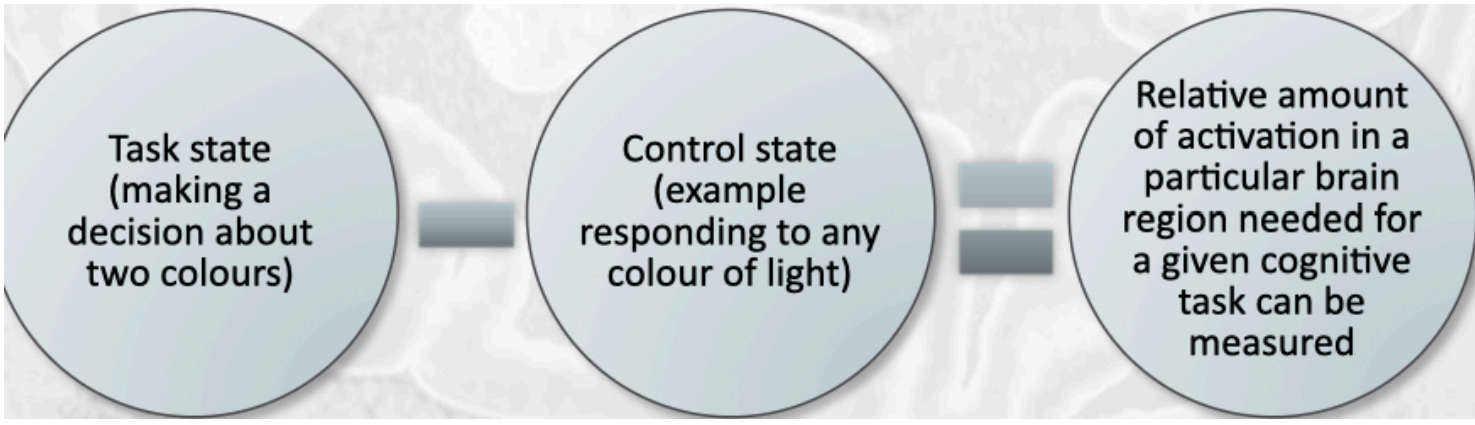
Donder's Subtractive Logic
In order to measure the time for a process to occur → need to measure and compare two reaction times or tasks (one with same component as the other and the process of interest)
This logic is also applied to the analysis of functional neural imaging data
Allows for isolation of regions of brain contributing to a given cog process
Choice RT – Simple RT = Time to make a decision
EEG – Electroencephalography
What it does: Detects electrical activity in the brain using electrodes placed on the scalp.
Used for:
Detecting different states of consciousness (awake, drowsy, asleep, coma).
Studying brain waves (alpha, beta, delta, etc.).
Pros:
Fast (great temporal resolution).
Non-invasive and safe.
Cons:
Poor spatial resolution (can’t pinpoint exact locations in the brain).
MEG – Magnetoencephalography
What it does: Measures magnetic fields created by brain activity (neurons firing).
Used for:
Similar to EEG but gives a more precise location of where activity is happening in the brain.
Pros:
Better spatial resolution than EEG.
Still has great temporal resolution.
Cons:
More expensive and less available than EEG.
ERP – Event-Related Potential
What it does: A type of EEG that measures brain response to a specific event or stimulus.
Used for:
Seeing how the brain responds before and after something happens (e.g., seeing an image, hearing a sound).
Setup:
Electrodes on scalp + repeated presentation of stimuli.
Pros:
Good for studying attention, perception, and decision-making in real-time.
Cons:
Still limited spatial accuracy.
PET – Positron Emission Tomography
Dynamic? but slow
What it does: Injects a radioactive tracer (A radioactive tracer is injected — often a form of glucose (since the brain uses glucose for energy)) that travels through the blood; detects which brain areas use the most energy.
Used for:
Showing which brain areas are active during specific tasks.
Used in research and medical diagnosis (e.g., Alzheimer’s, tumors).
Pros:
Provides a visual map of active brain areas.
Cons:
Invasive (involves radioactive injection).
Slow compared to EEG/MEG (poor temporal resolution).